Thoroughly analyzing TS Inter 2nd Year Zoology Model Papers and TS Inter 2nd Year Zoology Question Paper May 2017 helps students identify their strengths and weaknesses.
TS Inter 2nd Year Zoology Question Paper May 2017
Time: 3 Hours
Maximum Marks: 60
Instructions:
Note : Read the following instructions carefully.
- Answer all questions of Section ‘A’. Answer any six questions in Section ‘B’ and answer any two questions out of three in Section ‘C’.
- In Section A’ questions from Sr. Nos. 1 to 10 are of Very Short Answer Type. Each question carries two marks. Every answer may be limited to 5 lines. Answer all these questions at one place in the same order.
- In Section ‘B’, questions from Sr. Nos. 11 to 18 are of Short Answer Type. Each question carries four marks. Every answer may be limited to 20 lines.
- In Section ‘C’, questions from Sr. Nos. 19 to 21 are of Long Answer Type. Each question carries eight marks. Every answer may be limited to 60 lines.
- Draw labelled diagrams wherever necessary in Section ‘B’ and ‘C’
Section – A (10 × 2 = 20)
Note : Answer all the questions in 5 lines each:
Question 1.
What is Chyme ?
Answer:
Semi fluid mass of partly digested acidic food formed in the stomach is called chyme.
Question 2.
Define,glomerular filtration.
Answer:
The process of filtration of blood, which occur between glomerulus and lumen of the Bowman’s capsule due to difference in net pressure is called glomerular filtration. The filtered fluid which entered the Bowman’s capsule is primary urine or glomerular filtrate which is hypotonic.
Question 3.
Distinguish between the blind spot and the yellow spot.
Answer:
Blind Spot: The region of the retina where the optic nerve exists the eyeball and devoid of rods and cones is called blind spot.
Yellow spot: The centre of the posterior portion of the retina is called yellow spot.
![]()
Question 4.
What is corpus callosum ?
Answer:
Two cerebral hemispheres are internally connected by a transverse, wide and flat bundle of myelinated fibres beneath the cortex is called corpus callosum.
Question 5.
Distinguish between diabetes insipidus and diabetes mellitus.
Answer:
Diabetes insipidus : Deficiency of Vasopressin causes a disease called diabetes insipidus. It does not involve loss of sugar in urine.
Diabetes mellitus : Under secretion of insulin by the pancreatic gland increases the level of glucose in blood is called hyperglycemia. Prolonged hyperglycemia leads to a disease called diabetes mellitus, associated with loss of glucose through urine and formation of ketone bodies.
Question 6.
“Colostrum is very much essential for the new bora infants”. Justify.
Answer:
The colostrum secreted by the mother during the initial days of lactation has abdundant IgA antibodies to protect infant from initial sources of infection.
Question 7.
Mention the names of any four connecting links that you have studied.
Answer:
Connecting links clearly explain the path of evolution.
- Peripatas between annelida and arthropoda.
- Prototherians between reptilia and mamnalia.
Question 8.
Define atavism with an example.
Answer:
Sudden appearance of some vestigial organs in a better developed condition is called atavism. Eg: Tailed human baby.
Question 9.
Mention any four fish by-products.
Answer:
- Shark and cod liver oils
- Fish guano
- Shagreen
- Jsing lass.
Question 10.
List any four features of cancer cells.
Answer:
- Loss of contact inhibition
- Reduced intra cellular adhesion
- Immortalization
- Loss of anchorage dependence
Section – B (6 × 4 = 24)
Note : Answer any six questions in 20 lines each:
Question 11.
Draw a neat labelled diagram of L.S. of a tooth.
Answer:
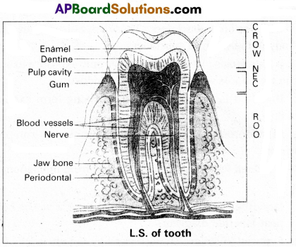
![]()
Question 12.
Describe disorders of respiratory system.
Answer:
Disorders of respiratory system.
1) Asthma : Asthma is a difficulty in breathing caused due to inflammation of bronchi and bronchioles. Symptoms include coughing, difficulty in breathing and wheezing.
2) Emphysema : It is a chronic disorder in which alveolar walls are damaged and their walls coalesce due to which respiratory surface area of exchange of gases is decreased. One of the major causes of this is smoking of tobacco.
3) Bronchitis: Bronchitis is the inflammation of the bronchi, resulting in the swelling of mucus lining of bronchi, increased mucus production and decrease in the diameter of bronchi. Symptoms include chronic cough with thick sputum.
4) Pneumonia : The infection of lungs caused by Strep-tococcus pneumoniae and also by certain Virus, Fungi, Protozoans and Mycoplasmas. Symptoms include inflammation of lungs, accumulation of mucus in alveoli and impaired exchange of gases, leading to death if untreated.
Occupational dissorders : These are caused by exposure of the body to the harmful substances.
E.g. : i) Asbestosis : It occurs due to chronic exposure to asbestos dust in the people working in asbestos industry.
ii) Silicosis : It occurs because of long term exposure to silica dust.
iii) Siderosis : It occurs due to deposition of iron particles in tissues.
iv) Black lung disease : It develops from inhalation of coal dust.
Question 13.
Describe the structure of synovial joint with the help of a neat labelled diagram.
Answer:
Synovial joints are characterised by the presence of a fluid filled synovial cavity between the articulating surfaces of the two bones.
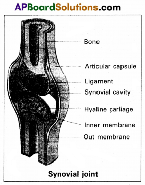
Structure of synovial joint: Synovial joint is covered by a double layered synovial capsule. The outer layer consist of dense fibrous irregular connective tissue with more collagen fibers. This layer is continuous with the periosteum and resists stretchihg and prevents the dislocation of joints. Some fibres of these membranes are arranged in bundles called ligaments.
The inner layer of synovial capsule is formed of areolar tissue and elastic fibers. It secretes a viscous synovial fluid which contains hyaluronic acid, phagocytes etc., and acts as a lubricant for the free movement of the joints.
Question 14.
Give an account of the secretions of pituitary gland.
Answer:
The pituitary gland is also called hypophysis. Anatomically pituitary gland is divided Into anterior and posterior pituitary.
I. Anterior Pituitary : It produces six important peptides. They are ;
- Growth hormone (GH) Somatotropin : They promote growth of the entire body by increasing protein synthesis, cell division and cell differentiation.
- Prolactin: It causes enlargement of the mammary glands of the breast and initiate the maintenance of lactation in mammals. Prolactin also promote the growth of corpus luteum and stimulate the production of progesterone.
- Thyroid stimulating hormone (TSH) : It stimulates the production of thyroid hormones from thyroid gland.
- Adreno corticotropic hormone (ACTH) : Controls the production of steroid hormones called glucocorticoids, by the adrenal cortex.
- Follicle stimulating hormone (FSH) : It stimulates growth the development of the ovarian follicles in females. In males FSH along with the androgens, regulates spermatogenesis.
- Luteinizing hormone (LH) : In males it stimulates production of androgens. In females it stimulates the ovaries to produce estrogens and progesterone and it maintains corpus luteum.
II. Posterior pituitary : It stores and releases two hormones called oxytocin and vasopressin.
Oxytocin : In females it stimulates contraction of pregnant uterus during child birth and ejection of milk from the mammary gland.
Vasopressin (ADH) : Affects the kidney and stimulates reabsorption of water and electrolytes by the DCT and collecting duct.
Question 15.
Write a short note on B-cells.
Answer:
The lymphocytes capable of producing antibodies and can capture circulating antigens are called B-cells. They are produced from the stem cells in the bone marrow, liver of foetus and bursa offabricius in birds. Mature B-cells express or display Ig M and Ig D antibodies on their membrane surfaces. As these antibodies can take antigens, the mature B-cells are also called immunocompetent B-cells. In secondary lymphoid organs these immunocompetent B-cells develop into functional immune cells which later differentiate into long lived memory cells and effector plasma cells. The plasma cells produce antibodies specific to the antigen to which they are exposed. Memory cells store information about the specific antigens and show quick response, when the same type of antigen invades the body later.
Question 16.
A colour-blind man married a woman who is the daughter of a colour-blind father and mother homozygous normal vision. Whattis the probability of their daughter being a colour-blind ?
Answer:
A colourblind man married a woman, who is daughter of a colourblind father and mother homozygous normal vision that means the woman is carrier i.e., the genotype is X+X–.
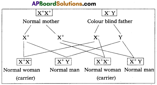
Here all women (daughters) are carriers. i.e., X+X–
A cross between colour blind man a woman from the above result
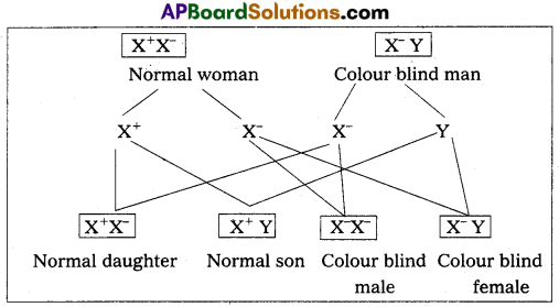
From the above cross the probability of their daughter being colour blind is 50% or 1/2 among the daughters or 1/4 among their child’s.
![]()
Question 17.
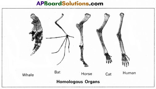
Distinguish between homologous and analogous organs.
Answer:
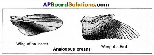
1. Homologous organs : The organs which have similar structure and origin but not necessarily the same function are called homologous organs. Eg: The appendages of vertebrates such as the flippers of whale, wings of bat, forelimbs of horse, paw of cat and hands of man have a common pattern in the arrangement of bones even though their external form and functions may vary to suit their mode of life.
2. Analogous organs : The organs which have dissimilar structure and origin but perform the same function are called the analogous organs. Eg : Wings of butterfly and wings of a bird.
Question 18.
Discuss briefly about the different waves and intervals in an E.C.G.
Answer:
ECG (electrocardiography) is commonly used, non-invasive procedure fro recording electrical changes in the heart.
The graphic record which is called an electrocardiogram, shows the series of waves that occur during each cardiac cycle.
The normal ECG consists of
(i) Waves
(ii) Intervals
(iii) Segments
(iv) Complexes.
i) Waves :
- The waves in a normal record are named P, Q, R, S and T in that order.
- A typical ECG tracing of a normal heartbeat consists of (I) a ‘P’ wave (II) a ‘QRS complex of waves’ (III) a T Wave.
- P wave : It represents the atrial systole and shows that the impulse is passing through atria. The duration of P. Wave is 0.1 sec.
- QRS complex of wave : It represents ventricular systole. Q wave is small negative., R-wave is tall positive and S wave is a negative wave. Its duration is 0.08 to 0.1 sec.
- T wave : It represents the ventricular repolarizction. It is a positive wave, its duration is 0.2 sec.
ii) Intervals :
P – R intervals : P – R intervals is the interval between the onset of p wave and the onset of Q wave. P – R interval is normally. 0.12 – 02 sec.
Q – T intervals : The interval between the onset of Q wave and the end of the T-wave. It represents the electrical activity in muscle of the ventricles. It lasts for about 0.4 seconds.
R – R intervals : It signifies the duration of one cardiac cycle and lasts for about 0.8 sec
Segments : S – T segment is the time period between the end of the ‘S’ wave and the onset of the T-wave. It is an isoelectric or zero voltage period.
Section – C (2 × 8 = 16)
Note : Answer any two questions in 60 lines each:
Question 19.
Describe the structure of the heart of a man with the help of a neat labelled diagram.
Answer:
Human heart is a hallow muscular, cone shaped, and pulsating organ situated between lungs. It is about the size of a closed fist.
The heart is covered by double walled pericardium, which consists of outer fibrous pericardium and inner serous pericardium. The serous pericardium is double layered, outer parietal layer and inner visceral layer. These two layers are separated by pericardial space, which is filled with pericardial fluid. This fluid reduces friction between the two membranes and allow free movement of the heart.
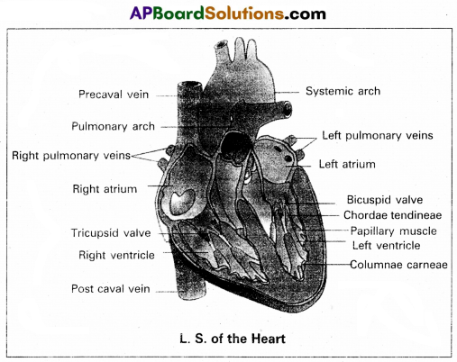
Human heart has four chambers with two smaller upper chambers called atria and two larger lower chambers called ventricles. Atria and ventricles are separated by a deep transverse groove called coronary sulcus.
i) Atria :
- Atria are thin walled receiving chambers. The right one is larger than the left.
- The two atria are separated by thin inter-atrial septum. It has a small pore known as Foramen Ovale’ in fetal stage.
Later it is closed and appears as depression (oval patch) known as ‘Fossa ovale’. If, the foramen ovale does not close properly it is called a patent foramen ovale. - The right atrium receives deoxygenated blood from different parts of the body, through three caval veins like two precaval veins and one post caval vein.
- The right atrium also receives blood from wall of the heart. through coronary sinus, whose opening into the right atrium is guarded by the valve of Thebesius.
- Opening of the post caval vein is guarded by the Eusta-chian valve. It is functional in fetal stage and directs the blood from post caval vein into left atrium through foramen ovale. But it is non-functional in adult.
- The openings of the precaval veins into the right atrium have no valves.
- Left atrium receives oxygenated blood from lungs through a pair of pulmonary veins, which open into the left atrium through a common pore.
- Atrio-ventricular septum separates atria and ventricles. It has right and left atrio-venticular apertures.
- Tricuspid valve guards the right atrio-ventricular aperture. Bicuspid valve guards the left atrio-ventricular aperture.
ii) Ventricles:
These are the thick walled blood pumping chambers, separated by an interventricular septum. The wall of the left ventricle is thicker than that of the right ventricle as . the left ventricle must force the blood to all the parts of the body.
The inner surface of the ventricles is raised into muscular ridges called columnae cameae. Some of them are large and conical and known as papillary muscles. Collagenous cords are known as chordae tendinae are present between atrio-ventricular valves and papillary muscles. They prevent the cusps of valves from bulging too far into atria during ventricular systole.
Nodal tissue : A specialized cardiac musculature called the nodal tissue is also distributed in the heart.
1) Sino-artrial node (SAN) – Present in the right upper comer of right atrium.
2) Atrio-ventricular node (AVN) – Present in the lower left corner of right atrium.
iii) Aortic arches ; Human heart has two aortic arches.
1) Pulmonary arch : Arises from the.left anterior angle of the right ventricle. It carries deoxygenated blood to lungs. It’s opening from right ventricle is guarded by pulmonary valve made with 3 semiluminar valves.
2) Left systemic arch : Arises from the left ventricle to distribute oxygenated blood to various parts in the body. Its opening is also guarded by aortic valve made with a set of 3 semilunar valves.
A fibrous strand, known as ligamentum arteriosm is present at the point of contact of the systemic and pulmonary arches. It is the remnant of the ductus arteriosus, which connects the systemic and pulmonary arches in the embryonic stage.
![]()
Question 20.
Describe female reproductive system of a woman with the help of a labelled diagram.
Answer:
The female reproductive system consists of a pair of ovaries long with a pair of oviducts, uterus, vagina and the external etalia located in the pelvic region. These parts of the system long with a pair of mammary glands are integrated structurally functionally to support the processes of ovulation, lization, pregnancy, birth and child care.
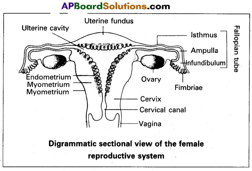
1) Ovaries : Ovaries are the primary female sex organs that produce the female gametes (ova) and several steroid hormones (ovarian hormones). A pair of ovaries is located on each side of lower abdomen. The double layered fold of peritoneum connecting the ovary with the wall of abdominal cavity is known as the meso ovarium.
The Ovaries are covered by a layer of ‘germinal (ovarian) epithelium. Underneath this layer, there is a dense connective tissue capsule called,’tunica albuginea’. The ovarian stroma is distinctly divided into an outer cortex and an inner medulla. The cortex appears more dense and granular due to numerous ovarian follicles. The medulla is a loose connective tissue with abundant blood vessels, lymphatic vessels and nerve fibres.
2) Fallopian tubes (oviducts): Each fallopian tube extends from the periphery of each ovary to the uterus and it bears a funnel shaped infundibulum, with finger like projections called ‘fimbriae’, which help in collection of ovum after ‘ovulation’. The infundibulum leads to a wider part of the oviduct called ‘ampulla’. The last part of the oviduct, ’isthmus’ has a narrow lumen and it joins the uterus. Fallopian tube is the site of fertilization. It conducts the ovum or zygote towards the uterus by peristalsis. The fallopian tube is attached to the abdominal wall by a peritoneal fold called ‘meso salpinx’.
3) Uterus: The uterus is a single and it is also called womb. It is a large, muscular, highly vascular and inverted pear-shaped structure present in the pelvis between the bladder and rectum. The lower narrow part through which the uterus opens into the vagina is called ‘cervix’. The cavity of the cervix is called ‘cervical canal’ which along with the vagina forms the ‘birth canal’.
The wall of the uterus has three layers of tissue. The external thin membranous ‘perimetrium’, the middle ‘ thick layer of ‘myometrium’ and inner glandulas lining layer called ‘endometrium’. The endometrium undergoes cyclic changes during menstrual cycle while myometrium exhibits strong contractions during parturition.
4) Vagina : The vagina is a large, median, fibromuscular tube that extends from the cervix to the vestibule (the space between labia minora). It is lined by non-keratinised stratified squamous epithelium. It is highly vascular and opens into the vestibule by the vaginal orifice.
5) Vulva: Vulva or pudendum refers to the external genitals of the female. The vestibule has two apertures – the upper external urethral orifice of the urethra and the lower vaginal orifice of vagina. Vaginal orifice is covered by a mucous membrane ’hymen’, vestibule is bound by two pairs of fleshly folds of tissue called inner ‘labia minora’ and outer larger ‘labia majora’. Clitoris is a sensitive, erectile structure, that lies at the upper junction of the two labia minora above the urethral opening. There is a cushion of fatty tissue covered by skin and pubic hair present above the labia major, called mons pubis.
Accessory reproductive glands of female : These include;
a) Bartholin’s glands: These are two glands located slightly posterior and to the left and right of the opening of the vagina. They secrete mucus to lubricate the vagina and are homologous to the bulbourethral glands of the male reproductive system.
b) Skene’s glands : These are located on the anterior wall of vagina, around the lower end of the urethra. They secrete a lubricating fluid when stimulated. The skene’s glands are homologous to the prostate gland of the male reproductive system.
c) Mammary glands : These are paired structures that contain glandular tissue and fat. The alveoli cells present in the mammary lobes of each glandular tissue secrete milk, which is stored in cavities of alveoli. The alveoli open into mammary tubes and then to mammary ducts, from there to mammary ampulla and finally connected to lactiferous duct through which milk is sucked out by the baby.
![]()
Question 21.
Describe chromosomal theory of sex-determination.
Answer:
Chromosomal sex determination : The chromosomes, which determine the somatic characters of an individual are known as autosomes. These chromosomes do not differ in morphology and number in male and female sex. Those chromosomes, which differ in morphology and number in male and female sex and contain genes responsible for the determination of sex are known as allosomes or sex chromosomes. There are two types of sex chromo somal mechanisms :
a) Heterogametic male and
b) Heterogametic female
a) Heterogametic male : In this mechanism, the female sex has two ‘X’ chromosomes, while the male sex has only a single X chromosome.
The heterogametic male may be of the following two types :
i) XX – XO
ii) XX – XY
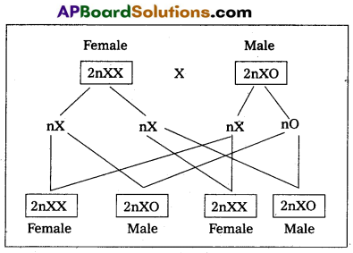
i) XX – XO type : In certain insects belonging to orders Hemiptera (true bugs), Orthoptera (grass hoppers) and Dictyo- ptera (cockroaches) female has two ‘X” chromosomes (XX) and are, thus homogametic, while male has only single ‘X” chromosome (XO). The male being heterogametic sex produces two types of sperms, half with X chromosome and half without X chromosome in equal proportions. The sex of the offspring depends upon the sperm that fertilises the egg, each of which carries a single X chromosome. Thus fertilisation between male and female gametes always produced zygotes with one ‘X’ chromosome from the female, but only 50% of the zygotes have an additional X Chromosome from the male. In this way, ‘XO’ and ‘XX’ types would be formed in equal proportions, the former being males and the latter being females.
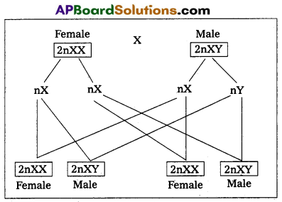
ii) XX – XY type : In man, other mammals, certain insects including Drosphila, the females possess two X chromosomes (XX) and are thus homogametic, produce one kind of eggs, each one with one X chromosome. While the males possess one X and one Y chromosome (XY) and are hence, heterogametic. They produce two kinds of sperms, half with X chromosome and half with Y chromosome. The sex of embryo depends on the kind of sperm. An egg fertilised by a X bearing sperm, produces a female, but if fertilised,by a Y bearing sperm, a male is produced.
b) Heterogametic female: In this method of sex determina-tion, the male produces similar type of gametes, while female produce dissimilar gametes. The heterogametic females may be of following two types.
(i) ZO – ZZ
(ii) ZW – ZZ.
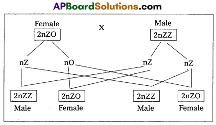
i) ZO – ZZ : This mechanism is found in certain moths and butterflies. In this case, female possesses one single ‘Z’ chromosome and hence is heterogametic, producing two kinds of eggs half with Z chromosome and another half without any Z chromosome. Male possesses two Z chromosomes and thus homogametic, producing single type of sperms, each carries single Z chromosome. The sex of the offspring depends on the kind of egg.
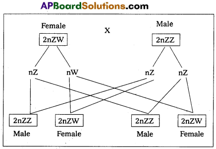
ii) ZW – ZZ : This system is found in certain insects (gypsy moth) and vertebrates such as fishes, reptiles and birds. In this system, the female is heterogametic and produces two types of gametes, one with ‘Z’ chromosome and the other with ‘W’ chromosome. On the other hand, male is homogametic and produces all sperms of same type carrying one Z’ chromosome. The sex of the offspring depends on the kind of egg being fertilised. The ‘Z’ chromosome bearing eggs produce males, but the ‘W chromosome bearing eggs produces females.