Thoroughly analyzing TS Inter 2nd Year Zoology Model Papers and TS Inter 2nd Year Zoology Question Paper March 2017 helps students identify their strengths and weaknesses.
TS Inter 2nd Year Zoology Question Paper March 2017
Time: 3 Hours
Maximum Marks: 60
Instructions:
Note : Read the following instructions carefully.
- Answer all questions of Section – A. Answer any six questions in Section – B and answer any two questions in Section – C.
- In Section – A, questions from Sr. Nos. 1 to 10 are of “Very Short Answer Type”. Each question carries two marks. Every answer may be limited to 5 lines. Answer all these questions at one place in the same order.
- In Section – B, questions from Sr. Nos. 11 to 18 are of “Short Answer Type”. Each question carries four marks. Every answer may be limited to 20 lines.
- In Section – C, questions from Sr. Nos. 19 to 21 are of “Long Answer Type”. Each question carries eight marks. Every answer may be limited to 60 lines.
- Draw labelled diagrams wherever necessary for questions in Sections – B and C.
Section – A (10 × 2 = 20)
Note : Answer all the questions in 5 lines each:
Quetsion 1.
What is chyme ?
Answer:
Semi fluid mass of partly digested acidic food formed in the stomach is called chyme.
Quetsion 2.
What are columns of Bertini ?
Answer:
Columns of Bertin are the medullary extensions of the renal cortex in between the renal pyramids.
Quetsion 3.
What is triad system ?
Answer:
In a skeletal muscle each transverse tubule (T-Tubule) is flanked on either side by several cistemae of the sarcoplasmic reticulum. T-tubule and the two terminal cisternae at its sides form the triad system.
![]()
Quetsion 4.
Why is the sympathetic division called thoracolumbar division ?
Answer:
The preganglionic sympathetic neurons have their cell bodies in the grey matter of thoracic and lumber regions of the spinal cord. So, sympathetic division is called thoraco-lumbar division.
Quetsion 5.
What is acromegaly ? Name the hormone responsible for this disorder.
Answer:
Acromegaly is a hormonal disorder that results when the pituitary gland produces excess growth hormone (GH). This disease is characterised by enlargement of the bones of the jaw, hand and feet, thickended nose, lips, eyelids and wide finger tips and gorilla like appearance of the person affected.
Quetsion 6.
“Colostrum is very much essential for the new bom infants.” Justify.
Answer:
The colostrum secreted by the mother during the initial days of lactation has abdundant.
Quetsion 7.
List out two Indian Carps and two Exotic Carps.
Answer:
Indian carps :
- Catla catla (catla)
- Cirrhinus mrigala (mrigal)
Exotic carps :
- Grass carp
- Silver carp
Quetsion 8.
What is apiculture ?
Answer:
Apiculture is the maintenance of hives of honeybees for the production of honey and wax.
Apiculture is an age-old cottage industry.
Quetsion 9.
What are panspermia ?
Answer:
According to Cosomozoic theory, life might have existed all over the universe in the form of resistant spores called panspermia. They might have reach the earth accidentally.
Quetsion 10.
Define Biogenetic law.
Answer:
It was proposed by Ernst Haeckel. It states that ontogeny repeats phylogeny which means the development history of an organism repeats the evolutionary history of its ancestor Eg : Tadpole larva of frog, it resembles fish both externally and internally. It possesses a tail, gills and two chambered heart like ‘ that of fish. Later it metamorphoses into adult frog.
![]()
Section – B (6 × 4 = 24)
Note : Answer any six questions in 20 lines each:
Quetsion 11.
What are the major transport mechanisms for CO2 ? Explain.
Answer:
Carbondioxide is transported in three ways.
1. In dissolved state : 7% of CO2 is transported in dissolved . state through plasma.
CO2 + H2O → H2CO3.
2. As Carbamino compounds : About 20-25% of CO2 combine directly with free amino group of haemoglobin and forms Carbamino haemoglobin in a reversible manner.
Hb – NH2 + CO2 → Hb – NHCOO– + H+.
pCO2 and pO2 could affect the binding of CO2 to haemoglobin.
- when pCO2 is high and pO2 is low as in the tissues, binding of more CO2 occurs.
- when pCO2 is low and pO2 is high as in the alveoli, dissociation of CO2 carbamino – haemoglobin takes place, (i.e., CO2 which is bound to haemoglobin from the tissues is delivered at the alveoli)
3. As Bicarbonates : About 70% of CO2 is transported as bicarbonate. RBCs contain a very high concentration of the enzyme, carbonic anhydrase and a minute quantity of the same is present in plasma too. This enzyme facilitates the following
reaction in both the directions.
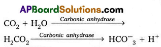
At the tissues where partial pressure of CO2 is high due to catabolism, CO2 diffuses into the blood and forms carbonic acid which dissociates into HCO–O3 + H+
At the alveolar site where pCO2 is low, the reaction proceeds in the opposite direction leading to the formation of CO2 and water. Thus CO2 is mostly trapped as bicarbonate at the tissues and transported to the alveoli where it is dissociated out as CO2.
Every 100 ml of deoxygenated blood delivers approximately 4 mZ of CO2 to the alveolar air.
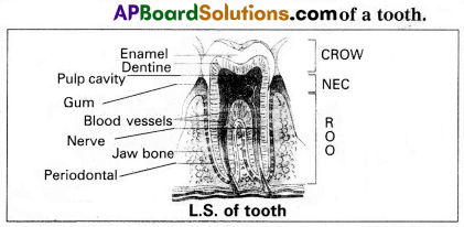
Quetsion 12.
Draw a neat labelled diagram of L.S. of a tooth.
Answer:
Quetsion 13.
Give an account of synaptic transmission.
Answer:
A nerve impulse is transmitted from one neuron to another through junction called synapses.
There are two types of synapses.
1) Electrical synapses
2) Chemical synapses.
Electrical synapses : These synapses are electrically conductive links between two neurons and are also called “gap junctions”. Impulses transmission across an electrical synapses is always faster than that across a chemical synapses.
Chemical synapses : Chemicals called neuro transmitters are involved in the transmission of impulses at those synapses. When an impulse arrives at the axon terminal, it depolarizes the membrane opening voltage gated calcium channels. Calcium ions stimulate the release of neurotransmitters in the cleft by exocytosis. The released neurotransmitters bind to their specific receptors, present on the post synaptic membrane.
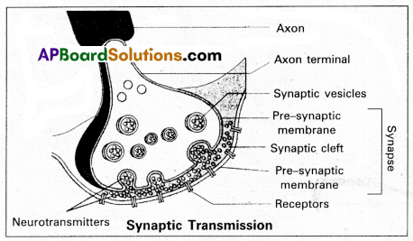
The post synaptic membrane has ligand gated channels. They are ion channels which respond to chemical signals, rather than to changes in the membrane potential. The entry of ions can generate a new potential in the post synaptic neuron. The new potential developed may be either excitatory (or) inhibitory.
Excitatory post synaptic potentials cause depolarisation, where as inhibitory post synaptic potentials cause hyper polarisation of post synaptic membrane.
Quetsion 14.
Write a note on Addison’s disease and Cushing’s syndrome.
Answer:
Addison’s disease : It is caused due to hyposecretion of glucocorticoids by the adrenal cortex. This disease is characterised by loss of weight, muscle weakness, fatigue and reduced blood pressure. Sometimes darkening of the skin in both exposed and non-exposed parts of the body occurs in this disorder.
Cushing’s Syndrome : It results due to over production of glucocorticoids. This condition is characterised by breakdown of muscle proteins and redistribution of body fat resulting in spindly arms and legs, a round moon-face, buffalo hump on the back and pendulous abdomen is also observed. Wound healing is poor. The elevated level of cortisols causes hyperglycemia, over deposition of glycogen in liver and rapid gain of weight.
![]()
Quetsion 15.
Describe the various types of barriers of innate immunity.
Answer:
Innate immunity is a non-specific type of defence mechanism which provides the first line of defence mechanism against infections. This is executed by providing different types of barriers like;
a) Physiological: Skin and mucus membranes are the main physical barriers. Skin prevents the entry of micro-organism, whereas the mucus membranes help in trapping the microbes entering our body.
b) Phyloigical barriers : Secretions of the body like HCZ in the stomach, saliva in the mouth, tears from the eyes are the main physiological barriers against microbes.
c) Cellular barriers : Certain types of cells like polymor-phonuclear leucocytes, monocytes, and natural killer cells in the blood as well as macrophages in the tissues are the main cellular barriers. They phagocytose and destroy the microbes.
d) Cytokine barriers: The cytokines secreted by the immune cells like interleukins and interferons are involved in differentiation of cells of immune system and protect the non-infected cells from further infection.
Quetsion 16.
Describe erythroblastosis foetalis.
Answer:
Erythroblastosis foetalis develops in an Rh positive foetus, whose father is Rh positive and mother is Rh negative. In Rh positive person rhesus antigens are present on the surface of blood cells where as in Rh negative person Rhesus antigens are absent.
During the process of delivery, the foetal blood cells may pass through the ruptured placenta into the Rh negative maternal blood. The mother’s immune system recognises the Rh antigens and gets sensitized and produces Rh antibodies. These antibodies are Ig G type they can pass through placenta. Generally first child is not effected because child is delivered by the time of the mother gets sensitized and produce antibodies.
During second pregnancy, if the second child is Rh positive, these antibodies cross the placenta, enter the foetal blood circulation and destroy the Rh positive blood cell of foetus (haemolysis), leads to haemolytic anemia and jaundice. To compensate the haemolysis of blood cells there is a rapid production of RBC’s from the bone marrow, and but also from liver and spleen. Now many large and immature blood cells in erythroblast stage are released into circulation. Because of this disease is called erythroblastosis foetalis.
Quetsion 17.
Explain Darwin’s theory of natural selection with industrial melanism as an experimental proof.
Answer:
Darwin’s theory of natural selection does not explain what exactly evolution is, but explains how evolution might have occurred in nature. A classical example for natural selection is industrial melanism, exhibited by peppered moth-Biston betularia. These moths were available in two colours grey and black. Grey moths were abundant before industrial revolution in all over England. The reason for the existence of large number of grey moths during that period was camouflage on the trunks of trees. But after the establishment of industries in England, black coloured moths were more and grey forms were less. This is due to pollution from industries in the form of soot turned barks of trees into black. So grey moths were easily identified and were more predated by birds. Thus grey moths decreased in number, black moths increased in the population.
Thus natural selection favoured the melanic moths (black) to reproduce more successfully. Natural selection of darker forms in response to industrial pollution is known as industrial melanism.
Quetsion 18.
Explain the different types of Cancers.
Answer:
Based on the origin Cancers are classified into :
1) Carcinomas : These are malignant tumor of epithelial cells. They are originating from the epithelial tissues of skin, lining of the respiratory, digestive, urinary and general systems or cells from various glands breast and nervous tissue etc. 85% of Cancers are Carcinomas.
2) Sarcomas : These are malignant tumors of connective tissues or organs that originate from mesoderm. About 2% of tumors are Sarcomas.
3) Leukemias : These are malignant tumors of stem cells of hematopoietic tissues, resulting in unrestrained production of WBC. They are liquid tumors. About 4% of Cancers are Leukemias.
4) Lymphomas : These are malignant tumors of secondary lymphoid organs like spleen, and lymphnodes. About 4% of Cancers are Lymphomas.
Section – C (6 × 4 = 24)
Note : Answer any six questions in 20 lines each:
Quetsion 19.
Describe the male reproductive system of human. Draw a labelled diagram.
Answer:
The male reproductive system or male genital system consists of a number of sex organs that are a part of the human reproductive process. The sex organs which are located in the pelvic region include a pair of testes, accessory ducts, glands and external genitalia.
1) Testes : The testes are a pair of oval pinkish male sex organs suspended in abdominal cavity within a pouch called scrotum. The scrotum helps in maintaining the low temperature of the testes (2 – 2.5°C) necessary for spermatogenesis. The cavity of scrotal sac is connected to the abdominal cavity through the ‘inguinal canal’. Testes is held in position in the scrotum of the ‘gubernaculum’, a fibrous cord that connects the testis with the bottom of scrotum and a ‘spermatic cord’, formed by the vas deferens, nerves, blood vessels and other tissues that run from abdomen down to each testicle, through inguinal canal. Each testis is enclosed in a fibrous envelope, ‘tunica albuginea’, which extends inwards into testis and divide it into lobules. Each lobule contains 1 to 3 highly coiled seminiferous tubules. A pouch of serous membrane ‘tunica vaginalis’ covers the testis.
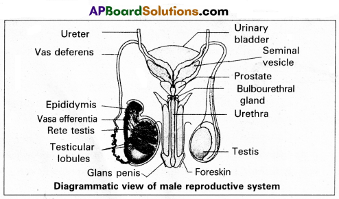
Miniferous tubules : Each seminiferous tubule is lined by ‘germinal epithelium’ which consists of undifferentiated male gum cells called ‘spermatogonial mother cells’ and it also bears nourishing cells’ called ‘sertoli cells’.
- Spermatogonial cells (or) primary spermatocytes undergo meiotic division, producing spermatozoa or sperms by a process spermatogenesis.
- Sertoli cells provide nutrition to spermatozoa and produce a hormone ‘inhibin’, which inhibits secretion of FSH.
The region outside the tubules, contain interstitial cells of ‘Leydig cells’. They produce androgens, the most important in testosterone. It controls the development of secondary sexual characters and spermatogenesis. The seminiferous tubules open into vasa effemtia through the rete testis. Rete testis is a network of tubules is of the testis carrying spermatozoa from the seminiferous tubules to the vasa efferentia.
2) Epididymis : The vasa efferntia leave the testis and open into a narrow, tightly coiled tube called ‘epididymis’ located along the posterior surface of each testis. The epididymis provides a storage space for sperms and gives them time to nature.
It is differentiated into three regions.
a) Caput epididymis
b) Corpus epididymis
c) Cauda epididymis
The caput epididymis receives spermatozoa via the vasa efferntia of the mediastinum testis. It is mass of a connective tissue at the back of the testis that encloses the rete testis.
3) Vasa deferentia : The vas deferens or ductus deferens is a long, narrow mascular tube. The mucosa of the ductus deferens consists of a pseudo stratified columnar epithelium and lamina propia. It starts from the tail of epididymis, passes through the inguinal canal into the abdomen and loops over the urinary bladder. It receives a duct from seminal vesicle.
The vas deferens and the duct of the seminal vesicle units to form a’short ejaculatory duct’ or ‘ductus ejaculatorius’. The two ducts, carrying spermatozoa and the fluid secreted by the seminal vesicles, converge in the centre of prostate and open into urethra, which transports the sperms to outside.
4) Urethra : In male, Urethra is the shared terminal duct of the reproductive and urinary systems. The urethra originates from urinary bladder and extends through the penis to its external opening called ’urethral meatus’. The urethra provides an exit for urine as well as semen during ejaculation.
5) Penis : Urethra opens into the major copulatory organ of male, the ‘penis’. The penis and scrotum constitute the male external genitalia. The penis serves as a urinal duct and intromittent organ the transfers spermatozoa to the vagina of a female.
The penis is made up of three columns of tissue : two upper Corpora cavernosa on the dorsal aspect and one Corpus spongiosum on the ventral side. Skin and a subcutaneous layer encloses all three columns, which consists of special tissue that helps in erection of penis. The enlarged and bulbous end of penis is called ‘glans penis’, which is covered by a loose fold of skin (foreskin) called prepuce.
Male accessory glands : Male accessory glands are :
(a) Seminal vesicles
(b) Prostate glands
(c) Bulbourethral glands
a) Seminal vesicles : These are a pair of simple tubular glands present postero-inferior to the urinary bladder in the pelvis. Each seminal vesicle enters prostate gland through vas deferens. The vesicles produce seminal fluid rich is fructose, proteins, citric acid, in organic phosphorus, potassium and prostaglandins. All these serve sperm cells.
b) Prostate gland : It is located directly beneath the urinary bladder. The gland surrounds the ‘Prostatic urethra’, and sends its secretions through prostatic ducts. The prostatic secretion activates spermatozoa and provides nutrition. In man, the prostate contributes 15 – 30% of the semen.
c) Bulbourethral glands : These are also called cowper’s glands located beneath the prostate gland at the beginning of the internal portion of the penis. They add an alkaline fluid to semen and the fluid secreted by them lubricates urethra. It acts as flushing agent washing out the acidic urinary residues that remain in the urethra, before the semen is ejaculated.
![]()
Quetsion 20.
Describe the structure of the Heart of human with the help of neat labelled diagram.
Answer:
Human heart is a hallow muscular, cone shaped, and pulsating organ situated between lungs. It is about the size of a closed fist.
The heart is covered by double walled pericardium, which consists of outer fibrous pericardium and inner serous pericardium. The serous pericardium is double layered, outer parietal layer and inner visceral layer. These two layers are separated by pericardial space, which is filled with pericardial fluid. This fluid reduces friction between the two membranes and allow free movement of the heart.
Human heart has four chambers with two smaller upper chambers called atria and two larger lower chambers called ventricles. Atria and ventricles are separated by a deep transverse groove called coronary sulcus.
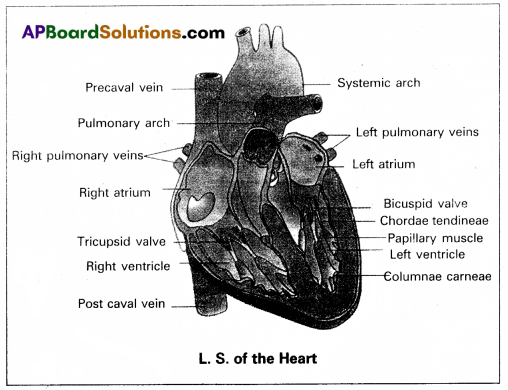
i) Atria :
- Atria are thin walled receiving chambers. The right one is larger than the left.
- The two atria are separated by thin inter-atrial septum. It has a small pore known as Foramen Ovale’ in fetal stage. Later it is closed and appeal’s as depression (oval patch) known as ‘Fossa ovale’. If, the foramen ovale does not close properly it is called a patent foramen ovale.
- The right atrium receives deoxygenated blood from different parts of the body, through three caval veins like two precaval veins and one post caval vein.
- The right atrium also receives blood from wall of the heart through coronary sinus, whose opening into the right atrium is guarded by the valve of Thebesius.
- Opening of the post caval vein is guarded by the Eustachian valve. It is functional in fetal stage and directs the blood from post caval vein into left atrium through foramen ovale. But it is non-functional in adult.
- The openings of the precaval veins into the right atrium have no valves.
- Left atrium receives oxygenated blood from lungs through a pair of pulmonary veins, which open into the left atrium through a common pore.
- Atrio-ventricular septum separates atria and ventricles.
- It has right and left atrio-venticular apertures.
- Tricuspid valve guards the right atrio-ventricular aperture. Bicuspid valve guards the left atrio-ventricular aperture.
ii) Ventricles :
These are the thick walled blood pumping chambers, separated by an interventricular septum. The wall of the left ventricle is thicker than that of the right ventricle as the left ventricle must force the blood to all the parts of the body.
The inner surface of the ventricles is raised into muscular ridges called columnae carneae. Some of them are large and conical and known as papillary muscles. Collagenous cords are known as chordae tendinae are present between atrio-ventricular valves and papillary muscles. They prevent the cusps of valves from bulging too far into atria during ventricular systole.
Nodal tissue : A specialized cardiac musculature called the nodal tissue is also distributed in the heart.
1) Sino-artrial node (SAN) – Present in the right upper comer of right atrium.
2) Atrio-ventricular node (AVN) – Present in the lower left comer of right atrium.
iii) Aortic arches : Human heart has two aortic arches.
1) Pulmonary arch : Arises from the left anterior angle of the right ventricle. It carries deoxygenated blood to lungs. It’s opening from right ventricle is guarded by pulmonary Valve made with 3 semiluminar valves.
2) Left systemic arch : Arises from the left ventricle to distribute oxygenated blood to various parts in the body. Its opening is also guarded by aortic valve made with a set of 3 semilunar valves.
A fibrous strand, known as ligamentum arteriosm is present at the point of contact of the systemic and pulmonary arches. It is the remnant of the ductus arteriosus, which connects the systemic and pulmonary arches in the embryonic stage.
Question 21.
Describe chromosomal theory of Sex determination.
Answer:
Chromosomal sex determination : The chromosomes, which determine the somatic characters of an individual are known as autosomes. These chromosomes do not differ in morphology and number in male and female sex. Those chromosomes, which differ in morphology and number in male and female sex and contain genes responsible for the determination of sex are known as allosomes or sex chromosomes. There are two types of sex chromosomal mechanisms :
a) Heterogametic male and
b) Heterogametic female
a) Heterogametic male : In this mechanism, the female sex has two ‘X’ chromosomes, while the male sex has only a single ‘X’ chromosome.
The heterogametic male may be of the following two types :
(i) XX – XO
(ii) XX – XY
i) XX – XO type : In certain insects belonging to orders Hemiptera (true bugs), Orthoptera (grass hoppers) and Dictyoptera (cockroaches) female has two ‘X” chromosomes (XX) and are, thus homogametic, while male has only single ‘X” chromosome (XO). The male being heterogametic sex produces two types of sperms, half with X chromosome and half without X chromosome in equal proportions. The sex of the offspring depends upon the sperm that fertilises the egg, each of which carries a single X chromosome. Thus fertilisation between male and female gametes always produced zygotes with one X’ chromosome from the female, but only 50% of the zygotes have an additional X Chromosome from the male. In this way, XO’ and ‘XX’ types would be formed in equal proportions, the former being males and the latter being females.
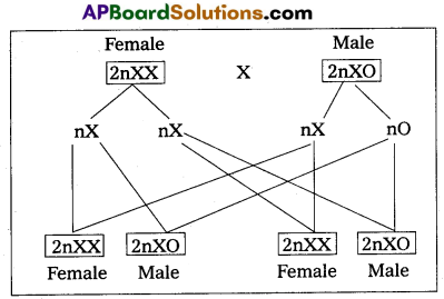
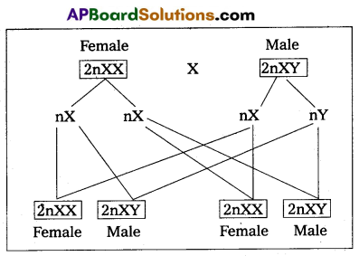
ii) XX – XY type : In man, other mammals, certain insects including Drosphila, the females possess two X chromosomes (XX) and are thus homogametic, produce one kind of eggs, each one with one X chromosome. While the males possess one X and one Y chromosome (XY) and are hence, heterogametic. They produce two kinds of sperms, half with X chromosome and half with Y chromosome. The sex of embryo depends on the kind of sperm. An egg fertilised by a X bearing sperm, produces a female, but if fertilised by a Y bearing sperm, a male is produced.
b) Heterogametic female : In this method of sex deter-mination, the male produces similar type of gametes, while female produce dissimilar gametes. The heterogametic females may be of following two types,
(i) ZO – ZZ
(ii) ZW – ZZ.
i) ZO – ZZ : This mechanism is found in certain moths and butterflies. In this case, female possesses one single ‘Z’ chromosome and hence is. heterogametic, producing two kinds of eggs half with Z chromosome and another half without any Z chromosome. Male possesses two Z chromosomes and thus homogametic, producing single type of sperms, each carries single Z chromosome. The sex of the offspring depends on the kind of egg.
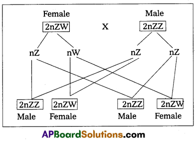
![]()
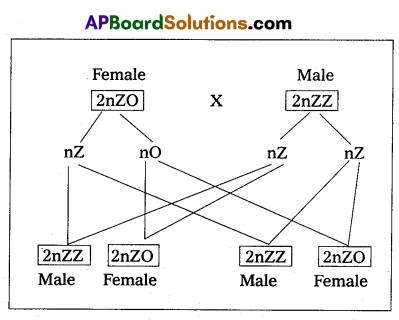
ii) ZW – ZZ : This system is found in certain insects (gypsy moth) and vertebrates such as fishes, reptiles and birds. In this system, the female is heterogametic and produces two types of gametes, one with ‘Z’ chromosome and the other with ‘W’ chromosome. On the other hand, male is homogametic and produces all sperms of same type carrying one ‘Z’ chromosome. The sex of the offspring depends on the kind of egg being fertilised. The ‘Z’ chromosome bearing eggs produce males, but the W chromosome bearing eggs produces females.