Thoroughly analyzing AP Inter 2nd Year Zoology Model Papers and AP Inter 2nd Year Zoology Question Paper May 2019 helps students identify their strengths and weaknesses.
AP Inter 2nd Year Zoology Question Paper May 2019
Time: 3 Hours
Maximum Marks: 60
Instructions:
Note : Read the following instructions carefully.
- Answer all questions of Section ‘A’. Answer ANY SIX questions in Section ‘B’ and answer ANY TWO questions in Section ‘C’.
- In Section ‘A’, questions from Sr Nos. 1 to 10 are of ‘Very Short Answer Type’. Each question carries TWO marks. Every answer may be limited to 5 lines. Answer all these questions at the one place in the same order.
- In ‘Section ‘B’, questions from Sr. Nos. 11 to 18 are of ‘Short Answer Type’. Each question carries FOUR marks. Every answer may be limited to 20 lines.
- In Section ‘C’, questions from Sr. Nos. 19 to 21 are of’Long Answer Type’. Each question carries EIGHT marks. Every answer may be limited to 60 tines.
- Draw labelled diagrams, wherever necessary in Sections ‘B’ and ‘C’.
Section – A (10 × 2 = 20)
Note : Answer all the questions in 5 lines each:
Question 1.
Name different types of papillae present on the tongue of man.
Answer:
The upper surface of the tongue has small projections called papillae. In humans the tongue bears 3 (three) types of papillae namely
- fungi form
- filiform
- Circumvallate papillae.
Question 2.
Where is the valve of Thebesius in the heart of man ?
Answer:
Opening of coronary sinus into left precaval vein is bound by a crescentic fold known as valve of Thebesius.
Question 3.
What is all-or-none principle ?
Answer:
The action potential occurs in response to a threshold stimulus (or) supra threshold stimulus but does not occur at subthreshold stimuli. It means the nerve impulse is either conducted totally (or) not conducted at all and this called all-or-none principle.
Question 4.
Distinguish between the blind spot and the yellow spot.
Answer:
Blind Spot : The region of the retina where the optic nerve exists the eyeball and devoid of rods and cones is called blind spot.
Yellow spot : The centre of the posterior portion of the retina is called yellow spot.
![]()
Question 5.
Which hormone is called anti-diuretic hormone ? Write the name of the gland that secretes it.
Answer:
Vasopressin is also called as anti – diuretic hormone which is secreted by posterior pituitary
Question 6.
What are androgens ? Which cells secrete them ?
Answer:
Androgens are male sex hormones usually steroid hormones.
E. g. : testosterone.
Androgens are produced by the Leydig cells of the testes and to a minor extent by the adrenal glands in both sexes.
Question 7.
What are the major components of the seminal fluid ?
Answer:
Seminal fluid is an alkaline, viscous fluid that contains fructose, proteins, citric acid, inorganic phosphorus, potassium and prostaglandins. It is produced by seminal vesicles present postero inferior to the urinary bladder in the pelvis.
Question 8.
What is “Amniocentesis’ ? Name any two disorders that can be detected by amniocentesis.
Answer:
Amniocentesis is a diagnostic procedure to detect genetic defects in the unborn baby, in which amniotic fluid is collected from foetus and diagnosed for abnormalities. Down’s syndrome, Turner’s syndrome and Edward’s syndrome can be detected by amniocentesis.
Question 9.
Define the terms layer and broiler.
Answer:
Layer: The birds which are raised exclusively for the production of eggs are called layers.
Broiler : The birds which are raised only for their meat are called broilers.
Question 10.
List out any four features of cancer cells.
Answer:
- Loss of contact inhibition
- Reduced intra cellular adhesion
- Immortalization
- Loss of anchorage dependence.
Section – B (6 × 4 = 24)
Note : Answer any six questions in 20 lines each:
Question 11.
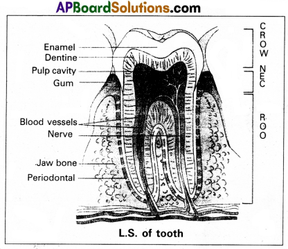
Draw a neat labelled diagram of L.S. of a tooth.
Answer:
Question 12.
What are the major transport mechanisms for CO2 ? Explain.
Answer:
Carbondioxide is transported in three ways.
1. In dissolved state : 7% of CO2 is transported in dissolved state through plasma. –
CO2 + H2O → H2CO3.
2. As Carbamino compounds : About 20 – 25% of CO2 combine directly with free amino group of haemoglobin and forms Carbamino haemoglobin in a reversible manner.
Hb – NH2 + CO2 Hb – NHCOO– + H+.
pCO2 and pO2 could affect the binding of C02 to haemoglobin.
- when pCO2 is high and pO2 is low as in the tissues, binding of more CO2 occurs.
- when pCO2 is low and pO2 is high as in the alveoli, dissocia- . tion of CO2 carbamino – haemoglobin takes place, (i.e., CO2 which is bound to haemoglobin from the tissues is delivered at the alveoli)
3. As Bicarbonates : About 70% Of CO2 is transported as bicarbonate. RBCs contain a very high concentration of the enzyme, carbonic anhydrase and a minute quantity of the same is present in plasma too. This enzyme facilitates the following reaction in both the directions.
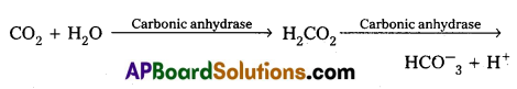
At the tissues where partial pressure of CO2 is high due to catabolism, CO2diffuses into the blood and forms carbonic acid which dissociates into HCO–3 + H+
At the alveolar site where pCO2 is low, the reaction proceeds in the opposite direction leading to the formation of CO2 and water. Thus CO2 is mostly trapped as bicarbonate at the tissues and transported to the alveoli where it is dissociated out as CO2.
Every 100 ml of deoxygenated blood delivers approximately 4 ml of CO2 to the alveolar air.
![]()
Question 13.
Describe the structure of synovial joint with the help of a neat labelled diagram.
Answer:
Synovial joints are characterised by the presence of a fluid filled synovial cavity between the articulating surfaces of the two bones.
Structure of synovial joint : Synovial joint is covered by a double layered synovial capsule. The outer layer consist of dense fibrous irregular connective tissue with more collagen fibers. This layer is continuous with the periosteum and resists stretching and prevents the dislocation of joints. Some fibres of these membranes are arranged in bundles called ligaments.
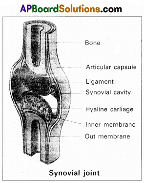
The inner layer of synovial capsule is formed of areolar tissue and elastic fibers. It secretes a viscous synovial fluid which contains hyaluronic acid, phagocytes etc., and acts as a lubricant for the free movement of the joints.
Question 14.
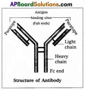
Write short notes on immunoglobulins.
Answer:
Whenever pathogen enters our body, the B-lymphocytes produce an army of proteins called antibodies to fight with them. They are highly specialised for binding with specific antigens. The part of an antibody that recognises an antigen is called the paratope antigen binding site.,
Based on their mobility, antibodies are of two types.
- Circulating or free antibodies : These are present in the body fluids like serum, lymph etc.
- Membrane bound antibodies : These are present on the surface of the mature B-cells as well as the memory cells.
Structure : Immunoglobulin is a ‘Y’ shaped molecule with four polypeptide chains of which two are long identical heavy chains (H) and two are small, identical light chains (L). These two chains are linked by disulfide bonds. One end of the antibody molecule is called Fab end (Fragment-antigen binding) and the other end is called Fc end (Fragment-Crystaline). Based on the structure, the antibodies are of five types namely Ig G, Ig A, Ig M, Ig D and Ig E.
Question 15.
Describe the genetic basis of ABO blood grouping.
Answer:
Bernstein proposed the genetic basis of ABO blood grouping. The genetic basis of ABO blood grouping is mainly dependent on the three alleles IA, IB and IO of the gene I, located in chromosome 9. The alleles IA and IB are responsible for the production of the respective antigens A and B on the surface of RBC. The allele IO does not produce any antigen on the surface of RBC. The alleles IA and IB are dominant to the allele IO, but co-dominant to each other (IA = IB > IO).
A child receives one of the three alleles from each parent, giving rise to six possible genotypes and four possible blood types. The genotypes are IAIA, IAIO , IBIB, IBIO , IAIB, IOIO.
The phenotypic expression of IAIA and IAIO are A-type blood, The phenotypic expression of IBIB and IBIO are B-type blood, and that of IAIB is AB blood type. The phenotype IOIO is ‘O’ – type blood.
Question 16.
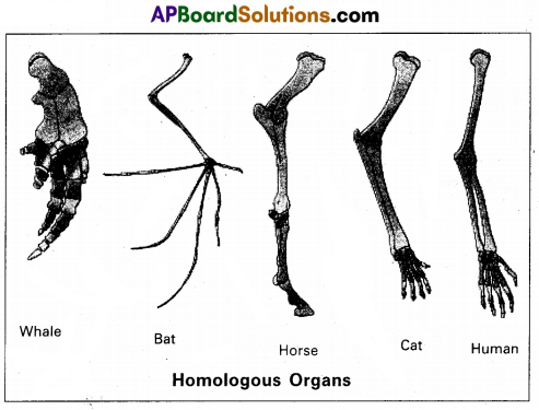
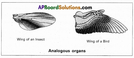
Distinguish between homologous and analogous organs.
Answer:
1. Homologous organs : The organs which have similar structure and origin but not necessarily the same function are called homologous organs. Eg : The appendages of vertebrates such as the flippers of whale, wings of bat, forelimbs of horse, paw of cat and hands of man have a common pattern in the arrangement of bones even though their external form and functions may vary to suit their mode of life.
2. Analogous organs : The organs which have dissimilar struc¬ture and origin but perform the same function are called the analo¬gous organs. Eg : Wings of butterfly and wings of a bird.
Question 17.
Write a short note on Neo-Darwinism.
Answer:
It was proposed by Fischer, Sewall Wright, Mayr. According to this theory, five basic factors are involved in the process of Organic
evolution. They are :
1. Gene mutations
2. Chromosomal mutations
3. Genetic recombinations
4. Natural selection
5. Reproductive isolation.
1) Gene mutations : Heritable changes in the structure of a gene are called gene mutations or point mutations. They alter the phenotypic character of the individuals. Thus, gene mutations tend to produce Variations in the offspring.
2) Chromosomal mutations: Heritable changes in the structure of chromosomes called chromdsomal mutations. They also bring about variations in the phenotype of organism which lead to the occurrence of variations in the offspring.
3) Genetic recombinations : Recombinations of genes due to crossing over during meiosis are responsible for bringing genetic variability among the individuals of the same species.
4) Natural selection : Natural selection does not produce any genetic changes, but it favours some genetic change while rejecting others.
5) Reproductive isolation : The absence of gene exchange between population is called the reproductive isolation. It plays a great role in giving rise to new species and preserving the species integrity.
![]()
Section – C (2 × 8 = 16)
Note : Answer any two questions in 60 lines each:
Question 18.
List out the various steps involved in MOET.
Answer:
The following are the steps involved in Multiple Ovulation and Embryo Transfer (MOET) :
- A cow is administrated hormones, with FSH like activity.
- This induces follicular maturation and super ovulation.
- In Super ovulation instead of one egg, which they produce per cycle, they produce 6 – 8 eggs.
- The cow is either mated with elite bull or artificially insemi-nated.
- The embryos are at 8-32 called stages are recovered non – surgically and transferred to surrogate mother, when the embryo develops into complete animal.
Now the genetic mother is ready for another round of super ovulation. This technology is in use for cattle, sheep, rabbits, buffaloes etc. to produce high yielding ones.
Question 19.
Describe the excretory system of man, giving the structure pf a nephron.
Answer:
In humans, the excretory system consists of a pair of kidney, a pair of ureters, a urinary bladder and urethra.
Kidney : Kidneys are reddish brown, bean shaped structures, situated on either side of the vertebral column between the levels of last thoracic and third lumbar vertebrae in a retroperitoneal position. The right kidney is slightly lower than the left one due to the presence of liver.
The outer surface of the kidney is convex and the inner surface is concave, where it has a deep notch called hilum, the point at which the renal artery and nerves enter and renal vein and ureter leave. Each-kidney is surrounded by a tough, fibrous tissue, called renal capsule.
Ureter: These are slender whitish tubes, which emerges from the pelvis of the kidney. The ureter rundown and open into the urinary bladder.
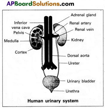
Urinary bladder Urethra
Urinary bladder : Urinary bladder is a pear shaped like muscular organ. It tempirarily stores the urine, situated in the lower abdominal cavity. The neck of the bladder leads into the urethra. Urethra opens near the vaginal orifice in the female and through the penis in the male.
Structure of nephron : Each kidney has nearly one million nephrons. These are structural and functional units of kidney, embedded in the loose connective tissue of cortex and medulla. Nephron consist of malpighian body and renal tubule.
I) Malphigian body : It begins in the cortex of the kidney. It contains Bowman’s capsule and glomerulus.
a) Bowman’s capsule : It is a thin walled, double layered cup. The inner wall of the Bowman’s capsule has certain unique cells called podocytes.
b) Glomerulus : It is a dense network of capillaries in the cup of Bowman’s capsule. Afferent arteriole of renal artery enter the cavity of Bowman’s capsule and split into five branches. They unite and come out of the Bowman’s capsule as an afferent arteriole.
The podocytes of inner wall of Bowman’s capsule wrap around each capillary. The podocytes are arranged in an intricate manner so as to leave some minute spaces called filteration slits. The endothelium cells of capillaries have numerous pores called fenestrations.
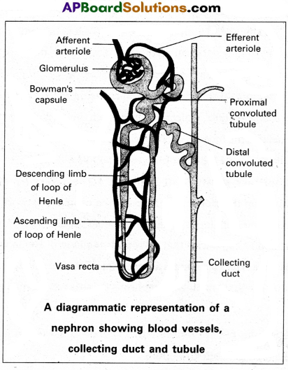
II) Renal tubule : It is narrow, delicate tubule arises from the posterior part of Bowman’s capsule known as neck. It opens into along narrow convoluted tubule with three parts like proximal convoluted tubule, Loop of Henle and Distal convoluted tubule.
a) Proximal convoluted tubule : It is a lined by simple cuboidal epithelium with brush border to increase area of absorption.
b) Loop of Henle: It is a hairpin like tubule present in medulla region. It consist of a descending limb and qn ascending limb. The proximal part of the ascending limb is thin and the distal part is thick. The thick ascending limb continuous into the distal convoluted tubule.
c) Distal convoluted tubule (DCT) : It is present in cortex. It is lined by simple cuboidal epithelium. The DCT continuous as the initial collecting duct in the cortex.
Collecting system : Some initial collecting ducts unite to form straight collecting duct, which passes through the medullary pyramid. In the medulla, the tubes of each pyramid join and form duct of Bellini, which finally opens into tip of the renal papilla.
Capillary network of nephron : The efferent arteriole emerging from the glomerulus forms a fine capillary network called the peritubular capillaries, around the renal tubule. The portion of the peritubular capillaries that surrounds the loop of Henle is called the vasa recta. The vasa recta is absent or highly reduced in the cortical nephrons. The juxta medullary nephrons possess well developed vasa recta.
![]()
Question 20.
Describe female reproductive system of a woman with the help of a labelled diagram.
Answer:
The female reproductive system consists of a pair of ovaries along with a pair of oviducts, uterus, vagina and the external genetalia located in the pelvic region. These parts of the system along with a pair of mammary glands are integrated structurally and functionally to support the processes of ovulation; fertilization, pregnancy, birth and child care.
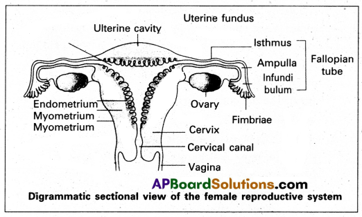
1) Ovaries : Ovaries are the primary female sex organs that produce the female gametes (ova) and several steroid hormones (ovarian hormones). A pair of ovaries is located on each side of lower abdomen. The double layered fold of peritoneum connecting the ovary with the wall of abdominal cavity is known as the meso ovarium.
The Ovaries are covered by a layer of ‘germinal (ovarian) epithelium. Underneath this layer, there is a dense connective tissue capsule called, ‘tunica albuginea’. The ovarian stroma is distinctly divided into an outer cortex and an inner medulla. The cortex appears more dense and granular due to numerous ovarian follicles. The medulla is a loose connective tissue with abundant blood vessels, ‘ lymphatic vessels and nerve fibres.
2) Fallopian tubes (oviducts) : Each fallopian tube extends from the periphery of each ovary to the uterus and it bears a funnel shaped infundibulum, with finger like projections called ‘fimbriae’, which help in collection of ovum after ‘ovulation’. The infundibulum leads to a wider part of the oviduct called ‘ampulla’. The last part of the oviduct, ‘isthmus’ has a narrow lumen and it joins the uterus. Fallopian tube is the site of fertilization. It conducts the ovum or zygote towards the uterus by peristalsis. The fallopian tube is attached to the abdominal wall by a peritoneal fold called ‘meso salpinx’.
3) Uterus : The uterus is a single and it is also called womb. It is a large, muscular, highly vascular and inverted pear¬shaped structure present in the pelvis between the bladder and rectum. The lower narrow part through which the uterus opens into the vagina is called ‘cervix’. The cavity of the cervix is called ‘cervical canal’ which along with the vagina
forms the ‘birth canal’.
The wall of the uterus has three layers of tissue, The external thin membranous ‘perimetrium’, the middle thick layer of ‘myometrium’ and inner glandulas lining layer called ‘endometrium’. The endometrium undergoes cyclic changes during menstrual cycle while myometrium exhibits strong contractions during parturition.
4) Vagina : The vagina is a large, median, fibromuscular tube that extends from the cervix to the vestibule (the space between labia minora). It is lined by non-keratinised stratified squamous epithelium. It is highly vascular and opens into the vestibule by the vaginal orifice.
5) Vulva : Vulva or pudendum refers to the external genitals of the female. The vestibule has two apertures – the upper external urethral orifice of the urethra and the lower vaginal orifice of vagina. Vaginal orifice is covered by a mucous membrane ‘hymen’, vestibule is bound by two pairs of fleshly folds of tissue called inner ‘labia minora’ and outer larger ‘labia majora’. Clitoris is a sensitive, erectile structure, that lies at the upper junction of the two labia minora above the urethral opening. There is a cushion of fatty tissue covered by skin and pubic hair present above the labia major, called mons pubis.
Accessory reproductive glands of female : These include;
a) Bartholin’s glands : These are two glands located slightly posterior and to the left and right of the opening of the vagina. They secrete mucus to lubricate the vagina and are homologous to the bulbourethral glands of the male reproductive system.
b) Skene’s glands : These are located on the anterior wall of ‘ vagina, around the lower end of the urethra. They secrete a lubricating fluid when stimulated. The skene’s glands are homologous to the prostate gland of the male reproductive system.
c) Mammary glands : These are paired structures that contain glandular tissue and fat. The alveoli cells present in the mammary lobes of each glandular tissue secrete milk, which is stored in cavities of alveoli. The alveoli open into mammary tuhes and then to mammary ducts, from there to mammary ampulla and finally connected to lactiferous duct through which milk is sucked out by the baby.
Question 21.
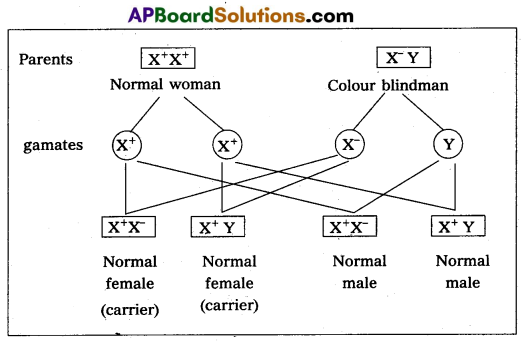
What is Criss-cross inheritance ? Explain the inheritance of one sex linked recessive character in human beings.
Answer:
The X-linked genes are represented twice in female (because female has two ‘X’-chromosomes) and once in males. (Because male has one X-chromosome). In male single X-linked recessive gene express it phenotypically, in contrast to female in which two ‘X’ linked recessive genes are necessary for the determination of a single phe-notypic trait related to sex.
The recessive X-Hnked genes have chracteristic crisscross in-heritance.
Crisscross inheritance : The inheritance of X-Unked recessive trait (genes) to his grandson (F2) through his daughter carrier) is called crisscross inheritance. Crisscross inheritance can be explained in humans by sex-linked recessive disorder, colour blindness.
Colourblindness : Colour blindness is a particular trait in human beings render them unable to differentiate between red and green colour. The gene for this colour blindness is located on X- chromosome. Colour blindness is recessive to normal vision so that if colour blind man marries a normal vision (homozygous) woman, all the sons and daughters are normal but daughter are heterozygous, which means that these daughters would be carrier for this trait. If such carrier woman marries a man with normal vision all the daughters and half of the sons have normal vision and half of sons are colour blind.
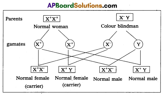
Colour blind trait is inhereted from a male parent to his grandson through carrier daughter i.e., this trait shows,crisscross inheri-tance.
![]()
If carrier female is married to normal male
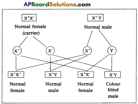
Characteristics of X-linked recessive traits :
- They never passed from father to son.
- Males are much more likely to be affected because they need only one copy of the mutant allele to express the phenotype.
- Affected males get the disease from their carrier mother only.
- Sons of heterozygous female (i.e., carrier female) have 50% chance of receiving mutant alleles. These disorders are typically passed from an affected grandfather to 50% of his grandsons.
- The X-linked recessive traits shows Crisscross pattern of inhertance. Eg : Colourblindness, Hemophilia, Muscular dystrophy etc.