Thoroughly analyzing AP Inter 2nd Year Zoology Model Papers and AP Inter 2nd Year Zoology Question Paper March 2019 helps students identify their strengths and weaknesses.
AP Inter 2nd Year Zoology Question Paper March 2019
Time: 3 Hours
Maximum Marks: 60
Instructions:
Note : Read the following instructions carefully.
- Answer all questions of Section ‘A’. Answer ANY SIX questions in Section B’ and answer ANY TWO questions in Section ‘C.
- In Section A’, questions from Sr. Nos. 1 to 10 are of ‘Very Short Answer Type’. Each question carries TWO marks. Every answer may be limited to 5 lines. Answer all these questions at the one place in the same order.
- ’In Section ‘B’, questions from Sr. Nos. 11 to 18 are of ‘Short Answer – Type’. Each question carries FOUR marks. Every answer may be limited to 20 lines.
- In Section ‘C’, questions from Sr. Nos. 19 to 21 are of ‘Long Answer Type’. Each question carries EIGHT marks. Every answer may be limited to 60 lines.
- Draw labelled diagrams, wherever necessary in Sections ‘B’ and ‘C’.
Section – A (10 × 2 = 20)
Note : Answer all the questions in 5 lines each:
Question 1.
Name different types of papillae present on the tongue of man.
Answer:
There are three pairs of Salivary glands in man.
- Parotid glands : Present below the pinna / inner surface of the cheeks.
- Sub maxillary (or) Sub mandibular glands : Located at the angles of lower jaw.
- Sublingual glands : Present below the tongue.
Question 2.
What are the coloumns of ‘Bertin’ ?
Answer:
Columns of Bertin are the medullary extensions of the renal cortex in between the renal pyramids.
Question 3.
Write the difference betweeh actin and myosin.
Answer:
| Actin | Myosin |
| 1. Actin is a thin contractile protein. | 1. Myosin is a thick contractile protein. |
| 2. It is present in light bands and is called an isotropic band. | 2. It is present in dark bands and is called an anisotropic band. |
| 3. Each actin filament is made of two ‘F’ actin molecules helically wound around each other, tropomyosin and a complex protein called troponin. | 3. Each myosin is made up of monomeric protein called meromyosins. Each mero-myosin has two parts namely head, and arm (or) neck. |
Question 4.
What is organ of Corti ?
Answer:
The hearing apparatus that is present in the middle canal of the cochlea is called organ of corti. The organ of corti contains hair cells that act as auditoiy receptors.
![]()
Question 5.
Name the gland that increases in size during childhood and decreases in size during adulthood. What important role does it play in case of infection ?
Answer:
Thymus is small at birth, it increases in size during childhood and reaches maximum size at puberty. During adulthood, it shrinks to its size at birth.
In old person thymus gland is degenerated, resulting in a de-creased production of thymosin. Thymosin plays an important role in immune developments. Thus, immune response against infections of old people becomes weak.
Question 6.
What is erythropoietin ? What is its function ?
Answer:
Erythropoietin is a hormone secreted the juxtaglomerular cells of the kidney. It plays an important role in the erythropoiesis i.e., in formation of RBC. Erythropoietin controls the formation of RBC by regulating the differentiation and proliferation of erythroid progenitor cells in the bone marrow.
Question 7.
It is true that MTP is not meant for population control’. Then why did the Government of India legalize MTP ?
Answer:
Medical Termination Pregnancy’ (MTP) or induced abortion is the procedure to terminate pregnancy with the help of medications. Government of India legalized MTP in 1971 to avoid its misuse, this is necessary to keep a check on indiscriminate and illegal female foeticides.
Question 8.
Mention the advantages of ‘Lactational amenorrhea method.
Answer:
Lactational amenorrhea is the absence of menstruation as long as mother breast feeds her baby.
The advantages of ‘lactational amenorrhea’ are
- As long as the mother fully breast feeds her child, chances of conception are almost zero.
- Breast feeding babies will have enhanced immunity, protection against allergies.
Question 9.
Mention any four fish by-products.
Answer:
- Shark and cod liver oils
- Fish guano
- Shagreen
- Isinglass.
Question 10.
How many amino acids and polypeptide chains are present in insulin ?
Answer:
Human insulin is made up of 51 aminoacids arranged in two polypeptides.
- polypeptide chain A with 21 aminoacids
- Polypeptide chain B with 30 aminoacids.
Which are held together by disulphide linkages.
Section – B (6 × 4 = 24)
Note : Answer any six questions in 20 lines each:
Question 11.
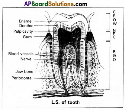
Draw a neat labelled diagram of L.S. of a tooth.
Answer:
Question 12.
What are the major transport mechanisms for CO2 ? Explain.
Answer:
Carbondioxide is transported in three ways.
1. In dissolved state : 7% of CO2 is transported in dissolved state through plasma. –
CO2 + H2O → H2CO3.
2. As Carbamino compounds : About 20 – 25% of CO2 combine directly with free amino group of haemoglobin and forms Carbamino haemoglobin in a reversible manner.
Hb – NH2 + CO2 Hb – NHCOO– + H+.
pCO2 and pO2 could affect the binding of C02 to haemoglobin.
- when pCO2 is high and pO2 is low as in the tissues, binding of more CO2 occurs.
- when pCO2 is low and pO2 is high as in the alveoli, dissocia- . tion of CO2 carbamino – haemoglobin takes place, (i.e., CO2 which is bound to haemoglobin from the tissues is delivered at the alveoli)
3. As Bicarbonates : About 70% Of CO2 is transported as bicarbonate. RBCs contain a very high concentration of the enzyme, carbonic anhydrase and a minute quantity of the same is present in plasma too. This enzyme facilitates the following reaction in both the directions.
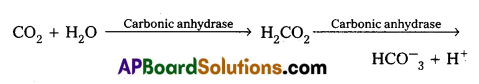
At the tissues where partial pressure of CO2 is high due to catabolism, CO2diffuses into the blood and forms carbonic acid which dissociates into HCO–3 + H+
At the alveolar site where pCO2 is low, the reaction proceeds in the opposite direction leading to the formation of CO2 and water. Thus CO2 is mostly trapped as bicarbonate at the tissues and transported to the alveoli where it is dissociated out as CO2.
Every 100 ml of deoxygenated blood delivers approximately 4 ml of CO2 to the alveolar air.
![]()
Question 13.
Describe the structure of synovial joint with the help of a neat labelled diagram.
Answer:
Synovial joints are characterised by the presence of a fluid filled synovial cavity between the articulating surfaces of the two bones.
Structure of synovial joint : Synovial joint is covered by a double layered synovial capsule. The outer layer consist of dense fibrous irregular connective tissue with more collagen fibers. This layer is continuous with the periosteum and resists stretching and prevents the dislocation of joints. Some fibres of these membranes are arranged in bundles called ligaments.
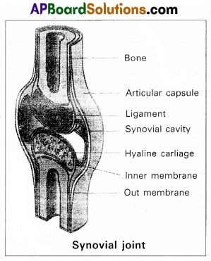
The inner layer of synovial capsule is formed of areolar tissue and elastic fibers. It secretes a viscous synovial fluid which contains hyaluronic acid, phagocytes etc., and acts as a lubricant for the free movement of the joints.
Question 14.
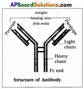
Write short notes on immunoglobulins.
Answer:
Whenever pathogen enters our body, the B-lymphocytes produce an army of proteins called antibodies to fight with them. They are highly specialised for binding with specific antigens. The part of an antibody that recognises an antigen is called the paratope antigen binding site.,
Based on their mobility, antibodies are of two types.
- Circulating or free antibodies : These are present in the body fluids like serum, lymph etc.
- Membrane bound antibodies : These are present on the surface of the mature B-cells as well as the memory cells.
Structure : Immunoglobulin is a ‘Y’ shaped molecule with four polypeptide chains of which two are long identical heavy chains (H) and two are small, identical light chains (L). These two chains are linked by disulfide bonds. One end of the antibody molecule is called Fab end (Fragment-antigen binding) and the other end is called Fc end (Fragment-Crystaline). Based on the structure, the antibodies are of five types namely Ig G, Ig A, Ig M, Ig D and Ig E.
Question 15.
Describe erythroblastosis foetalis.
Answer:
Erythroblastosis foetalis develops in an Rh positive foetus, whose father is Rh positive and mother is Rh negative. In Rh positive person rhesus antigens are present on the surface of blood cells where as in Rh negative person Rhesus antigens are absent.
During the process of delivery, the foetal blood cells may pass through the ruptured placenta into the Rh negative maternal blood. The mother’s immune system recognises the Rh antigens and gets sensitized and produces Rh antibodies. These antibodies are Ig G type they can pass through placenta. Generally first child is not effected because child is delivered by the time of the mother gets sen¬sitized and produce antibodies.
During second pregnancy, if the second child is Rh positive, these antibodies cross the placenta, enter the foetal blood circulation and destroy the Rh positive blood cell of foetus (haemolysis), leads to haemolytic anemia and jaundice. To compensate the haemolysis of blood cells there is a rapid production of RBC’s from the bone marrow, and but also from liver and spleen. Now many large and immature blood cells in erythroblast stage are released into circulation. Because of this disease is called erythroblastosis foetalis.
Question 16.
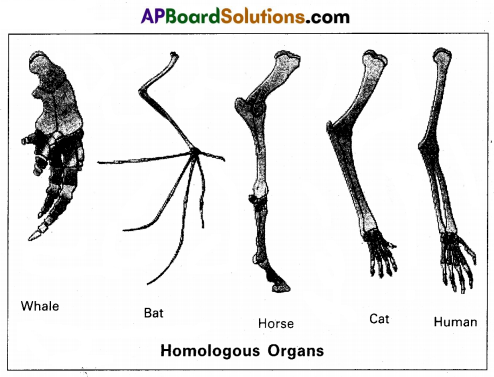
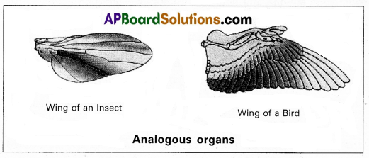
Distinguish between homologous and analogous organs.
Answer:
1. Homologous organs : The organs which have similar structure and origin but not necessarily the same function are called homologous organs. Eg : The appendages of vertebrates such as the flippers of whale, wings of bat, forelimbs of horse, paw of cat and hands of man have a common pattern in the arrangement of bones even though their external form and functions may vary to suit their mode of life.
2. Analogous organs : The organs which have dissimilar struc¬ture and origin but perform the same function are called the analo¬gous organs. Eg : Wings of butterfly and wings of a bird.
Question 17.
What is meant by genetic drift ? Explain genetic drift , citing the example of Founder Effect.
Answer:
The change in the frequency of a gene that occurs merely by chance and not by selection, in small proportion is called genetic drift.
Suppose, for a gene with two alleles, the frequency of a particular allele is 1% (e = 0.01) the probability of losing that allele by chance from small population is more. The end result is either fixation or loss of that allele.
Genetic drift tend to reduce the amount of genetic variation within the population mainly by removing the alleles with low frequencies. It can examplified by Founder and Bottleneck effect.
Founder effect: If a small group of individuals from a population starts a new colony in an isolated region, those individuals are called the founders of the new population. The allelic frequency of their descendants are similar those of the founders rather than to their ancestral parent population.
Eg : O+ve blood group is present in nearly 100% of the red Indians. It means the forefathers of the Red Indians tribe were predominantly O+ve and they isolated themselves reproductively from other population.
![]()
Question 18.
Write briefly about different waves and intervals in an ECG. Ans. ECG (electrocardiography) is commonly used, non-invasive – procedure fro recording electrical changes in the heart.
The graphic record which is called an electrocardiogram, shows the series of waves that occur during each cardiac cycle.
The normal ECG consists of :
i) Waves
ii) Intervals
iii) Segments
iv) Complexes,
i) Waves :
- The waves in a normal record are named P, Q, R, S and T in that order.
- A typical ECG tracing of a normal heartbeat consists of (I) a ‘P’ wave (II) a ‘QRS complex of waves’ (HI) a t Wave.
- P wave : It represents the atrial systole and shows that the impulse is passing through atria. The duration of P. Wave is 0.1 sec.
- QRS complex of wave : It represents ventricular systole. Q wave is small negative., R – wave is tall positive and S wave is a negative wave. Its duration is 0.08 to 0.1 sec.
- T wave : It represents the ventricular repolarization. It is a positive wave, its duration is 0.2 sec.
ii) Intervals :
P – R intervals: P – R intervals is the interval between the onset of p wave and the onset of Q wave. P – R interval is normally. 0.12 – 02 sec.
Q – T intervals : The interval between the onset of Q wave and the end of the T-wave. It represents the electrical activity in muscle of the ventricles. It lasts for about 0.4 seconds.
R – R intervals: It signifies the duration of one cardiac cycle and lasts for about 0.8 sec
Segments : S -T segment is the time period between the end of the ‘S’ wave and the onset of the T-wave. It is an isoelectric or zero voltage period.
Section – C (2 × 8 = 16)
Note : Answer any two questions in 60 lines each:
Question 19.
Write notes on the working of the heart of man.
Answer:
Human heart is a hallow muscular, cone shaped, and pulsating organ situated between lungs. It is about the size of a closed fist.
The heart is covered by double walled pericardium, which con-sists of outer fibrous pericardium and inner serous pericardium. The serous pericardium is double layered, outer parietal layer and inner visceral layer. These two layers are separated by pericardial space, which is filled with pericardial fluid. This fluid reduces friction between the two membranes and allow free movement of the heart.
Human heart has four chambers with two smaller upper cham-bers called atria and two larger lower chambers called ventricles. Atria and ventricles are separated by a deep transverse groove called coronary msulcus.
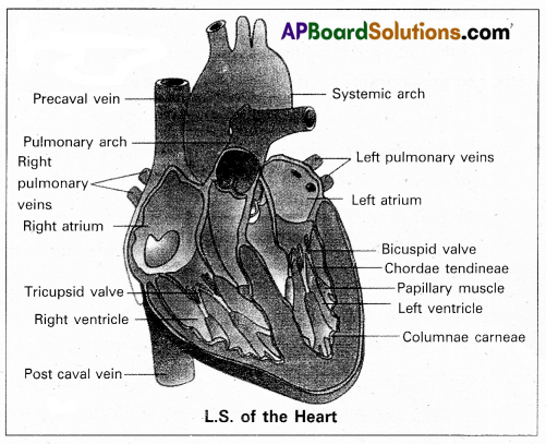
Atria :
- Atria are thin walled receiving chambers. The right one is larger than the left.
- The two atria are separated by thin inter-atrial septum. It has a small pore known as Foramen Ovale’ in fetal stage. Later it is closed and appears as depression (oval patch) known as ‘Fossa ovale’. If, the foramen ovale does not close properly it is called a patent foramen ovale.
- The right atrium receives deoxygenated blood from dif-ferent parts of the body, through three caval veins like two precaval veins and one post caval vein.
- The right atrium also receives blood from wall of the heart through coronary sinus, whose opening into the right atrium is guarded by the valve of Thebesius.
- Opening of the post caval vein is guarded by the Eustachian valve. It is functional in fetal stage and directs the blood from post caval vein into left atrium through foramen ovale. But it is non-functional in adult.
- The openings of the precaval veins into the right atrium have no valves.
- Left atrium receives oxygenated blood from lungs through a pair of pulmonary veins, which open into the left atrium through a common pore.
- Atrio-ventricular septum separates atria and ventricles. It has right and left atrio-venticular apertures.
- Tricuspid valve guards the right atrio-ventricular aper-ture. Bicuspid valve guards the left atrio-ventricular ap-erture.
ii) Ventricles :
These are the thick walled blood pumping chambers, separated by an interventricular septum. The wall of the left ventricle is thicker than that of the right ventricle as the left ventricle must force the blood to all the parts of the body.
The inner surface of the ventricles is raised into mus cular ridges called columnae carneae. Some of them are large and conical and known as papillary muscles. Collagenous cords are known as chordae tendinae are present between atrio-ventricular valves and papillary muscles. They prevent the cusps of valves from bulging too far . into atria during ventricular systole.
Nodal tissue : A specialized cardiac musculature called the nodal tissue is also distributed in the heart.
- Sino-artrial node (SAN) – Present in the right upper comer of right atrium.
- Atrio-ventricular node (AVN) – Present in the lower left comer of right atrium.
iii) Aortic arches : Human heart has two aortic arches.
1) Pulmonary arch : Arises from the left anterior angle of the right ventricle. It carries deoxygenated blood to lungs. It’s opening from right ventricle is guarded by pulmonary valve made with 3 semiluminar valves.
2) Left systemic arch : Arises from the left ventricle to dis-tribute oxygenated blood to various parts in the body. Its opening is also guarded by aortic valve made with a set of 3 semilunar valves.
A fibrous strand, known as ligamentum arteriosm is present at the point of contact of the systemic and pulmonary arches. It is the remnant of the ductus arteriosus, which connects the systemic and pulmonary arches in the embryonic stage.
![]()
Question 20.
Describe male reproductive system of a man. Draw a labelled diagram of it.
Answer:
The male reproductive system or male genital system consists of a number of sex organs that are a part of the human reproductive process. The sex organs which are located in the pelvic region include a pair of testes, accessory ducts, glands and external genitalia.
1) Testes : The testes are a pair of oval pinkish male sex organs suspended in abdominal cavity within a pouch called scrotum. The scrotum helps in maintaining the low temperature of the testes (2 – 2.5°C) necessary for spermatogenesis. The cavity of scrotal sac is connected to the abdominal cavity through the ‘inguinal canal’. Testes is held in position in the scrotum of the gubernacu- lum’, a fibrous cord that connects the testis with the bottom of scrotum and a ‘spermatic cord1, formed by the vas deferens, nerves, blood vessels and other tissues that run from abdomen down to each testicle, through inguinal canal. Each testis is enclosed in a fibrous envelope, ‘tunica albuginea’, which extends inwards into testis and di¬vide it into lobules. Each lobule contains 1 to 3 highly coiled seminiferous tubules. A pouch of serous membrane ‘tunica vaginalis’ covers the testis.
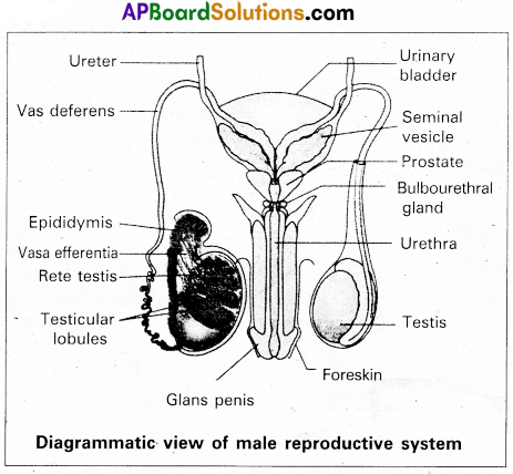
Miniferous tubules : Each seminiferous tubule is lined by ‘germinal epithelium’ which consists of undifferentiated male gum cells called ‘spermatogonial mother cells’ and it also bears ‘nourishing cells’ called ‘sertoli cells’.
- Spermatogonial cells (or) primary spermatocytes undergo meiotic division, producing spermatozoa or sperms by a process spermatogenesis.
- Sertoli cells provide nutrition to spermatozoa and produce a hormone ‘inhibin’, which inhibits secretion of FSH.
The region outside the tubules, contain interstitial cells of ‘Leydig cells’. They produce androgens, the most important in testosterone. It controls the development of secondary sexual characters and spermatogenesis. The seminiferous tubules open into vasa efferntia through the rete testis. Rete testis is a network of tubules is of the testis carrying spermatozoa from the seminiferous tubules to the vasa efferentia.
2) Epididymis : The vasa efferntia leave the testis and open into a narrow, tightly coiled tube called epididymis’ located along the posterior surface of each testis. The epididymis provides a storage space for sperms and gives them time to nature.
It is differentiated into three regions.
a) Caput epididymis
b) Corpus epididymis
c) Cauda epididymis.
The caput epididymis receives spermatozoa via the vasa effemtia of the mediastinum testis. It is mass of a connective tissue at the back of the testis that encloses the rete testis.
3) Vasa deferentia : The vas deferens or ductus deferens is a long, narrow mascular tube. The mucosa of the ductus deferens consists of a pseudo stratified columnar epithelium and lamina propia. It starts from the tail of epididymis, passes through the inguinal canal into the abdomen and loops over the urinary bladder. It receives a duct from seminal vesicle.
The vas deferens and the duct of the seminal vesicle units to form a ’short ejaculatory duct’ or ‘ductus ejaculatorius’. The two ducts, carrying spermatozoa and the fluid secreted by the seminal vesicles, converge in the centre of prostate and open into urethra, which transports the sperms to outside.
4) Urethra : In male, Urethra is the shared terminal duct of the reproductive and urinary systems. The urethra originates from urinary bladder and extends through the penis to its external opening called ‘urethral meatus’. The urethra provides an exit for urine as well as semen during ejaculation.
5) Penis : Urethra opens into the major copulatory organ of male, the ‘penis’. The penis and scrotum constitute the male external genitalia. The penis serves as a urinal duct and intromittent organ the transfers spermatozoa to the vagina of a female.
The penis is made up of three columns of tissue : two upper Corpora cavernosa on the dorsal aspect and one Corpus spongiosum on the ventral side. Skin and a subcutaneous layer encloses all three columns, which consists of special tissue that helps in erection of penis.. The enlarged and bulbous end of penis is called ‘glans penis’, which is-covered by a loose fold of skin (foreskin) called prepuce.
Male accessoiy glands :
Male accessory glands are :
a) Seminal vesicles
b) Prostate glands
c) Bulbourethral glands.
a) Seminal vesicles: These are a pair of simple tubular glands present postero-inferior to the urinary bladder in the pelvis. Each seminal vesicle enters prostate gland through vas deferens. The vesicles produce seminal fluid rich is fructose, proteins, citric acid, in organic phosphorus, potassium and prosta- glandins. All these serve sperm cells.
b) Prostate gland : It is located directly beneath the urinary bladder. The gland surrounds the ‘Prostatic urethra’, and sends its secretions through prostatic ducts. The prostatic secretion activates spermatozoa and provides nutrition. In man, the prostate contributes 15-30% of the semen.
c) Bulbourethral glands : These are also called cowper’s glands located beneath the prostate gland at the beginning of the internal portion of the penis. They add an alkaline fluid to semen and the fluid secreted by them lubricates urethra. It acts as flushing agent washing out the acidic urinary residues that remain in the urethra, before the,semen is ejaculated.
![]()
Question 21.
What is Criss-cross inheritance ? Explain the inheritance of one sex linked recessive character in human beings.
Answer:
The X-linked genes are represented twice in female (because female has two ‘X’-chromosomes) and once in males, (because male has one X-chromosome). In male single X-linked recessive gene express it phenotypically, in contrast to female in which two ‘X’ linked recessive genes are necessary for the determination of a single phenotypic trait related to sex.
The recessive X-linked genes have chracteristic crisscross inheritance.
Crisscross inheritance : The inheritance of X-linked recessive trait (genes) to his grandson (F2 through his daughter(carrier) is called crisscross inheritance. Crisscross inheritance can be explained in humans by sex-linked recessive disorder, colour blindness.
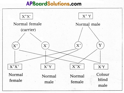
Colourblindness : Colour blindness is a particular trait in human beings render them unable to differentiate between red and green colour. The gene for this colour blindness is located on X-chromosome. Colour blindness is recessive to normal vision so that if colour blind man marries a normal vision (homozygous) woman, all the sons and daughters are normal but daughter are heterozygous, which means that these daughters would be carrier for this trait. If such carrier woman marries a man with normal vision all the daughters and half of the sons have normal vision and half of sons the colour blind.
Colour blind trait is inhereted from a male parent to his grandson through carrier daughter i.e., this trait shows criss-cross inheritance.
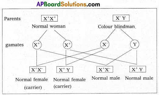
If carrier female is married to normal male
Characteristics of X-linked recessive traits :
- They never passed from father to son.
- Males are much more likely to be affected because they need only one copy of the mutant allele to express the phenotype.
- Affected males get the disease from their carrier mother only.
- Sons of heterozygous female (i.e., carrier female) have 50% chance of receiving mutant alleles. These disorders are typi-cally passed from an affected grandfather to 50% of his grandsons.
- The X-linked recessive traits shows Crisscross pattern of inhertance.
Eg : Colourblindness, Hemophilia, Muscular dystrophy etc.,