Thoroughly analyzing AP Inter 2nd Year Zoology Model Papers and AP Inter 2nd Year Zoology Question Paper May 2018 helps students identify their strengths and weaknesses.
AP Inter 2nd Year Zoology Question Paper May 2018
Time: 3 Hours
Maximum Marks: 60
Instructions:
Note : Read the following instructions carefully.
- Answer all the questions of Section ‘A’ Answer any six questions in Section ‘B’ and answer any two questions in Section ‘C’.
- In Section A’, questions from Sr. Nos. 1 to 10 are of Very Short Answer Type. Each question carries two marks. Every answer may be limited to 5 lines. Answer all these questions at one place in the same order.
- In Section ‘B’, questions from Sr. Nos. 11 to 18 are of Short Answer Type. Each question carries four marks. Every answer may be limited to 20 lines.
- In Section ‘C’, questions from Sr. Nos. 19 to 21 are of Long Answer Type. Each question carries eight marks. Every answer may be limited to 60 lines.
- Draw labelled diagrams wherever necessary in Sections ‘B’ and ‘C’.
Section – A (10 × 2 = 20)
Note : Answer all the questions in 5 lines each:
Question 1.
What is meant by chloride shift ?
Answer:
Chloride shift : It refers to the exchange of chloride and bicarbonate ions between erythrocytes and plasma. It is also called Hamburger’s phenomenon:
Question 2.
Distinguish between the enzymes renin and rennin.
Answer:
Renin : Renin is an enzyme produced by the JG cells. This enzyme catalyses the conversion of angiotensinogen into angioten – sin – I.
Rennin: Rennin is also an enzyme found in the gastric juice of infants. It acts on the milk protein casein in the presence of calcium ions and convert it into calcium para caseinate and proteoses.
Question 3.
Name two cranial sutures and their locations.
Answer:
- Coronal suture – between the frontal and parietal bones.
- Lambdoid suture – between the parietal and occipital bones.
Question 4.
What is triad system ?
Answer:
In a skeletal muscle each transverse tubule (T-Tubule) is flanked on either side by several cistemae of the sarcoplasmic reticulum. T-tubule and the two terminal cistemae at its sides form the triad system.
![]()
Question 5.
What is acromegaly ? Name the hormone responsible for this disorder.
Answer:
Acromegaly is a hormonal disorder that results when the pituitary gland produces excess growth hormone (GH). This disease is characterised by enlargement of the bones of the jaw, hand and feet, thickended nose, lips, eyelids and wide finger tips and gorilla like appearance of the person affected.
Question 6.
What is erythropoietin ? What is its function ?
Answer:
Erythropoietin is a hormone secreted the juxtaglomerular cells of the kidney. It plays an important role in the erythro- poiesis i.e., in formation of RBC. Erythropoietin controls the formation of RBC by regulating the differentiation and proliferation of erythroid progenitor cells in the bone marrow.
Question 7.
What are the functions of Sertoli cells of the seminiferous tabules and the Leydig cells in man ?
Answer:
- Sertoli cells : Also known as ‘nourishing cells’ helps in the nourishment of spermatozoa and produce a hormone ‘inhibin’, which inhibits the secretion of FSH.
- Leydig cells : Produce Testosterone that controls the secondary sexual characters and spermatogenesis.
Question 8.
Mention the advantages of ‘Lactational amenorrhea method’.
Answer:
Lactational amenorrhea is the absence of menstruation as long as mother breast feeds her baby.
The advantages of ‘lactational amenorrhea’ are
- As long as the mother fully breast feeds her child, chances of conception are almost zero.
- Breast feeding babies will have enhanced immunity, protection against allergies.
Question 9.
Explain the term hypophysation.
Answer:
Making the fishes to breed artificially to meet the demand of carpseed as called hypophysation.
Question 10.
What is electrocardiography and what are the normal components of ECG ?
Answer:
Electrocardiography is a commonly used, non invasive procedure for recording electrical changes in the heart.
Normal components of ECG
- Waves
- Intervals
- Segments
- Complexes.
Section – B (6 × 4 = 24)
Note : Answer any six questions in 20 lines each:
Question 11.
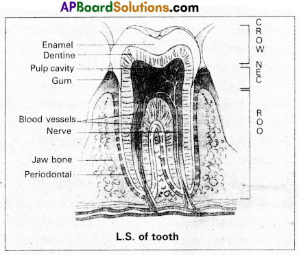
Draw a neat labelled diagram of L.S. of a tooth.
Answer:
Question 12.
Explain the process of inspiration and expiration under normal conditions.
Answer:
Inspiration: Intake of atmospheric air into the lungs is called inspiration. It is an active process, as it takes place by the contraction of the muscles of the diaphragm and the external inter-costal muscles which extend in between the ribs. The contraction of diaphragm increases the volume of thoracic chamber in the anterio posterior axis. The contraction of external inter costal muscles lifts up the ribs and sternum causing an increase in the dorso-ventral axis. The overall increase in the thoracic volume causes a similar increase in the pulmonary volume. An increase in the pulmonary volume decreases the intra-pulmonary pressure to less than that of the atmosphere, which forces the air from the outside to move into the lungs, that is inspiration.
Expiration : Release of alveolar air to the exterior is called expiration. It is a passive process. Relaxation of the diaphragm and external inter-costal muscles returns the diaphragm and sternum to their normal positions, and reduces the thoracic volume and thereby the pulmonary volume. This leads to an increase in the intra-pulmonary pressure to slightly above that of the atmospheric pressure, causing the expulsion of air from the lungs, that is called expiration.
Question 13.
Give an account of the retina of the human eye.
Answer:
Retina is the inner coat of the eye. It consist of a pigmented epithelium and a neural portion. The pigmented epithelium . is a sheet of melanin containing epithelial cells. The neural portion has three layers namely photoreceptor layer, bipolar cell layer and ganglion cell layer.
Photoreceptor layer consist of rods and cones. Rods contain a protein called rhodopsin. Rods are concerned with dim light. Cones contain a visual pigment called iodopsin and they are important in daylight vision and colour vision. There are three types of cones and are response to red, green and blue colours.
The centre of the posterior portion of the retina is called yellow spot. A depression present in the yellow spot is called ‘Forea’ contractile and it contains only cones. Forea is responsible for sharp vision. The region of retina which is devoid of rods and cones is known as blind spot (or) optic disc, which form the optic nerve called 2nd cranial nerve.
![]()
Question 14.
Write short notes on B-cells.
Answer:
The lymphocytes capable of producing antibodies and can capture circulating antigens are called B-cells. They are produced from the stem cells in the bone marrow, liver of foetus and bursa offabricius in birds. Mature B-cells express or display Ig M and Ig D antibodies on their membrane surfaces. As these antibodies can take antigens, the mature B-cells are also called immuno-competent B-cells. In secondary lymphoid organs these immuno-competent B-cells develop into functional immune cells which later differentiate into long lived memory cells and effector plasma cells. The plasma cells produce antibodies specific to the antigen to which they are exposed. Memory cells store information about the specific antigens and show quick response, when the same type of antigen invades the body later.
Question 15.
Describe erythroblastosis foetalis.
Answer:
Erythroblastosis foetalis develops in an Rh positive foetus, whose father is Rh positive and mother is Rh negative. In Rh positive person rhesus antigens are present on the surface of blood cells where as in Rh negative person Rhesus antigens are absent.
During the process of delivery, the foetal blood cells may ‘ pass through the ruptured placenta into the Rh negative maternal blood. The mother’s immune system recognises the Rh antigens and gets sensitized and produces Rh antibodies. These antibodies are Ig G type they can pass through – placenta. Generally first child is not effected because child is delivered by the time of the mother gets sensitized and produce antibodies.
During second pregnancy, if the second child is Rh positive, these antibodies cross the placenta, enter the foetal blood circulation and destroy the Rh positive blood cell of foetus (haemolysis), leads to haemolytic anemia and jaundice. To compensate the haemolysis of blood cells there is a rapid production of RBC’s from the bone marrow, and but also from liver and spleen. Now many large and immature blood cells in erythroblast stage are released into circulation. Because of this disease is called erythroblastosis foetalis.
Question 16.
Write a short note on the theory of mutations.
Answer:
Theory of mutation was proposed by Hugo de Varies, who coined the term mutation.
Mutations are sudden, random inheritable changes that occur in organisms. Hugo de Varies observed this phenomenon in the evening primrose plant Oenothera. lamarckiana, which shows different forms like ;
- O. brevistylis – with small style.
- O. levifolia – with smooth leaves.
- O. gigas – with the giant form.
- O. nanella – with dwarf form.
These characters are inherited to the progeny.
- Darwin called mutations as sports of nature or saltations.
- Bateson called them as discontinuous variations.
Salient features of mutation theory :
- Mutations occur from time to time in naturally breeding population.
- Mutants differ from their parents.
- Mutations are inheritable.
- Mutations occur in any direction i.e., they are random.
- They are discontinuous and not accumulated over generations.
- They are full-fledged and so there are no intermediate forms.
- They are subjected to natural selection.
Question 17.
Explain Darwin’s theory of Natural Selection with industrial melanism as an experimental proof.
Answer:
Darwin’s theory of natural selection does not explain what exactly evolution is, but explains how evolution might have occurred in nature. A classical example for natural selection is industrial melanism, exhibited by peppered moth-Biston betularia. These moths were available in two colours grey and black. Grey moths were abundant before industrial revolution in all over England. The reason for the existence of large number of grey moths during that period was camouflage on the trunks of trees. But after the establishment of industries in England, black coloured moths were more and grey forms were less. This is due to pollution from industries in the form of soot turned barks of trees into black. So grey moths were easily identified and were more predated by birds. Thus grey moths decreased in number, black moths increased in the population.
Thus natural selection favoured the melanic moths (black) to reproduce more successfully. Natural selection of darker forms in response to industrial pollution is known as industrial melanism.
Question 18.
Discuss in brief about ‘A vian Flu’.
Answer:
AvianFlu or birdFlu is an important disease affecting poultry birds and man.
Causative organism : AvianFlu or birdFlu is caused by an “avianFlu virus” the H5NI. The virus that causes the bird infection infects humans too. It is a pandemic disease.
Mode of infection : Infection may be spread simply by touching contaminated surfaces. Birds infected by this type of influenza, continue to release the virus as in their faeces and saliva for as long as 10 days.
Symptoms : In humans it causes typical-flu-like symptoms, include cough, diarrhoea difficulty in breathing, fever, headache, malaise, muscle aches and sore throat.
Prevention :
- Avoiding consumption of under cooked chicken.
- People who work for poultry birds should use protective clothing and special breathing masks.
- Complete culling of infected flock by burying or burning them.
![]()
Question 19.
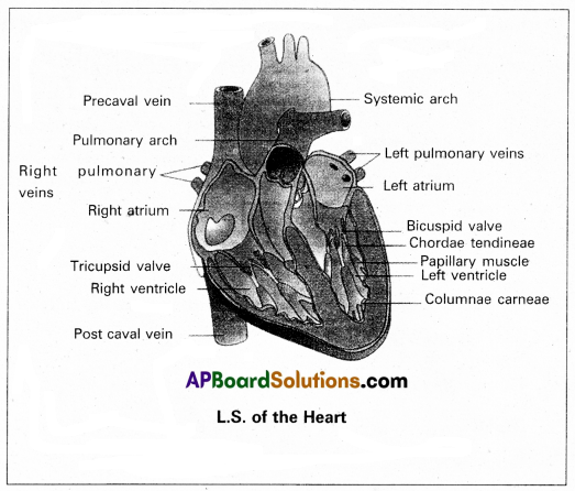
Discribe the structure of the heart of man with the help of neat labelled diagram.
Answer:
Human heart is a hallow muscular, cone shaped, and pulsating organ situated between lungs. It is about the size of a closed fist.
The heart is covered by double walled pericardium, which consists of outer fibrous pericardium and inner serous pericardium. The serous pericardium is double layered, outer parietal layer and inner visceral layer. These two layers are separated by pericardial space, which is filled with pericardial fluid. This fluid reduces friction between the two membranes and allow free movement of the heart.
Human heart has four chambers with two smaller upper chambers called atria and two larger lower chambers called ventricles. Atria and ventricles are separated by a deep transverse groove called coronary sulcus.
i) Atria :
- Atria are thin walled receiving chambers. The right one is larger than the left.
- The two atria are separated by thin inter-atrial septum. It has a small pore known as Foramen Ovale’ in fetal stage. Later it is closed and appears as depression (oval patch) known as ‘Fossa ovale’. If, the foramen ovale does not close properly it is called a patent foramen ovale.
- The right atrium receives deoxygenated blood from different parts of the body, through three caval veins like two precaval veins and one post caval vein.
- The right atrium also receives blood from wall of the heart through coronary sinus, whose opening into the right atrium is guarded by the valve of Thebesius.
- Opening of the post caval vein is guarded by the Eustachian valve. It is functional in fetal stage and directs the blood from post caval vein into left atrium through foramen ovale. But it is non-functional in adult.
- The openings of the precaval veins into the right atrium have no valves.
- Left atrium receives oxygenated blood from lungs through a pair of pulmonary veins, which open into the left atrium through a common pore.
- Atrio-ventricular septum separates atria and ventricles. It has right and left atrio-venticular apertures.
- Tricuspid valve guards the right atrio-ventricular aperture. Bicuspid valve guards the left atrio-ventricular aperture,
ii) Ventricles:
- These are the .thick walled blood pumping chambers, separated by an interventricular septum. The wall of the left ventricle is thicker than that of the right ventricle as the left ventricle must force the blood to all the parts of the body.
- The inner surface of the ventricles is raised into muscular ridges called columnaecameae. Some of them are large and conical and known as papillary muscles. Collagenous cords are known as chordae tendinae are present between atrioventricular valves and papillary muscles.
They prevent the cusps of valves from bulging too far into atria during ventricular systole.
Nodal tissue : A specialized cardiac musculature called the nodal tissue is also distributed in the heart.
- Sino-artrial node (SAN) – Present in the right upper comer of right atrium.
- Atrio-ventricular node (AVN) – Present in the lower left comer of right atrium.
iii) Aortic arches : Human heart has two aortic arches.
1) Pulmonary arch : Arises from the left anterior angle of the right ventricle. It carries deoxygenated blood to lungs. It’s opening from right ventricle is guarded by pulmonary valve made with 3 semiluminar valves.
2) Left systemic arch : Arises from the left ventricle to distribute oxygenated blood tovarious parts in the body. Its opening is also guarded by aortic valve made with a set of 3 semilunar valves.
A fibrous strand, known as ligamentum arteriosm is present at the point of contact of the systemic and pulmonary arches. It is the remnant of the ductus arteriosus, which connects the systemic and pulmonary arches in the embryonic stage.
Question 20.
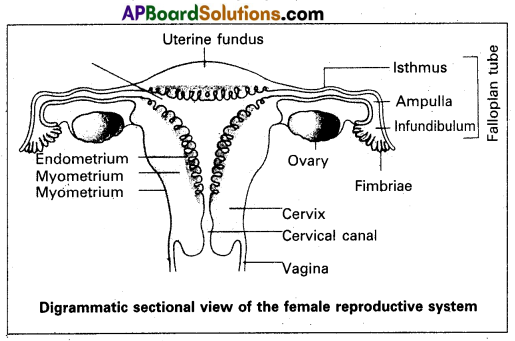
Describe female reproductive system of a human with the help of a labelled diagram.
Answer:
The female reproductive system consists of a pair of ovaries along with a pair of oviducts, uterus, vagina and the external genetalia located in the pelvic region. These parts of the system along with a pair of mammary glands are integrated structurally and functionally to support the processes of ovulation, fertilization, pregnancy, birth and child care.
1) Ovaries : Ovaries are the primary female sex organs that produce the female gametes (ova) and several steroid hormones (ovarian hormones). A pair of ovaries is located on each side of lower abdomen. The double layered fold of peritoneum connecting the ovary with the wall of abdominal cavity is known as the meso ovarium.
The Ovaries are covered by a layer of ‘germinal (ovarian) epithelium. Underneath this layer, there is a dense connective tissue capsule called, ‘tunica albuginea’. The ovarian stroma is distinctly divided into an outer cortex and an inner medulla. The cortex appears more dense and granular due to numerous ovarian follicles. The medulla is a loose connective tissue with abundant blood vessels, lymphatic vessels and nerve fibres.
2) Fallopian tubes (oviducts) : Each fallopian tube extends from the periphery of each ovary to the uterus and it bears a funnel shaped infundibulum, with finger like projections called ‘fimbriae’, which help in collection of ovum after ‘ovulation’. The infundibulumTeads to a wider part of the oviduct called ‘ampulla’. The last part of the oviduct, ‘isthmus’ has a narrow lumen and it joins the uterus. Fallopian tube is the site of fertilization. It conducts the ovum or zygote towards the uterus by peristalsis. The fallopian tube is attached to the abdominal wall by a peritoneal fold called ‘meso salpinx’.
3) Uterus : The uterus is a single and it is also called womb. It is a large, muscular, highly vascular and inverted pear-shaped structure present in the pelvis between the bladder and rectum. The lower narrow part through which the uterus opens into the vagina is called ‘cervix’. The cavity of the cervix is called ‘cervical canal’ which along with the vagina forms the ‘birth canal’.
The wall of the uterus has three layers of tissue. The external thin membranous ’perimetrium’, the middle thick layer of ’myometrium’ and inner glandulas lining layer called’endometrium’. The endometrium undergoes cyclic changes during menstrual cycle while myometrium exhibits strong contractions during parturition.
4) Vagina: The vagina is a large, median, fibromuscular tube that extends from the cervix to the vestibule (the space between labia minora). It is lined by non-keratinised stratified squamous epithelium. It is highly vascular and opens into the vestibule by the vaginal orifice.
5) Vulva : Vulva or pudendum refers to the external genitals of the female. The vestibule has two apertures – the upper external urethral orifice of the urethra and the lower vaginal orifice of vagina. Vaginal orifice is covered by a mucous membrane ‘hymen’, vestibule is bound by two pairs of fleshly folds of tissue called inner ‘labia minora’ and outer larger ‘labia majora’. Clitoris is a sensitive, erectile structure, that lies at the upper junction of the two labia minora above the urethral opening. There is a cushion of fatty tissue covered by skin and pubic hair present above the labia major, called mons pubis.
Accessory reproductive glands of female : These include;
a) Bartholin’s glands : These are two glands located slightly posterior and to the left and right of the opening of the vagina. They secrete mucus to lubricate the va/fina and are homologous to the bulbourethral glands of the male reproductive system.
b) Skene’s glands: These are located on the anterior vall of vagina, around the lower end of the urethra. They crete a lubricating fluid when stimulated. The skene’s ands are homologous to the prostate gland of the male reproductive system.
c) Mammary glands : These are paired structures that contain glandular tissue and fat. The alveoli cells present in the mammary lobes of each glandular tissue secrete milk, which is stored in cavities of alveoli. The alveoli open into mammary tubes and then to mammary ducts, from there to mammary ampulla and finally connected to lactiferous duct through which milk is sucked out by the baby.
![]()
Question 21.
Describe chromosomal theory of sex determination.
Answer:
Chromosomal sex determination: The chromosomes, which determine the somatic characters of an individual are known as autosomes. These chromosomes do not differ in mor-phology and number in male and female sex. Those chromosomes, which differ in morphology and number in male and female sex and contain genes responsible for the determination of sex are known as allosomes or sex chromosomes. There are two types of sex chromosomal mechanisms :
a) Heterogametic male and
b) Heterogametic female.
a) Heterogametic male : In this mechanism, the female sex has two ‘X’ chromosomes, while the male sex has only a single ‘X’ chromosome.
Tlhe heterogametic male may be of the following two types :
(i) XX – XO
(ii) XX – XY
(i) XX – XO type : In certain insects belonging to orders Hemiptera (true bugs), Orthoptera (grass hoppers) and Dictyoptera (cockroaches) female has two X” chromosomes (XX) and are, thus homogametic, while male has only single X” chromosome (XO). The male being heterogametic sex produces two types of sperms, half with X chromosome and half without X chromosome in equal proportions. The sex of the offspring depends Upon the sperm that fertilises the egg, each of which carries a single X chromosome. Thus fertilisation between male and female gametes always pro-duced zygotes with one ‘X’ chromosome from the female, but only 50% of the zygotes have an additional X Chromosome from the male. In this way, ‘XO’ and ‘XX’ types would be formed in equal proportions, the former being males and the latter being females.

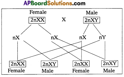
ii) XX – XY type : In man, other mammals, certain insects including Drosphila, the females possess two X chromosomes (XX) and are thus homogametic, produce one kind of eggs, each one with one X chromosome. While the males possess one X and one Y chromosome (XY) and are hence, hetero- gametic. They produce two kinds of sperms, half with X chromosome and half with Y chromosome. The sex of embryo depends on the kind of sperm. An egg fertilised by a X bearing sperm, produces a female, but if fertilised by a Y bearing sperm, a male is produced.
b) Heterogametic female : In this method of sex deter-mination, the male produces similar type of gametes, while female produce dissimilar gametes. The heterogametic females may be of following two types,
(i) ZO – ZZ
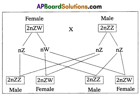
(ii) ZW – ZZ.
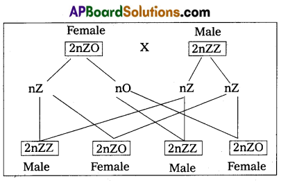
i) ZO – ZZ : This mechanism is found in certain moths and butterflies. In this case, female possesses one single ‘Z’ chromosome and hence is heterogametic, producing two kinds of eggs half with Z chromosome and another half without any Z chromosome. Male possesses two Z chromosomes and thus homogametic, producing single type of sperms, each carries single Z chromosome. The sex of the offspring depends on the kind of egg.
ii) ZW – ZZ : This system is found in certain insects (gypsy moth) and vertebrates such as fishes, reptiles and birds. In this system, the female is heterogametic and produces two types of gametes, one with ‘Z’ chromosome and the other with ‘W chromosome. On the other hand, male is homogametic and produces all sperms of same type carrying one ‘Z’ chromosome. The sex of the offspring depends on the kind of egg being fertilised. The ‘Z’ chromosome bearing eggs produce males, but the W chromosome bearing eggs produces females.