Thoroughly analyzing AP Inter 2nd Year Zoology Model Papers and AP Inter 2nd Year Zoology Question Paper March 2018 helps students identify their strengths and weaknesses.
AP Inter 2nd Year Zoology Question Paper March 2018
Time: 3 Hours
Maximum Marks: 60
Instructions:
Note : Read the following instructions carefully.
- Answer all the questions of Section – A. Answer any six questions in Section – B and answer any two questions in Section – C.
- In Section – A, questions from Sr. Nos. 1 to 10 are of ‘Very Short Answer Type”. Each question carries two marks. Every answer may be limited to 5 lines. Answer all these questions at one place in the same order.
- In Section – B, questions from Sr. Nos. 11 to 18 are of “Short Answer Type”. Each question carries four marks. Every answer may be limited to 20 lines.
- In Section-C, questions from Sr. Nos. 19 to 21 are of “Long Answer Type”. Each question carries eight marks. Every answer may be limited to 60 lines.
- Draw labelled diagrams, wherever necessary in Section – B and C.
Section – A 10 × 2 = 20
Note : Answer all the questions in 5 lines each:
Question 1.
Mention any two occupational respiratory disorders and their causes in human beings.
Answer:
Occupational respiratory disorders are caused by exposure of the body to the harmful substances.
E.g.:
- Asbestosis: It occurs due to chronic exposure to asbestos dust in the people working in asbestos factory.
- Silicosis : It occurs because of long term exposure to ‘silica dust’ in the people working in mining industries, quarries etc.,
Question 2.
What is Juxtaglomerular apparatus?
Answer:
Macula densa along with juxtaglomerular cells form juxta glomerular apparatus which releases an enzyme called renin.
![]()
Question 3.
Distinguish between red muscle fibres and white muscle fibres.
Answer:
| Red muscle fiber | White muscle fiber |
| 1. Red muscle fibers are thin and smaller in size. | 1. White muscle fibers are thick and larger in size. |
| 2. They are red in colour as they contain large amount of myoglobin. | 2. They are white in colour as they contain small amount of myoglobin. |
| 3. They contain numerous mitochondria. | 3. They contain less num¬ber of mitochondria. |
| 4. They carry out slow and sustained contractions for a long , period. | 4. They carry out fast work for short duration. |
Question 4.
Name two cranial sutures and their locations.
Answer:
- Coronal suture – between the frontal and parietal bones.
- Lambdoid suture – between the parietal and occipital bones.
Question 5.
What is’insulin shock’?
Answer:
Hyper secretion of insulin leads to decreased level of glucose in blood (hypoglycemia) resulting in insulin shock.
Question 6.
Distinguish between diabetes insipidus and diabetes mellitus.
Answer:
Diabetes insipidus: Deficiency ofVasopressin causes a disease called diabetes insipidus. It does not involve loss of sugar in urine.
Diabetes mellitus : Under secretion of insulin by the pancreatic gland increases the level of glucose in blood is called hyperglycemia. Prolonged hyperglycemia leads to a disease called diabetes mellitus, associated with loss of glucose through urine and formation of ketone bodies.
Question 7.
What are the measures one has to take to prevent contracting STDs ?
Answer:
The measures one has to be taken to prevent STDs are
a) Avoiding sex with unknown partners / multiple partners.
b) Using condoms compulsorily during coitus.
c) Consulting qualified doctor for early detection of STDs and getting complete treatment in case of infections.
Question 8.
What is ‘amniocentesis’ ? Name any two disorders that can be detected by amniocentesis.
Answer:
Amniocentesis is a diagnostic procedure to detect genetic defects in the unborn baby, in which amniotic fluid is collected from foetus and diagnosed for abnormalities. Down’s syndrome, Turner’s syndrome and Edward’s syndrome can be detected by amniocentesis.
![]()
Question 9.
List out any two Indian carps and two exotic carps.
Answer:
Indian carps:-
- Catla catla (catla)
- Cirrhinus mrigala. (mrigal)
Exotic carps: –
- Grass carp
- Silver carp.
Question 10.
MRI scan is harmless. Justify.
Answer:
MRI does not use ionizing radiation, as involved in X-rays, and is generally safe and harmless procedure.
Section – B (6 × 4 = 24)
Note : Answer any six questions in 20 lines each:
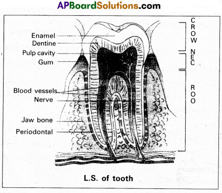
Question 11.
Draw a neat labelled diagram of L.S. of tooth.
Answer:
Question 12.
How is respiratory movements regulated in man ?
Answer:
In human beings the respiratory movements are regulated by neural system.
1. A special centre present in the medulla region of brain, called ‘respiratory rhythm centre’ is primarily responsible for this regulation.
2. Another centre present in the pons of the brain stem called ‘pneumotaxic centre’ can moderate the functions of the respiratory rhythm centre. Neural signal from and this centre can reduce the duration of inspiration and thereby alter the respiration rate.
3. A chemo-sensitive area is situated adjacent to the respiratory rhythm centre which is highly sensitive to C02 and H+. Increase in these substances can activate this centre, which inturn can send signals to the respiratory rhythm centre to make necessary adjustments in the respiratory process by which these substances can be eliminated.
4. Receptors associated with aortic arch and carotid artery also recognize changes in C02 and H+ concentration and send necessary signals to the respiratory rhythm centre for necessary actions.
The role of oxygen in the regulation of the respiratory rhythm is quite insignificant.
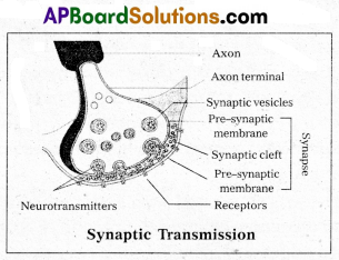
Question 13.
Give an account of synaptic transmission.
Answer:
A nerve impulse is transmitted from one neuron to another through junction called synapses.
There are two types of synapses.
1) Electrical synapses
2) Chemical synapses.
Electrical synapses : These synapses are electrically conductive links between two neurons and are also called “gap junctions “. Impulses transmission across an electrical synapses is always faster than that across a chemical synapses.
Chemical synapses: Chemicals called neuro transmitters are involved in the transmission of impulses at those synapses. When an impulse arrives at the axon terminal, it depolarizes the membrane opening voltage gated calcium channels. Calcium ions stimulate the release of neurotransmitters in the cleft by exocytosis. The released neurotransmitters bind to their specific receptors, present on the post synaptic membrane.
The post synaptic membrane has ligand gated channels. They are ion channels which respond to chemical signals, rather than to changes in the membrane potential. The entry of ions can generate a new potential in the post synaptic neuron. The new potential developed may be either excitatory (or) inhibitory.
Excitatory post synaptic potentials cause depolarisation, where as inhibitory post synaptic potentials cause hyper polarisation of post synaptic membrane.
Question 14.
Explain the mechanism by which HIV multiplies and leads to AIDS.
Answer:
AIDS is non-congential, transmissible, lethal, sexually transmitted disease caused by Human Immuno deficiency Virus (HIV). HIV is a retro virus with an envelop enclosing two ss RNA molecules as the genetic material.
Mechanism : After getting into the body of a person, the HIV enters the TH cells, macrophages or dendritic cells In these cells ss RNA of HIV synthesizes a DNA strand complementary to the viral RNA using the enzyme reverse transcriptase. The same enzyme responsible for formation of second DNA strand, complementary to the first strand forming the double stranded viral DNA. This dsDNA gets incorporated into the DNA of the host’s DNA by a viral enzyme called integrase and it is in the form of a provirus.
Transcription of DNA results in the production of RNA, which can act as the genome for new virus and it can be translated into viral proteins. The various components of the viral particles are assembled and the HIV particles are produced. The infected human cells continue to produce virus particles. New viruses bud off from the host cell and attack another TH cells. This leads to decrease CD4 receptors containing TH cells in the infected person leading to the immuno deficiency in him, finally causes AIDS.
Question15.
How is sex determined in human beings ?
Answer:
The sex determination in humans is XX – XY type. In human beings both females and males have same number of chromosomes i.e., 23 pairs. Out of 23 pairs, 22 pairs are exactly same in males and females. These are called autosomes. In addition to these (autosomes) female possesses two ‘X chromosomes while male possess one ‘X1 and one IT chromosome. During spermatogenesis among males, two types of gametes are produced. 50% of the total sperm produced carry the X-chromosome and the rest 50% has Y- chromosomes besides the autosomes. Females however, produce only one type of ovum with an ‘X’ chromosome.
There is equal probability of fertilisation of ovum with sperm carrying either X or Y chromosome. In case the ovum is fertilised with sperm carrying X-chromosome, the zygote develops into a female and the fertilisation of ovum with Y-chromosome carrying sperm results into male offspring. Thus, the sex of the child depends on the type of sperm involved in the fertilisation.
![]()
Question 16.
Explain Darwin’s theory of Natural Selection with industrial melanism as an experimental proof.
Answer:
Darwin’s theory of natural selection does not explain what exactly evolution is, but explains how evolution might have occurred in nature. A classical example for natural selection is industrial melanism, exhibited by peppered moth-Biston betularia. These moths were available in two colours grey and black. Grey moths were abundant before industrial revolution in all over England. The reason for the existence of large number of grey moths during that period was camouflage on the trunks of trees. But after the establishment of industries in England, black coloured moths were more and grey forms were less. This is due to pollution from industries in the form of soot turned barks of trees into black. So grey moths were easily identified and were more predated by birds. Thus grey moths decreased in number, black moths increased in the population.
Thus natural selection favoured the melanic moths (black) to reproduce more successfully. Natural selection of darker forms in response to industrial pollution is known as industrial melanism.
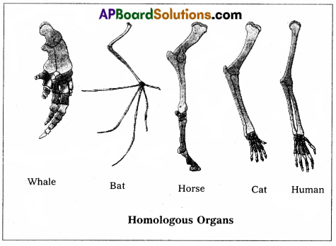
Question 17.
Distinguish between homologous and analogous organs.
Answer:
1. Homologous organs : The organs which have similar structure and origin but not necessarily the same function are called homologous organs. Eg : The appendages of vertebrates such as the flippers of whale, wings of bat, forelimbs of horse, paw of cat and hands of man have a common pattern in the arrangement of bones even though their external form and functions may vary to suit their mode of life.
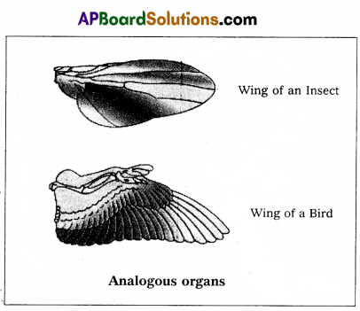
2. Analogous organs : The organs which have dissimilar structure and origin but perform the same function are called the analogous organs. Eg: Wings of butterfly and wings of a bird.
Question 18.
Explain the different types of cancers.
Answer:
Based on the origin Cancers are classified into :
1) Carcinomas: These are malignant tumor of epithelial cells’. They are originating from the epithelial tissues of skin, lining of the respiratory, digestive, urinary and genetal systems or cells from various glands breast and nervous tissue etc. 85% of Cancers are Carcinomas.
2) Sarcomas : These are malignant tumors of connective tissues or organs that originate from mesoderm. About 2% of tumors are Sarcomas.
3) Leukemias: These are malignant tumors of stem cells of hematopoietic tissues, resulting in unrestrained production of WBC. They are liquid tumors. About 4% of Cancers are Leukemias.
4) Lymphomas : These are malignant tumors of secondary lymphoid organs like spleen, and lymphnodes. About 4% of Cancers are Lymphomas.
Section – C (2 × 8 = 16)
Note : Answer any two questions in 60 lines each:
Question 19.
Describe the structure of the heart of man with the help of a neat labelled diagram.
Answer:
Human heart is a hallow muscular, cone shaped, and pulsating organ situated between lungs. It is about the size of a closed fist.
The heart is covered by double walled pericardium, which consists of outer fibrous pericardium and inner serous pericardium. The serous pericardium is double layered, outer parietal layer and inner visceral layer. These two layers are separated by pericardial space, which is filled with pericardial fluid. This fluid reduces friction between the two membranes and allow free movement of the heart.
Human heart has four chambers with two smaller upper chambers called atria and two larger lower chambers called ventricles. Atria and ventricles are separated by a deep transverse groove called coronary sulcus.
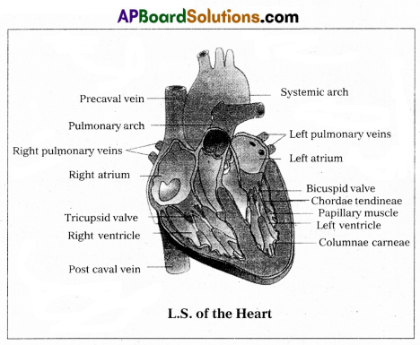
Precaval vein Pulmonary arch Right pulmonary veins Right atrium
i) Atria :
- Atria are thin walled receiving chambers. The right one is larger than the left.
- The two atria are separated by thin inter-atrial septum. It has a small pore known as Foramen Ovale’ in fetal stage. Later it is closed and appears as depression (oval patch) known as ‘Fossa ovale’. If, the foramen ovale does not close properly it is called a patent foramen ovale.
- The right atrium receives deoxygenated blood from different parts of the body, through three caval veins like two precaval veins and one post caval vein.
- The right atrium also receives blood from wall of the heart through coronary sinus, whose opening into the right atrium is guarded by the valve of Thebesius.
- Opening of the post caval vein is guarded by the Eustachian valve. It is functional in fetal stage and directs the blood from post caval vein into left atrium through foramen ovale. But it is non-functional in adult.
- The openings of the precaval veins into the right atrium have no valves.
- Left atrium receives oxygenated blood from lungs through a pair of pulmonary veins, which open into the left atrium through a common pore.
- Atrio-ventricular septum separates atria and ventricles. It has right and left atrio-venticular apertures.
- Tricuspid valve guards the right atrio-ventricular aperture. Bicuspid valve guards the left atrio-ventri- cular aperture.
ii) Ventricles:
- These are the thick walled blood pumping chambers, separated by an interventricular septum. The wall of the left ventricle is thicker than that of the right ventricle as the left ventricle must force the blood to all the parts of the body.
- The inner surface of the ventricles is raised into muscular ridges called columnae carneae. Some of them are large and conical and known as papillary muscles. Collagenous cords are known as chordae tendinae are present between atrio-ventricular valves and papillary muscles. They prevent the cusps of valves from bulging too far into atria during ventricular systole.
Nodal tissue : A specialized cardiac musculature called the nodal tissue is also distributed in the heart.
- Sino-Artrial Node (SAN) – Present in the right upper comer of right atrium.
- Atrio-Ventricular Node (AVN) – Present in the lower left comer of right atrium.
iii) Aortic arches : Human heart has two aortic arches.
- Pulmonary arch: Arises from the left anterior angle of the right ventricle. It carries deoxygenated blood to lungs. It’s opening from right ventricle is guarded by pulmonary valve made with 3 semiluminar valves.
- Left systemic arch : Arises from the left ventricle to distribute oxygenated blood to various parts in the body. Its opening is also guarded by aortic valve made with a set of 3 semilunar valves.
A fibrous strand, known as ligamentum arteriosm is present at the point of contact of the systemic and pulmonary arches. It is the remnant of the ductus arteriosus, which connects the systemic and pulmonary arches in the embryonic stage.
![]()
Question 20.
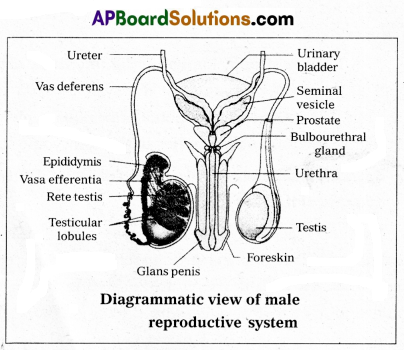
Describe male reproductive system of a man. Draw a labelled diagram of it.
Answer:
The male reproductive system or male genital system consists of a number of sex organs that are a part of the human reproductive process. The sex organs which are located in the pelvic region include a pair of testes, accessory ducts, glands and external genitalia.
1) Testes : The testes are a pair of oval pinkish male sex organs suspended in abdominal cavity within a pouch called scrotum. The scrotum helps in maintaining the low temperature of the testes (2 – 2.5°C) necessary for sper-matogenesis. The cavity of scrotal sac is connected to the abdominal cavity through the ‘inguinal canal’. Testes is held in position in the scrotum of the ‘gubernaculum’, a fibrous cord that connects the testis with the bottom of scrotum and a ‘spermatic cord’, formed by the vas deferens, nerves, blood vessels and other tissues that run from abdomen down to each testicle, through inguinal canal. Each testis is enclosed in a fibrous envelope, ‘tunica albuginea’, which extends inwards into testis and divide it into lobules. Each lobule contains 1 to 3 highly coiled seminiferous tubules. A pouch of serous membrane ‘tunica vaginalis’ covers the testis.
Miniferous tubules : Each seminiferous tubule is lined by ‘germinal epithelium’ which consists of undifferentiated male gum cells called ‘spermatogonial mother cells’ and it also bears ‘nourishing cells’ called ‘sertoli cells’.
- Spermatogonial cells (or) primary spermatocytes undergo meiotic division, producing spermatozoa or sperms by a process spermatogenesis.
- Sertoli cells provide nutrition to spermatozoa and produce a hormone ‘inhibin’, which inhibits secretion of FSH.
The region outside the tubules, contain interstitial cells of ‘Leydig cells’. They produce androgens, the most important in testosterone. It controls the development of secondary sexual characters and spermatogenesis. The seminiferous tubules open into vasa efferntia through the rete testis. Rete testis is a network of tubules is of the testis carrying spermatozoa from the seminiferous tubules to the vasa efferentia.
2) Epididymis : The vasa efferntia leave the testis and open into a narrow, tightly coiled tube called ‘epididymis’ located along the posterior surface of each testis. The epididymis provides a storage space for sperms and gives them time to nature.
It is differentiated into three regions.
a) Caput epididymis
b) Corpus epididymis
c) Cauda epididymis.
The caput epididymis receives spermatozoa via the vasa efferntia of the mediastinum testis. It is mass of a connective tissue at the back of the testis that encloses the rete testis.
3) Vasa deferentia: The vas deferens or ductus deferens is a long, narrow mascular tube. The mucosa of the ductus deferens consists of a pseudo stratified columnar epithelium and lamina propia. It starts from the tail of epididymis, passes through the inguinal canal into the abdomen and loops over the urinary bladder. It receives a duct from seminal vesicle.
The vas deferens and the duct of the seminal vesicle units to form a ‘short ejaculatory duct’ or ‘ductus ejaculatorius’. The two ducts, carrying spermatozoa and the fluid secreted by the seminal vesicles, converge in the centre of prostate and open into urethra, which transports the sperms to outside.
4) Urethra : In male, Urethra is the shared terminal duct of the reproductive and urinary systems. The urethra originates from urinary bladder and extends through the penis to its external opening called ‘urethral meatus’. The urethra provides an exit for urine as well as semen during ejaculation.
5) Penis : Urethra opens into the major copulatory organ of male, the ‘penis’. The penis and scrotum constitute the male external genitalia. The penis serves as a urinal duct and intromittent organ the transfers spermatozoa to the vagina of a female.
The penis is made up of three columns of tissue : two upper Corpora cavernosa on the dorsal aspect and one Corpus spongiosum on the ventral side. Skin and a subcutaneous layer encloses all three columns, which consists of special tissue that helps in erection of penis. The enlarged and bulbous end of penis is called ‘glans penis’, which is covered by a loose fold of skin (foreskin) called prepuce.
Male accessory glands: Male accessory glands are :
(a) Seminal vesicles
(b) Prostate glands
(c) Bulbourethral glands.
a) Seminal vesicles: These are a pair of simple tubular glands present postero-inferior to the urinary bladder in the pelvis. Each seminal vesicle enters prostate gland through vas deferens. The vesicles produce seminal fluid rich is fructose, proteins, citric acid, in organic phosphorus, potassium and prostaglandins. All these serve sperm cells.
b) Prostate gland : It is located directly beneath the urinary bladder. The gland surrounds the ‘Prostatic urethra’, and sends its secretions through prostatic ducts. The prostatic secretion activates spermatozoa and provides nutrition. In man, the prostate contributes 15 – 30% of the semen.
c) Bulbourethral glands : These are also called cowper’s glands located beneath the prostate gland at the beginning of the internal portion of the penis. They add an alkaline
fluid to semen and the fluid secreted by them lubricates urethra. It acts as flushing agent washing out the acidic urinary residues that remain in the urethra, before the semen is ejaculated.
![]()
Question21.
What are multiple alleles ? Describe multiple alleles with the help of ABO blood groups in man.
Answer:
Generally a gene has two alternative forms called allele. Sometimes a gene may have more than two alleles. These are referred to as multiple alleles. When more than two alleles exist in a population of a specific organism, the phenomenon is called multiple allelism. Multiple alleles cannot be observed in the genotype of a diploid individual, but can be observed in a population.
The number of genotypes that can occur for multiple alleles is given by the expression where ‘n’ = number of alleles.
ABO blood groups are the best example for multiple allelism in human beings.
The ABO blood group system was proposed by Karl Land- steiner. The blood groups A, B, AB and O types are characterised by the presence or absence of antigens on the surface of RBC. Blood type ‘A’ person have antigen A on their RBCs and anti-B antibodies in the plasma. Blood type ‘B’ person have antigen B. On their RBCs and anti-A antibodies in the plasma. Blood type ‘AB’ person have antigens A and B on the RBCs and no antibodies in the plasma. Blood type ‘O’ person have no antigens on their RBCs and both anti-A, and anti-B antibodies are present in the plasma.
| Blood group | Antigen on RBC | Antibodies in Plasma |
| A | A | b |
| B | B | a |
| AB | AB | — |
| O | — | a, b |
Bernstein discovered that these phenotypes were inherited by the interaction of three ‘autosomal alleles’ of the gene named T, located on chromosome 9. IA, IB and IO are the three alleles of the gene I. The alleles IA and IB are responsible for the production of the respective antigens A and B. The allele IO does not produce any antigen. The alleles IA and IB are dominant to the allele IO but co-dominant to each other (IA = IB > IO).
A child receives one of the three alleles from each parent, giving rise to six possible genotypes and four possible blood types. The genotypes are IA IA , IA IO , IB IB , IB IO , IA IB , IO IO . The phenotypic expressions of IA IA and IA IO , are A-type blood, the phenotypic expression of IBIB and IBIO are B-type blood, and that of IA IB , is AB blood type. The phenotype IO IO is ‘O-type’ blood.