Thoroughly analyzing AP Inter 2nd Year Zoology Model Papers and AP Inter 2nd Year Zoology Question Paper March 2017 helps students identify their strengths and weaknesses.
AP Inter 2nd Year Zoology Question Paper March 2017
Time: 3 Hours
Maximum Marks: 60
Instructions:
Note : Read the following instructions carefully.
- Answer all the questions of Section – A. Answer any six questions in Section – B and answer any two questions in Section – C.
- In Section – A, questions from Sr. Nos. 1 to 10 are of “Very Short Answer Type”. Each question carries two marks. Every answer may be limited to 5 lines. Answer all these questions at one place to the same order.
- In Section – B, questions from Sr. Nos. 11 to 1.8 are of “Short Answer Type”. Each question carries four marks. Every answer may be limited to 20 lines.
- In Section – C, questions from Sr. Nos. 19 to 21 are of “Long Answer Type”. Each question carries eight marks. Every answer may be limited to 60 lines.
- Draw labelled diagrams, wherever necessary in Sections – B and C.
Section – A (10 × 2 = 20)
Note : Answer all the questions in 5 lines each:
Question 1.
What is Chyme ?
Answer:
Semi fluid mass of partly digested acidic food formed in the stomach is called chyme.
Question 2.
Define Glomerular Filtration.
Answer:
The process of filteration of blood, which occur between glomerulus and lumen of the Bowman’s capsule due to difference in net pressure is called glomerular filteration. The filtered fluid which entered the Bowman’s capsule is primary urine or glomerular filtrate which is hypotonic.
Question 3.
What is Carpus Callosum ?
Answer:
Two cerebral hemispheres are internally connected by a transverse, wide and flat bundle of myelinated fibres beneath the cortex is called corpus callosum.
![]()
Question 4.
What is organ of Corti ?
Answer:
The hearing apparatus that is present in the middle canal of the cochlea Is called organ of corti. The organ of corti contains hair cells that act as auditory receptors.
Question 5.
What are Complement Proteins ?
Answer:
Complement proteins are a group of inactive plasma proteins and cell surface proteins. They are activated in cascade fashion. When activated, they form a membrane attack complex (MAC) that forms a pore in the plasma membrane, allowing ECF to enter the cell and make it swell and burst.
Question 6.
Differentiate between Perforins and Granzymes.
Answer:
Perforins : Perforins are the enzymes produced during the process of cell mediated immunity from cytotoxic T-lymphocytes. Perforins form pores in the cell membrane of the infected cells.
Granzymes : Granzymes are the enzymes produced during the process of cell mediated immunity from cytotoxic T-lymphocytes. Granzymes enter the infected Cells through the perfororations and activate certain proteins which help in distinction of the infected cell i.e., called apoptosis.
Question 7.
What is ‘Amniocentesis’ ? Name any two disorders that can be detected by Amniocentesis.
Answer:
Amniocentesis is a diagnostic procedure-to detect genetic defects in the unborn baby, in which amniotic fluid is collected from foetus and diagnosed for abnormalities. Down’s syndrome, Turner’s syndrome and Edward’s syndrome can be detected by amniocentesis.
Question 8.
What are the measures one has to take to prevent the contacting STDs ?
Answer:
The measures one has to be taken to prevent STDs are
a) Avoiding sex with unknown partners / multiple partners.
b) Using condoms compulsorily during coitus.
c) Consulting qualified doctor for early detection of STDs and getting complete treatment in case of infections.
Question 9.
Define the terms Layer and Broiler.
Answer:
Layer : The birds which are raised exclusively for the production of eggs are called layers.
Broiler : The birds which are raised only for their meat are called broilers.
![]()
Question 10.
What is apiculture ?
Answer:
Apiculture is the maintenance of hives of honeybees for the production of honey and wax.
Apiculture is an age-old cottage industry.
Section – B (6 × 4 = 24)
Note : Answer any six questions in 20 lines each:
Question 11.
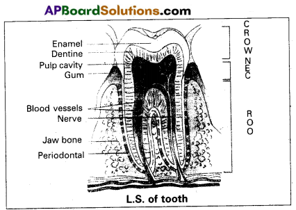
Draw a neat labelled diagram of L.S. of a tooth.
Answer:
Question 12.
Describe disorders of Respiratory System.
Answer:
Disorders of respiratory system.
1) Asthma : Asthma is a difficulty” in breathing caused due to inflammation of bronchi and bronchioles. Symptoms include coughing, difficulty in breathing and wheezing.
2) Emphysema : It is a chronic disorder in which alveolar walls are damaged and their walls coalesce due to which respiratory surface area of exchange of gases is decreased. One of the major causes of this is smoking of tobacco.
3) Bronchitis : Bronchitis is the inflammation of the bronchi, resulting in the swelling of mucus lining of bronchi, increased mucus production and decrease in the, diameter of bronchi. Symptoms include chronic cough with thick sputum.
4) Pneumonia : The infection of lungs caused by Strep-tococcus pneumoniae and also by certain Virus, Fungi, Protozoans and Mycoplasmas. Symptoms include inflammation of lungs, accumulation of mucus in alveoli and impaired exchange of gases, leading tq death if untreated.
Occupational dissorders : These are caused by exposure of the body to the harmful substances.
E.g. : i) Asbestosis : It occurs due to chronic exposure to asbestos dust in the people working in asbestos industiy.
ii) Silicosis : It occurs because of long term exposure to silica dust.
iii) Siderosis : It occurs due to deposition of iron particles in tissues.
iv) Black lung disease : It develops from inhalation of coal dust.
Question 13.
Describe the structure of a Synovial Joint with the help of neat labelled.
Answer:
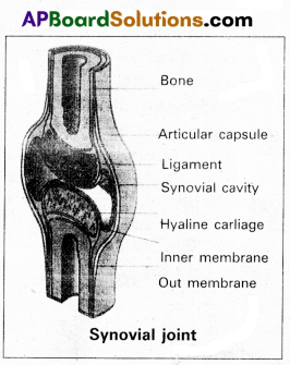
Synovial joints are characterised by the presence of a fluid filled synovial cavity between the articulating surfaces of the two bones.
Structure of synovial joint : Synovial joint is covered by a double layered synovial capsule. The outer layer consist of dense fibrous irregular connective tissue with more collagen fibers. This layer is continuous with the periosteum and resists stretching and prevents the dislocation of joints. Some fibres of these membranes are arranged in bundles called ligaments.
The inner layer of synovial capsule is formed of areolar tissue and elastic fibers. It secretes a viscous synovial fluid which contains hyaluronic acid, phagocytes etc., and acts as a lubricant for the free movement of the joints.
Question 14.
Explain how Hypothyroidism and Hyperthyroidism can effect the body.
Answer:
Hypothyroidism: Inadequate supply of iodine or impairment in the function of thyroid glands leads to decrease in production of thyroid hormones (T3 & T4) results in hypothyroidism and enlargement of the thyroid gland called Simple goiter.
During pregnancy due to hypothyroidism, defective development of the growing body leads to a disorder called Cretinism. Physical and mental growth gets severely stunted due to untreated congenital hypothyroidism, stunted growth, mental retardation, low IQ, deafness, and mutism are some characteristics features of this disease.
In adult women it may cause irregular menstrual cycles. Hypothyroidism in adult causes Myxoedema characterized by bagginess under the eyes, puffiness of face, dry skin, slowness in physical and mental activities.
Hyperthyroidism : Over activity of thyroid, cancer of the gland or development of nodule of thyroid lead to hyper thyroidism. In adults it causes an abnormal growth leads to a disease called Exophthalmic goiter with characteristically protruded eyeballs. Hyperthyroidism also affects the physiology of the body i.e., increased metabolic rate, nervousness, rapid heartbeat, sweating, increased appetite etc.
![]()
Question 15.
Describe Erythroblastosis foetalis.
Answer:
Erythroblastosis foetalis develops in an Rh positive foetus, whose father is Rh positive and mother is Rh negative. In Rh positive person rhesus antigens are present on the surface of blood cells where as in Rh negative person Rhesus antigens are absent.
During the process of delivery, the foetal blood cells may pass through the ruptured placenta into the Rh negative maternal blood. The mother’s immune system recognises the Rh antigens and gets sensitized and produces Rh antibodies. These antibodies are Ig G type they can pass through placenta. Generally first child is not effected because child is delivered by the time of the mother gets sensitized and produce antibodies.
During second pregnancy, if the second child is Rh positive, these antibodies cross the placenta, enter the foetal blood circulation and destroy the Rh positive blood cell of foetus (haemolysis), leads to haemolytic anemia and jaundice. To compensate the haemolysis of blood cells there is a rapid production of RBC’s from the bone marrow, and but also from liver and spleen. Now many large and immature blood cells in erythroblast stage are released into circulation. Because of this disease is called erythroblastosis foetalis.
![]()
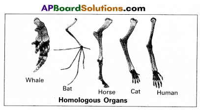
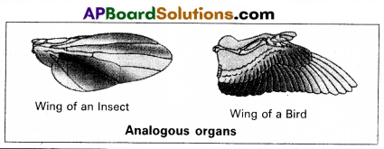
Question 16.
Distinguish between Homologous and Analogous organs.
Answer:
1. Homologous organs : The organs which have similar structure and origin but not necessarily the same function are called homologous organs. Eg : The appendages of vertebrates such as the flippers of whale, wings of bat, forelimbs of horse, paw of cat and hands of man have a common pattern in the arrangement of bones even though their external form and functions may vary to suit their mode of life.
2. Analogous organs : The organs which have dissimilar structure and origin but perform the same function are called the analogous organs. Eg : Wings of butterfly and wings of a bird.
Question 17.
Explain Darwin’s theory of Natural Selection with industrial melanism as an experimental proof.
Answer:
Darwin’s theory of natural selection does not explain what exactly evolution is, but explains how evolution might have occurred in nature. A classical example for natural selection is industrial melanism, exhibited by peppered moth-Biston betularia. These moths were available in two colours grey and black. Grey moths were abundant before industrial revolution in all over England. The reason for the existence of large number of grey moths during that period was camouflage on the trunks of trees. But after the establishment of industries in England, black coloured moths were more and grey forms were less. This is due to pollution from industries in the form of soot turned barks of trees into black. So grey moths were easily identified and were more predated by birds. Thus grey moths decreased in number, black moths increased in the population.
Thus natural selection favoured the melanic moths (black) to reproduce more successfully. Natural selection of darker forms in response to industrial pollution is known as industrial melanism.
Question 18.
Write about the procedure involved in MRI.
Answer:
MRI Scan is a diagnostic radiology technique that uses magnetism, radiowaves and a computer to produce images of body components.
Procedure : MRI Scanner is giant circular magnetic tube.
- The patient is placed on a movable bed that is inserted into the magnet.
- Human body is mainly composed of water which contains two protons.
- The magnet creates a strong magnetic field that makes these proton align with the direction of the magnetic field.
- A second radiofrequency electromagnetic field is then turned on for a brief period. The protons absorb some energy from these radio waves.
- When this second radio frequency emitting field is turned off, the protons release energy at a radio frequency which can be detected by the MRI scanner.
- Different types of tissues emit different quanta of energy. Abnormal tissues such as tumors can be detected because the protons in different types of tissues return to their equilibrium state at different rates.
- Tissues of bone with less water content look different in MRI, and pathological tissues also can be detected.
The information received is processed by computer and generated an image.
Section – C (2 × 8 = 16)
Note : Answer any two questions in 60 lines each:
Question 19.
Write notes on the working of the heart of human.
Answer:
The human heart is an organ that provides a continuous blood circulation through the cardiac cycle.
Special conducting tissues of heart : Human heart is myogenic. It contains a specialized cardiac musculature called the nodal tissue. A patch of this tissue called sino-atrial node (SAN), is present in the right upper comer of the right atrium. Another mass of this tissue, called the atrio-ventricular tissue (AVN), is present in the lower left corner of the right atrium. A bundle of nodel fibers called AV bundles/His bundles continues from the AVN into the inter-ventricular septum. It divides into right and left bundle branches. These branches give rise to minute fibers called purkinje fibers that extend throughout the ventricular musculature.
SAN has the ability to generate action potentials without any .external stimuli, hence called pacemaker. AV node is a relay point that relay the action potentials received from the SA node to the ventricular musculature.
Cardiac cycle : Cardiac cycle consists of the sequence of the cardiac events that occur from the beginning of one heart beat to the beginning of next. At beginning of cardiac cycle all the four chambers of the heart are in relaxed state. Cardiac cycle is divided into three phases, namely.
1) Atrial systole
2) Ventricular systole
3) Cardiac diastole
1) Atrial systole : It lasts about 0.1 seconds. SAN now stimulate an action potential which stimulates both the atria to contract simultaneously causing the atrial systole. This increases the flow of blood into the ventricles by about 30%, the remaining blood flows into the ventricle before the atrial systole.
2) Ventricle systole : It lasts about 0.3 seconds. The action potential from the SAN reaches the AVN, from where they are conducted through the bundle of His, its branches and the Purkinje fibers to entire ventricular musculature. This causes the simultaneous ventricular systole. The atria undergo relaxation coinciding with ventricular systole. Ventricular systole increases the pressure causing closure of AV valves preventing the back flow of blood, results in the production of first heart beat sound ‘Lub’. When pressure in ventricles exceeds the pressure in aortic arches, semilunar valves open. It results in the flow of blood from ventricles into aortic arches.
3) Cardiac diastole: It lasts about 0.4 seconds. The ventricles now relax and ventricular pressure falls causing the closure of the semilunar valves which prevent the back flow of blood. This results in the production of second heart sound known as ‘Dup’. When pressure in ventricles falls below atrial pressure, AV valves open and ventricular filling begins. All the chambers are now again in relaxed state. Soon another cardiac cycle sets in.
Question 20.
Describe male reproductive system of human. Draw a labelled diagram of it.
Answer:
The male reproductive system or male genital system consists of a number of sex organs that are a part of the human reproductive process. The sex organs which are located in the pelvic region include a pair of testes, accessory ducts, glands and external genitalia.
1) Testes : The testes are a pair of oval pinkish male sex organs suspended in abdominal cavity within a pouch called scrotum. The scrotum helps in maintaining the low temperature of the testes (2 – 2.5°C) necessary for sperma-togenesis. The cavity of scrotal sac is connected to the abdominal cavity through the ‘inguinal canal’. Testes is held in position in the scrotum of the ’guberna-culum’, a fibrous cord that connects the testis with the bottom of scrotum and a ’spermatic cord’, formed by the vas deferens, nerves, blood vessels and other tissues that run from abdomen down to each testicle, through inguinal canal. Each testis is enclosed in a fibrous envelope, ‘tunica albuginea’, which extends inwards into testis and divide it into lobules. Each lobule contains 1 to 3 highly coiled seminiferous tubules. A pouch of serous membrane ‘tunica vaginalis’ covers the testis.
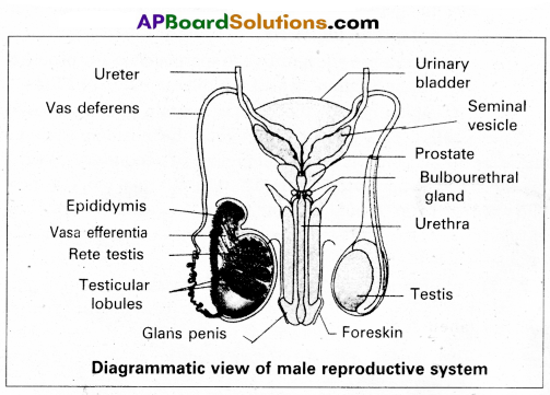
Miniferous tubules : Each seminiferous tubule is lined by ‘germinal epithelium’ which consists of undifferentiated male gum cells called ‘spermatogonial mother cells’ and it also bears ‘nourishing cells’ called ‘sertoli cells’.
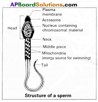
- Spermatogonial cells (or) primary spermatocytes undergo meiotic division, producing spermatozoa or sperms by a process spermatogenesis.
- Sertoli cells provide nutrition to spermatozoa and produce a hormone ‘inhibin’, which inhibits secretion of FSH.
The region outside the tubules, contain interstitial cells of ‘Leydig cells’. They produce androgens, the most important in testosterone. It controls the development of secondary sexual characters and spermatogenesis. The seminiferous tubules open into vasa efferntia through the rete testis. Rete testis is a network of tubules is of the testis carrying spermatozoa from the seminiferous tubules to the vasa efferentia.
2) Epididymis : The vasa efferntia leave the testis and open into a narrow, tightly coiled tube called ‘epididymis’ located along the posterior surface of each testis. The epididymis provides a storage space for sperms and gives them time to nature.
It is differentiated into three regions.
a) Caput epididymis
b) Corpus epididymis
c) Cauda epididymis
The caput epididymis receives spermatozoa via the vasa efferntia of the mediastinum testis. It is mass of a connective tissue at the back of the testis that encloses the rete testis.
3) Vasa deferentia : The vas deferens or ductus deferens is a long, narrow mascular tube. The mucosa of the ductus deferens consists of a pseudo stratified columnar epithelium and lamina propia. It starts from the tail of epididymis, passes through the inguinal canal into the abdomen and loops over the urinary bladder. It receives a duct from seminal vesicle.
The vas deferens and the duct of the seminal vesicle units to form a ‘short ejaculatory duct’ or ‘ductus ejaculatorius’. The two ducts, carrying spermatozoa and the fluid secreted by the seminal vesicles, converge in the centre of prostate and open into urethra, which transports the sperms to outside.
4) Urethra : In male, Urethra is the shared terminal duct of the reproductive and urinary systems. The urethra originates from urinary bladder and extends through the penis to its external opening called ‘urethral meatus’. The urethra provides an exit for urine as well as semen during ejaculation.
5) Penis : Urethra opens into the major copulatoiy organ of male, the ‘penis’. The penis and scrotum constitute the male external genitalia. The penis serves as a urinal duct and intromittent organ the transfers spermatozoa to the vagina of a female.
The penis is made up of three columns of tissue : two upper Corpora cavernosa on the dorsal aspect and one Corpus spongiosum on the ventral side. Skin and a subcutaneous layer encloses all three columns, which consists of special tissue that helps in erection of penis. The enlarged and bulbous end of penis is called ‘glarts penis’ , which is covered by a loose fold of skin (foreskin) called prepuce.
Male accessory glands : Male accessory glands are :
(a) Seminal vesicles
(b) Prostate glands
(c) Bulbourethral glands
a) Seminal vesicles : These are a pair of simple tubular glands present postero-inferior to the urinary bladder in the pelvis. Each seminal vesicle enters prostate gland through vas deferens. The vesicles produce seminal fluid rich is fructose, proteins, citric acid, in organic phosphorus, potassium and prostaglandins. All. these serve sperm cells.
b) Prostate gland: It is located directly beneath the urinary bladder. The gland surrounds the ‘Prostatic urethra’, and sends its secretions through prostatic ducts. The prostatic secretion activates spermatozoa and provides nutrition. In man, the prostate contributes 15 – 30% of the semen.
c) Bulbourethral glands : These are also called cowper’s glands located beneath the prostate gland at the beginning of the internal portion of the penis. They add an alkaline fluid to semen and the fluid secreted by them lubricates urethra. It acts as flushing agent washing out the acidic urinary residues that remain in the urethra, before the semen is ejaculated.
![]()
Question 21.
What is criss-cross Inheritance ? Explain the inheritance of one sex linked recessive character in human beings.
Answer:
The X-linked genes are represented twice in female (because female has two ‘X’-chromosomes) and once in males, (because male has one X-chromosome). In male single X-linked recessive gene express it phenotypically, in contrast to female in which two X’ linked recessive genes are necessary for the determination of a single phenotypic trait related to sex.
The recessive X-linked genes have chracteristic crisscross- inheritance.
Crisscross inheritance : The inheritance of X-liriked recessive trait (genes) to his grandson (F2) through his daughter (carrier) is called crisscross inheritance. Crisscross inheritance can be explained in humans by sex-linked recessive disorder, colour blindness.
Colourblindness : Colour blindness is a particular trait in human beings render them unable to differentiate between red and green colour. The gene for this colour blindness is located on X-chromosome. Colour blindness is recessive to normal vision so that if colour blind man marries a normal vision (homozygous) woman, all the sons and daughters are normal but daughter are heterozygous, which means that these daughters would be carrier for this trait. If such carrier woman marries a man with normal vision all the daughters and half of the sons have normal vision and half of sons are colour blind.
Colour blind trait is inhereted from a male parent to his grandson through carrier daughter i.e., this trait shows crisscross inheritance
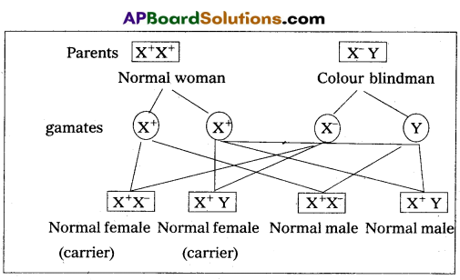
If carrier female is married to normal male
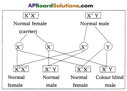
- They never passed from father to son.
- Males are much more likely to be affected because they need only one copy of the mutant allele to express the phenotype.
- Affected males get the disease from their carrier mother only.
- Sons of heterozygous female (i.e., carrier female) have 50% chance of receiving mutant alleles. These disorders are typically passed from an affected grandfather to 50% of his grandsons.
- The X-linked recessive traits, shows Crisscross pattern of inhertance.
Eg: Colourblindness, Hemophilia, Muscular dystrophy etc.,