Thoroughly analyzing AP Inter 2nd Year Zoology Model Papers Set 3 helps students identify their strengths and weaknesses.
AP Inter 2nd Year Zoology Model Paper Set 3 with Solutions
Time: 3 Hours
Maximum Marks: 60
Section – A (10 × 2 = 20)
Answer all the questions. (Very Short Answer Type)
Question 1.
What is meant by chloride shift ?
Answer:
Chloride shift : It refers to the exchange of chloride and bicarbonate ions between erythrocytes and plasma. It is also called Hamburger’s phenomenon.
Question 2.
Name the valves that guard the left and right atrio-ventricular opertures in man.
Answer:
Bicuspid valve (or) Mitral valve – Left atrio-ventricular aperture. Tricuspid valve – Right atrio-venticular aperture.
Question 3.
Write the difference between actin and myosin.
Answer:
| Actin | Myosin |
| 1) Actin is a thin contractile protein. | 1) Myosin is a thick contractile protein. |
| 2) It is present in light bands and is called an isotropic band. | 2) It is present in dark bands and is called an anisotropic band. |
| 3) Each actin filament is made of two ‘F’ actin molecules helically wound around each other, tropo-myosin and a complex protein called troponin. | 3) Each myosin is made up of monomeric protein called meromyosins. Each mer-myosin has two parts namely head, and arm (or) neck. |
Question 4.
What are Islets of langerhans ?
Answer:
The endocrine region of pancreas is called Islets of langerhans where it contain 1 to 2 millions Islets of langerhans. There are two main types of cells α-cells and β-cells.
α-cells produce the hormone glucagon, whereas β- cells produce insulin.
![]()
Question 5.
Define spermiogenesis and spermiation.
Answer:
Spermiogenesis : The process in which haploid spermatids are transformed into spermatozoa or sperms.
Spermiation : The process in which sperm head becomes embedded in the Sertoli cells and finally released from the seminiferous tubules.
Question 6.
What is erythroblastasis foetalis ?
Answer:
Erythroblastosis foetalsis is an alloimmune condition that develops in an Rh positive foetus whose father is Rh positive and mother is Rh negative.
In this disorder the antibodies developed against the Rh antigen in mother, cross, placenta and destroy the RBC cells of the Rh+ve foetus during second pregnancy.
Question 7.
Mention the names of any four connecting links that you have studied.
Answer:
Connecting links clearly explain the path of evolution.
- Peripatas between annelida and arthropoda.
- Prototherians between reptilia and mamnalia.
Question 8.
What is meant by the term osmoregulation ?
Answer:
The process of maintaining the quantity of water and dissolved solutes in balance is referred to as osmoregulation.
Question 9.
What is Lyonisation ?
Answer:
Lyonisation is a process by which one of two copies of X- chromosome present in the body cells of female mammals is inactivated. The inactive X-chromosome is transcriptionally inactive called heterochromatic body.
Question 10.
List out any four features of cancer cells.
Answer:
- Loss of contact inhibition
- Reduced intra cellular adhesion
- Immortalization
- Loss of anchorage dependence
Section – B (6 × 4 = 24)
Answer any six questions. (Short Answer Type)
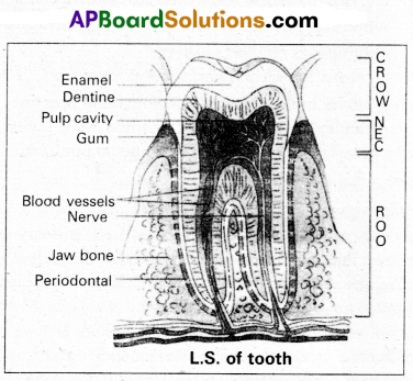
Question 11.
Draw a neat labelled diagram of L.S. of a tooth.
Answer:
Question 12.
Describe the events in a cardiac cycle, briefly.
Answer:
The cardiac events that occur from the beginning of on heart beat to the beginning of the next, is called cardiac cycle. Cardiac eye consists of three phases namely atrial systole, ventricular sys tie and cardiac diastole.
i) Atrial systole : It lasts about 0.1 seconds.
The SAN generate an action potential which stimulate contraction of atria, which helps in the flow of blood into ventricles by about 30%. The r emaining blood flows into the ventricles before the atrial systole.
ii) Ventricular systole : It lasts about 0.3 seconds .
- Ventricles contract and atria relax during this phase.
- Contraction of ventricles raises the pressure in ventricles due to which AV valves are closed. It causes the first heart sound “Lub”.
- When pressure in ventricles exceeds the pressure in aortic arches, semilunar valves open. It results the flow of blood from ventricles into aortic arches.
iii) Cardial diastole : It lasts about 0.4 seconds.
- The ventricles now relax, atria are also in diastolic condition.
- When pressure in ventricles falls below that in aortic arches, semilunar valves are closed.
- It causes the second heart sound “dup”.
When pressure in ventricles falls below atrial pressure, AV valves open and ventricular filling begins. The total cycle takes about 0.8 seconds. This gives a heart rate of about 75 beats per minute.
Question 13.
Describe the Graafian fallicle in woman.
Answer:
During ‘oogenesis’, the formed garnet mother cells or oogonia in each foetal ovary are called primary oocytes. Each primary oocyte gets sorrounded by a flattened layer of follicular cells. It is called ‘primordial follicle’. The follicles become cuboidal and proliferate to produce stratified epithelium made up of cells called granulosa cells. Follicles at this stage of development are called ’primary follicles’. A homogenous membrane, the ‘zona pellucida’ appears between primary oocyte and granulosa cells. The innermost layer of granulosa cells are firmly attached to zona pellucida forming ‘corona radiata’.
A cavity appears in membrane granulosa, it increases in size, wall of follicle becomes thin. As the follicle expands the stromal cells sorrounding the granulosa become condensed to form a covering called inner tneca interna’ and outer theca externa’. Now these follicles are called secondary follicles’.
The cells of theca interna secrete a hormone called Oestrogen. At this stage, the primary oocyte within the secondary follicles grows in size and completes ‘meiosis I’ forming a large haploid ‘Secondary oocyte’ and a small ‘first polar body’. Then the 2nd meoitic division starts but Stops at metaphase. The secondary follicle further changes into the nature follicle called ‘Grafian follicle’. The rupture of graafian follicle by LH results in the release of ovum, a process called ovulation.
![]()
Question 14.
Describe the placenta in a women.
Answer:
Placenta is a structural and functional unit of both chorionic villi and uterine tissue and it develops between the embryo (foetus) and the mother. The maternal and foetal blood do not mix with each other. They are seperated by the placental membrane.
The placenta consists of two essential portions : a maternal part of the placenta derived from the endometrium of the uterus and foetal membranes of the foetal part of the placenta.
The maternal components of the Placenta are :
a) Uterine epithelium
b) Uterine connective tissue
c) Uterine capillary endothelium.
The foetal components of the Placenta are :
a) Foetal capillary endothelium
b) Foetal connective tissue
c) Foetal chorionic epithelium.
The Placenta of human is called chorioallantoic placenta’as allantois fuse with chorion in the process of vascularisation. Placenta is discoidal as the Villi are restricted to the dorsal surface of- blastodisc. Placenta is haemochorial as the maternal blood comes into direct contact with foetal chorion. During parturition the placenta is cast off with the loss of embryonic membranes and the encapsulating maternal tissues (decidua) causing extensive haemorrhage and there by bleeding. So, it is also called deciduate placenta.
Functions of placenta :
- Supplies Oxygen and nutrients to the embryo.
- Removes CO2 and excretory materials produced by embryo.
- Secretes’ Progesterone which is essential for maintenance of pregnancy after 4th month.
- Secretes Oestrogens (mainly estradiol) that reach maternal blood and promote uterine growth and development of mammary glands.
- Secretes Human Chorionic Gonadotropin (HCG) that is similar to luteinizing hormone is its action. This hormone is also used as indicator in the detection of pregnancy is early stages.
- Somatomammotropin secreted by placenta has an anti-insulin effect on the mother leading to increased plasma levels of glucose and aminoacids in the maternal circulation. In this way it increases the availability of these materials to the foetus.
Question 15.
Write a note on the mechonism of action of harmones.
Answer:
Hormones are primary messengers which interacting with receptors and they generate secondary messengers. These secondary messengers regulate cellular metabolism in the target cells.
Mechanism of action of lipid insoluble hydrophillic hormone :
- The Hormone binds to a stimulatory membrane bound receptor, and stimulate ‘G’ protein.
- ‘G’ protein of the cell membrane binds to GTP and activates adenylate cyclase. .
- Adenylate Cyclase forms cAMP from ATP.
- cAMP activates the protein kinase, which activates the enzyme phosphorylase.
- Phosphorylase further phosphorylate the inactive enzyme and convert it to active form and involved in the metabolic process.
Eg : Epinephrine.
Mechanism of action of lipid soluble hormone Lipid soluble hormones easily diffuse through the cell membrane.
- It binds to a specific receptor in the cytoplasm forming hormone receptor complex molecule.
- This complex molecule enters the nucleus and binds to the DNA and stimulate the production of specific m-RNA molecule.
- The m-RNA passes into the cytoplasm, where it is involved in the translation process and synthesizes a protein. These proteins produced by the cell as a response of hormone and plays an important role in. their respective metabolism. Eg: Aldosterone
Question 16.
Explain Darwin’s theory of natural selection with industrial melanism as an experimental proof.
Answer:
Darwin’s theory of natural selection does not explain what exactly evolution is, but explains how evolution might have occurred in nature. A classical example for natural selection is industrial melanism, exhibited by peppered moth-Biston betularia. These moths were available in two colours grey and black. Grey moths were abundant before industrial revolution in all over England. The reason for the existence of large number of grey moths during that period was camouflage on the trunks of trees. But after the establishment of industries in England, black coloured moths were more and grey forms were less. This is due to pollution from industries in the form of soot turned barks of trees into black. So grey moths were easily identified and were more predated by birds. Thus grey moths decreased in number, black moths increased in the population.
Thus natural selection favoured the melanic moths (black) to reproduce more successfully. Natural selection of darker forms in response to industrial pollution is known as industrial melanism.
![]()
Question 17.
Write a short notes on controlled breeding experiments.
Answer:
Controlled breeding experiments are carried out using artificial insemination and multiple ovulation and embryo transfer technology.
- In this technique the semen is collected from superior bulls. This semen can be used immediately or can be frozen and used later period. It can be transported in a frozen form to place where a female is housed.
- Meanwhile a cow or animal is administered hormones, with FSH like activity.
- These hormones induces follicular maturation and super ovulation.
- Now the cow is artificially inseminated for fertilisation.
- The embryos are at 8 – 32 celled stages are recovered non-surgically and transferred to surrogate mother uterus for further development.
This technology is use for cattle, sheep, rabbits, buffaloes etc. By using this method we can produce high milk and meat yielding animals and also control the venereal diseases.
Question 18.
Describe atria of the heart of man.
Answer:
Atria are thin walled receiving chambers, form the anterior part of the heart. The right one is larger than the left, they are separated by inter-atrial septum. It has small pore in embryonic stage known as Foramen Ovale. Later it is closed and appears as a depression in the septum known as Fossa ovalis. If the foramen ovale does not close properly, it is galled a patent foramen ovale.
The right atrium receives deoxygenated blood from different parts ,of the body (except the lungs) through three caval veins like two precaval veins and one post caval vein. The right atrium also receives blood from the walls of the heart through the coronary sinus, whose opening into the right atrium is guarded by a crescentric fold, the valve of Thebesius. Opening of the post caval vein is guarded by the valve of inferior vena cavae or Eustachian valve. It directs the blood to the left atrium through the foramen ovale, in the fetal stage, but in the adults it becomes non functional.
The openings of the precaval veins into the right atrium have no valves. The left atrium receives oxygenated blood from lungs through a pair of pulmonary veins, which opens into the left atrium through a common pore. Atrio-ventricular septum separates atria and ventricles. It has right and left atrio-ventricular apertures.
Tricuspid valve guards the right atrio-ventricular aperture and bicuspid valve (mitral valve) guards the left atrio-ventricular aperture.
Section – C (2 × 8 = 16)
Answer any two questions. (Long Answer Type)
Question 19.
Explain the transmission of nerve impulse through a nerve fibre with the help of suitable diagram.
Answer:
Nerve impulse is tlje Combination of mechanical, chemical (or) electrical disturbances occur in neuron because of stimulus. The propagation of a impulse along nerve fibre is called transmission. In this process both physical and chemical changes are involved. The entire process is divided into stimulation, excitation, conduction and response.
Resting membrane potential : The resting membrane potential exists because of a small buildup of negative ions in the axoplasm along the inside of the membrane and an equal buildup of positive ions in the extra cellular fluid along the outer surface of the membrane. Such a separation of positive and negative electrical charges is a form of potential enerfsr. In neurons, the resting membrane potential ranges from -40 to – 90 mV. A typical value is-70 mV.
At resting phase, the axolemma is polarized. If the inner side becomes less negative, it is said to be depolarized. If the inner side becomes more negative, it is said to be hyperpolarized. During the resting phase the activation gates of sodium are closed, the inactivation gates of sodium are open and the activation gates of potassium are closed.
Sodium-potassium pump : Sodium and potassium ions diffuse inwards and outwards, respectively, down their concentration gradients through leakage channels. Such a movement of ions, if unchecked, would eventually disturb the resting membrane potential. These flows of ions are offset by sodium-potassium pumps (Na+/K+ ATPases) present in the axonal walls. These pumps expel three Na+ ions for each two K+ ions imported. As these pumps remove more positive charges from the axoplasm than they bring into it, they contribute to the negativity of the resting membrane potential i.e., -70mv.
Depolarization (Rising phase) : When a nerve fibre is stimulated, the plasma membrane becomes more permeable to Na+ ions than to K+ ions as the activation and inactivation voltage gates of sodium open and activation voltage gates of potassium close. As a result the rate of flow of Na+ into the axoplasm exceeds the rate of flow of K+ to the ECF. Hence, the axolemma is positively charged inside and negatively charged outside. This reversal of electrical charge is called “depolarization”.
Outer face of the point which is adjacent to the site of depolarization remains positively charged. The electrical potential difference between these two areas is called “action potential”. An action potential occurs in the membrane of the axon of a neuron when depolarization reaches a certain level called ‘threshold potential’ (-55 mV). The particular stimulus which is able to bring the membrane potential to threshold is called ‘threshold stimulus’.
Repolarization (Falling phase) : As the wave of depolarization passes away from its site of origin to the adjacent point, the activation gates of sodium remain open, inactivation gates of sodium close and activation gates of potassium open at the site of origin of depolarization. As a result the influx of Na+ ions into the axoplasm from the ECF is checked and ’efflux’ of K+ ions occurs, which leads to the returning of axolemma to the resting state (exit of potassium ions causes a reversal of membrane potential to negative inside). This is called ‘repolarization’.
Hyperpolarization (Undershoot) : The repolarization typically goes more negative than the resting potential to about -90 mV This is called ‘hyperpolarization’. This occurs because of the increased K+ permeability that exists while voltage gated K+ channels are open activation and inactivation gates of Na+ channels remain closed. The membrane potential returns to its original resting state as the K+ channels close completely. As the voltage falls below the -70 mV level of the resting state, it is called ‘undershoot’.
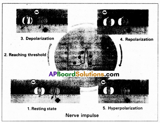
The refractory periods: The period of time after an action potential begins during which the neuron cannot generate another action potential in response to a normal threshold stimulus is called the ‘refractory period’. There are two kinds of refractory periods, namely absolute refractory period and relative refractory period. During the absolute refractory period, even a very strong stimulus cannot initiate a second action potential. The relative refractory period is the time during which a second action potential can be initiated by a larger than normal stimulus.
Conduction speed : The conduction speed of a nerve impulse depends on the diameter of the axon : the greater the axon’s diameter, the faster is the conduction. In a myelinated axon, the voltage-gated Na+ and K+ channels are concentrated at the nodes of Ranvier. As a result the impulse jumps’ from one Ranvier’s node to the next, rather than travelling the entire length of the nerve fibre. This mechanism of conduction is called Saltatory conduction. Saltatory conduction is faster (in myelinated fibres) than the continuous conduction (in nonmyelinated fibres).
![]()
Question 20.
Write notes on the working of heart of man.
Answer:
The human heart is an organ that provides a continuous blood circulation through the cardiac cycle.
Special conducting tissues of heart : Human heart is myogenic. It contains a specialized cardiac musculature called the nodal tissue. A patch of this tissue called sino-atrial node (SAN), is present in the right upper corner of the right atrium. Another mass of this tissue, called the a trio-ventricular tissue (AVN), is present in the lower left corner of the right atrium. A bundle of nodel fibers called AV bundles/His bundles continues from the AVN into the inter-ventricular septum. It divides into right and left bundle branches. These branches give rise to minute fibers called purkinje fibers that extend throughout the ventricular musculature.
SAN has the ability to generate action potentials without any external stimuli, hence called pacemaker. AV node is a relay point that relay the action potentials received from the SA node to the ventricular musculature.
Cardiac cycle : Cardiac cycle consists of the sequence of the cardiac events that occur from the beginning of one heart beat to the beginning of next. At beginning of cardiac cycle all the four chambers of the heart are in relaxed state. Cardiac cycle is divided into three phases, namely.
1) Atrial systole
2) Ventricular systole
3) Cardiac diastole
1) Atrial systole : It lasts about 0.1 seconds. SAN now stimulate an action potential which stimulates both the atria to contract simultaneously causing the atrial systole. This increases the flow of blood into the ventricles by about 30%, the remaining blood flows into the ventricle before the atrial systole.
2) Ventricle systole : It lasts about 0.3 seconds. The action potential from the SAN reaches the AVN, from where they are conducted through the bundle of His, its branches and the Purkinje fibers to entire ventricular musculature. This causes the simultaneous ventricular systole. The atria undergo relaxation coinciding with ventricular systole. Ventricular systole increases the pressure causing closure of AV valves preventing the back flow of blood, results in the production of first heart beat sound ‘Lub’. When pressure in ventricles exceeds the pressure in aortic arches, semilunar valves open. It results in the flow of blood from ventricles into aortic arches.
3) Cardiac diastole : It lasts about 0.4 seconds. The ventricles now relax and ventricular pressure falls, causing the closure of the semilunar valves which prevent the back flow of blood. This results in the production of second heart sound known as ‘Dup’. When pressure in ventricles falls below atrial pressure, AV valves open and ventricular filling begins. All the chambers are’ now again in relaxed state. Soon another cardiac cycle sets in.
![]()
Question 21.
Describe male reproductive system of a man. Draw a labelled diagram of it.
Answer:
The male reproductive system or male genital system consists of a number of sex organs that are a part of the human reproductive process. The sex organs which are located in the pelvic region include a pair of testes, accessory ducts, glands and external genitalia.
1) Testes : The testes are a pair of oval pinkish male sex organs suspended in abdominal cavity within a pouch called scrotum. The scrotum helps in maintaining the low temperature of the testes (2 – 2.5°C) necessary for spermatogenesis. The cavity of scrotal sac is connected to the abdominal cavity through the ‘inguinal canal’. Testes is held in position in the scrotum of the ‘gubemaculum’, a fibrous cord that connects the testis with the bottom of scrotum and a ‘spermatic cord’, formed by the vas deferens, nerves, blood vessels and other tissues that run from abdomen down to each testicle, through inguinal canal. Each testis is enclosed in a fibrous envelope, ‘tunica albuginea’, which extends inwards into testis and divide it into lobules. Each lobule contains 1 to 3 highly coiled seminiferous tubules. A pouch of serous membrane ‘tunica vaginalis’ covers the testis.
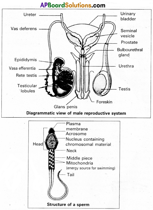
Miniferous tubules : Each seminiferous tubule is lined by ‘germinal epithelium’ which consists of undifferentiated male gum cells called spermatogonial mother cells’ and it also bears ‘nourishing cells’ called ’sertoli cells’.
- Spermatogonial cells (or) primary spermatocytes undergo meiotic division, producing spermatozoa or sperms by a process spermatogenesis.
- Sertoli cells provide nutrition to spermatozoa and produce a hormone ‘inhibin’, which inhibits secretion of FSH.
The region outside the tubules, contain interstitial cells of ‘Leydig cells’. They produce androgens, the most important in testosterone. It controls the development of secondary sexual characters and spermatogenesis .-The seminiferous tubules open into vasa effemtia through the rete testis. Rete testis is a network of tubules is of the testis carrying spermatozoa from the seminiferous tubules to the vasa efferentia.
2) Epididymis : The vasa effemtia leave the testis and open into a narrow, tightly coiled tube called’epididymis’ located along the posterior surface of each testis. The epididymis provides a storage space for sperms and gives them time to nature.
It is differentiated into three regions.
a) Caput epididymis
b) Corpus epididymis
c) Cauda epididymis
The caput epididymis receives spermatozoa via the vasa effemtia of the mediastinum testis. It is mass of a connective tissue at the back of the testis that encloses the rete testis.
3) Vasa deferentia: The vas deferens or ductus deferens is a long, narrow mascular tube. The mucosa of the ductus deferens consists of a pseudo stratified columnar epithelium and lamina propia. It starts from the tail of epididymis, passes through the inguinal canal into the abdomen and loops over the urinary bladder. It receives a duct from seminal vesicle.
The vas deferens and the duct of the seminal vesicle units to form a ‘short ejaculatory duct’ or ‘ductus ejaculatorius’. The two ducts, carrying spermatozoa and the fluid secreted by the seminal vesicles, converge in the centre of prostate and open into urethra, which transports the sperms tp outside.
4) Urethra : In male, Urethra is the shared terminal duct of the reproductive and urinary systems. The urethra originates from urinary bladder and extends through the penis to its external opening called ‘urethral meatus’. The urethra provides an exit for urine as well as semen during ejaculation.
5) Penis : Urethra opens into the major copulatory organ of male, the ‘penis’. The penis and scrotum constitute the male external genitalia. The penis serves as a urinal duct and intromittent organ the transfers spermatozoa to the vagina of a female.
The penis is made up of three columns of tissue : two upper Corpora cavernosa on the dorsal aspect and one Corpus spongiosum on the ventral side. Skin and a subcutaneous layer encloses all three columns, which consists of special tissue that helps in erection of penis. The enlarged and bulbous end of penis is called ’glans penis’, which is covered by a loose fold of skin (foreskin) called prepuce.
Male accessory glands : Male accessory glands are :
a) Seminal vesicles
b) Prostate glands
c) Bulbourethral glands
a) Seminal vesicles : These are a pair of simple tubular glands present postero-inferior to the urinary bladder in the pelvis. Each seminal vesicle enters prostate gland through vas deferens. The vesicles produce seminal fluid rich is fructose, proteins, citric acid, in organic phosphorus, potassium and prostaglandins. All these serve sperm cells.
b) Prostate gland : It is located directly beneath the urinary bladder. The gland surrounds the ‘Prostatic urethra’, and sends its secretions through prostatic ducts. The prostatic secretion activates spermatozoa and provides nutrition. In man, the prostate contributes 15 – 30% of the semen.
c) Bulbourethral glands : These are also called cowper’s glands located beneath the prostate gland at the beginning of the internal portion of the penis. They add an alkaline fluid to semen and the fluid secreted by them lubricates urethra. It acts as flushing agent washing out the acidic urinary residues that remain in the urethra, before the semen is ejaculated.