Thoroughly analyzing AP Inter 2nd Year Zoology Model Papers and AP Inter 2nd Year Zoology Question Paper June 2015 helps students identify their strengths and weaknesses.
AP Inter 2nd Year Zoology Question Paper June 2015
Time: 3 Hours
Maximum Marks: 60
Instructions:
- Answer all questions of Section – A. Answer ANY SIX questions in Section – B and answer ANY TWO questions in Section – C.
- In Section – A questions from SI. Nos. 1 to 10 are of Very Short Answer Type. Each question carries TWO marks. Every answer may be limited to 5 lines. Answer all these questions at one place in the same order.
- In Section – B, questions from SI. Nos. 11 to 18 are of Short Answer Type. Each question carries FOUR marks. Every answer may be limited to 20 lines.
- In Section – C, questions from SI. Nos. 19 to 21 are of Long Answer Type. Each question carries EIGHT marks. Every answer may be limited to 60 lines.
- Draw labelled diagrams wherever necessary in Sections – B and C.
Section – A
Note : Answer ALL the questions. (10 × 2 = 20)
Question 1.
Name the muscles that help in normal breathing movements.
Answer:
Normal breathing movements are aided by
- Phrenic muscles of diaphragm,
- External and internal costal muscles of ribs.
Question 2.
What are the Columns of Bertini ?
Answer:
The renai pyramids are separated by the projections of the cortex called Columns of Bertin (renal column).
Question 3.
Distinguish between Red muscle fibres and White muscle fibres.
Answer:
a) Red muscle fibres : Myoglobin of these fibres is high which give a reddish appearance. Such muscle fibres are called red fibres. They also contain plenty of mitochondria.
b) White muscle fibres: Some of the muscle fibres possess very less quantity of myoglobin in their muscle fibres and therefore appear pale or whitish. Hence called white muscle fibres.
![]()
Question 4.
What is Corpus Callosum ?
Answer:
In brain of man the two cerebral hemispheres are internally connected by a transverse, wide and flat bundle of myelinated fibres beneath the cortex called Corpus Callosum.
Question 5.
“Colostrum is very much essential for the new born infants”. Justify.
Answer:
The colostrum secreted by the mother during the initial days of . lactation has abundant Ig-A antibodies, to protect the infant. It is called natural passive acquired immunity.
Question 6.
Write the names of any four mononuclear phagocytes.
Answer:
Mono nuclear phagocytes are
- Histocytes
- Kupffer cells
- Microglia
- Osteo clasts
- Synovial cells.
Question 7.
Define Gestation period. What is the duration of gestation period in human beings ?
Answer:
Intra uterine development of the embryo or foetus is called gestation period. In humanbeing gestation period is 266 days or 38 weeks.
Question 8.
Mention the advantages of “Lactational amenorrhea method”.
Answer:
Ovulation generally will not occur during the period of intense lactation by the mother following parturition (delivery). This is known as lactation amenorrhea. Some couples utilize the contraceptive benefit of this method.
As long as the mother fully breast feeds her child, chances of conception are almost zero. In addition breast feeding offers many benefits to the infant such as enhanced immunity, protection against allergies.
Question 9.
What is popularly called ‘Guardian Angel of Cell’s Genome’ ?
Answer:
The protein P53 plays an important role with reference to the G1 check point in the regulation of cell division cycle. It guards the integrity of the DNA. Hence it is often called the Guardian Angel of Cell’s Genome.
Question 10.
What is Tomogram ?
Answer:
The X-ray detector of the CT-scanner can see hundreds of different levels of density and tissues in a solid organ. The data is transmitted to a computer which builds up 3 – D cross sectional picture of the part of the body and displays the picture on the screen. This recorded image is called Tomogram.
![]()
Section – B (6 × 4 = 24)
Note : Answer ANY SIX questions.
Question 11.
What are the functions of Liver ?
Answer:
Functions of the liver :
Liver performs a variety of functions such as synthesis, storage and secretion of various substances. There are as follows :
- Liver secretes bile juice. It does not contain enzymes, but it contains bile salts such as glycocholates and taurocholates of sodium and potassium and ‘bile pigments’ the bilirubin and biliverdin.
- Liver plays the ‘key role’ in carbohydrate metabolism (glyco genesis, glycogenolysis, gluconeogenesis and lipogenesis).
- Liver also plays a role in lipid metabolism (synthesis of cholesterol and production of triglycerides).
- Deamination of proteins (removal of NH2 group from the amino acids) and conversion of ammonia into to urea – via the ornithine cycle).
- The lactic acid formed during anaerobic muscle contraction is converted into glycogen (gluconeogenesis) in the liver by Cori cycle.
- Liver is the chief organ of detoxification of toxic substances that enter the gut along with food.
- Liver acts as thermoregulatory organ (like skeletal muscle, liver too takes part in thermogenesis as it has high glucose at its disposal).
- Liver acts as a haemopoietic organ in the foetus and erythroclastic organ in the adult.
- The liver synthesizes the plasma proteins such as albumins, globulins, blood clotting factors such as fibrinogen, prothrombin, etc., and the anticoagulant, called heparin.
- Kupffer’s cells/ Kupffer cells are the large phagocytic cells which remove unwanted substances and microbes that attack the liver by phagocytosis. They are present in the sinusoids that lie in between hepatic cords and they are also called hepatic macrophages.
Question 12.
What are the major transport mechanisms for CO2 ? Explain.
Answer:
Transport of Carbon Dioxide : CO2 is transported in three ways.
i) In dissolved state : 7 per cent of CO2 is carried in a dissolved state (physical solution) through plasma.
CO2 + H2O → H2CO3
ii) As carbamino compounds : About 20 – 25 per cent of CO2 combines directly with free amino group of the haemoglobin and forms carbamino-haemoglobin in a reversible manner.
Hb – NH2 + CO2 → Hb – NHCOO– + H+
This binding of CO2 is related to the partial pressure of CO2. pO2 is a major factor which could affect this binding. When pCO2 is high and pO2 is low as in the tissues, binding of more carbon dioxide occurs. When pCO2 is low and pO2 is high as in the alveoli, dissociation of CO2 from carbamino – haemoglobin takes place, i.e., CO2 which is bound to haemoglobin from the tissues is delivered at the alveoli. Carbamino compounds are also formed by the union of CO2 with plasma proteins.
iii) As Bicarbonates: About 70 per cent of C02 is transported as bicarbonate. RBCs contain a very high concentration of the enzyme, carbonic anhydrase and a minute quantity of the same is present in the plasma too. This enzyme facilitates the following reaction in both the directions.
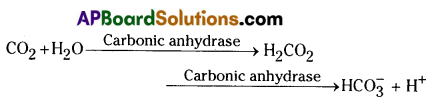
At the tissues where partial pressure of CO2 is high due to catabolism, CO2 diffuses into the blood (RBC and Plasma) and forms carbonic acid which dissociates into HCO3– and H+. At the alveolar site where pCO2 is low, the reaction proceeds in the opposite direction leading to the formation of CO2 and water. Thus CO2 is mostly trapped as bicarbonate at the tissues and transported to the alveoli where it is released out as CO2.
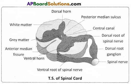
Question 13.
Draw a labelled diagram of the T.S. of the spinal cord of man.
Answer:
Question 14.
Write note on Addison’s disease and Cushing’s syndrome.
Answer:
Addison’s disease is caused due to hyposecretion of glucocorticoids by the adrenal cortex. This disease is characterised by loss of weight, muscle weakness, fatigue and reduced blood pressure. Sometimes darkening of the skin in both exposed and nonexposed parts of the body occurs in this disorder This disorder does not allow an individual to respond to stress.
Cushing’s syndrome : It results due to over production of gluco-corticoids. This condition is characterized by breakdown of muscle proteins and redistribution of body fat resulting in spindly arms and legs accompanied by a round moon face, buffalo hump on the back and pendulous abdomen. Wound healing is poor. The elevated level of cortisols causes hyperglycemia, over deposition of glycogen in liver and rapid gain of weight.
![]()
Question 15.
How is sex determined in human beings ?
Answer:
Sex determination in Humans : It has already been mentioned that the sex determining mechanism in case of humans is XX – XY type. Out of 23 pairs of chromosomes present, 22 pairs are exactly same in both males and females; these are the autosomes. A pair of X – chromosomes is present in the female, where as the presence of an X and Y – chromosome are determinant of the male characteristic. During spermatogenesis among males, two types of gametes are produced. 50 percent of the total sperm produced carry the X – chromosome and the rest 50 percent has Y – chromosome besides the autosomes. Females, however, produce only one type of ovum with an X – chromosome.
There is an equal probability of fertilisation of the ovum by the sperm carrying either X or Y chromosome. In case the ovum is fertilised by a sperm carrying X – chromosome, the zygote develops into a female and the fertilisation of ovum with Y – chromosome carrying sperm results into a male offspring. Thus, it is evident that it is the genetic makeup of the sperm that determines the sex of the child. It is also evident that in each pregnancy there is always 50 percent probability of either a male or a female child.
Question 16.
Write a short note on the theory of Mutation.
Answer:
Mutation theory : It was proposed by Hugo de Vries, a Dutch botanist who coined the term ‘mutation’. Mutations are sudden, random inheritable changes that occur in organisms. He found four different forms in Oenothera lamarckiana (commonly called ‘evening primrose’) such as O. brevistylis-small style, O. levifolia- smooth leaves. O. gigas- the giant form, O. nanella- the dwarf form (mutant varieties). T.H. Morgan studied the inheritance pattern of mutations in Drosophila melanogaster. Darwin called mutations (large variations) sports of nature or saltations, whereas Bateson called them discontinuous variations.
Salient Features of Mutation theory:
- Mutations occur from time to time in naturally breeding populations.
- They are discontinuous and are not accumulated over generations.
- They are full-fledged, and so there are no ‘intermediate forms’.
- They are subjected to Natural Selection.
Question 17.
What is meant by Genetic drift ? Explain genetic drift citing the example of founder effect.
Answer:
Experimental verification of Natural Selection – Industrial melanism : An important practical proof for the operation of Natural Selection is the classical case of industrial melanism, exhibited by peppered moth – Bistort betularia. These moths were available in two colours, grey and black. Prior to industrial revolution, the grey moths were abundant. During the industrial revolution, the black forms were more and the grey forms were less in the industrial cities like Birmingham. Biologists proposed that with the industrial revolution, more soot was released due to the burning of coal, which resulted in the darkening of the barks of trees.
Grey moths on the dark bark were easily identified and predated more by birds. Hence the number of grey moths decreased and that of the black moths increased in the population. It means Nature offered ‘positive selection’ pressure to the black (melanic) forms. Bernard Kettlewell, a British ecologist, tested this hypothesis experimentally. He collected both the grey and the black forms of Bistort betularia for his experiment. He released them in two sets of equal numbers; one set in Birmingham, a polluted urban area, and the other set in Dorset, an unpolluted rural area.
After a few days he recaptured them. Of those moths recaptured from Birmingham, there were more black forms. Among those recaptured from Dorset there were more grey forms. The reason for such a difference is : the melanic forms could not be easily spotted by predator birds as their body colour merged with the dark colour of the bark of trees in Birmingham area. In the rural areas (Dorset) the grey forms had better survival chance as their body colour merged with the light coloured surroundings. This explains the differential survival of the moths due to Natural Selection. It will be interesting to know that there was a reversal in the selection process after the introduction of pollution check laws in the urban areas.
Question 18.
List out the various steps involved in MOET.
Answer:
Multiple Ovulation and Embryo Transfer (MOET) :
The following are the steps involved in MOET.
- A cow is administered hormones, with FSH-like activity.
- This induces follicular maturation and super ovulation (In super ovulationinstead of one egg, which they normally produce per cycle, they produce 6 – 8 eggs).
- The animal (cow) is either mated with an elite bull or artificially inseminated.
- The embryos are at 8 – 32 celled stages are recovered non- surgically and transferred to surrogate mother (an animal that develops the offspring of another animal in its womb).
Now the genetic mother is ready for another round of super ovulation.
Section – C (2 × 8 = 16)
Note : Answer ANY TWO of the following questions.
Question 19.
Write notes on the working of the heart of man.
Answer:
The cardiac events that occur from the beginning of one heart beat to the beginning of the next constitute a cardiac cycle. This cardiac cycle consists of three phases, namely atrial systole, ventricular systole and cardiac diastole.
To begin with, all the four chambers of the heart are in a relaxed state / joint diastole stage. Blood from the pulmonary veins and venae cavae flows into the respective atria. As the A – V valves are in open condition, blood flows into the left and right ventricles, through the left and right atrioventricular apertures. The semilunar valves of the pulmonary and aortic arches are closed at this stage.
Atrial systole : The SAN now generates an action potential which stimulates both the atria to contract simultaneously causing the ‘atrial systole’. It lasts about 0.1 sec. This increases the flow of blood into the ventricles by about 30%. It means atrial systole accounts for about 30% of the filling of the ventricles, the remaining blood flows into the ventricles before the atrial systole.
Ventricular systole : The action potentials from the SAN reach the AVN from where they are conducted through the bundle of His, its branches and the Purkinje fibres to the entire ventricular musculature. This causes the simultaneous ventricular systole. It lasts for about 0.3 sec. The atria undergo relaxation coinciding with the ventricular systole. Ventricular systole increases the pressure causing the closure of the AV valves preventing the ‘backflow’ of blood. It results in the production of the first heart sound known as ‘Lub’ . As the ventricular pressure increases further, the semilunar valves guarding the pulmonary artery and the aorta are forced open. This allows the blood in the ventricles to flow into the aortic arches and enter the circulatory pathway.
Cardiac diastole : The ventricles now relax and the ventricular pressure falls causing the closure of the semilunar valves which prevents the back flow of blood. This result in the production of the second heart sound known as ‘Dup’. As the ventricular pressure declines further, the AV valves are pushed open by the pressure in the atria exerted by the blood, which flowed into them through the larger veins. The blood now once again flows freely into the ventricles. All the heart chambers are now again in a relaxed state (joint diastolic phase). Soon, another cardiac cycle sets in.
Cardiac output : The volume of blood pumped out by each ventricle, for each heart beat, is known as the stroke volume. The volume of blood pumped out by the heart from each ventricle per minute is termed cardiac output.
Cardiac output = Stroke volume × No. of beats per minute = 70 ml / beat × 72 beats / minute = 5040 ml/ mtn. or approximately 5 litres.
![]()
Question 20.
Describe female reproductive system of a woman with the help of a labelled diagram.
Answer:
The female reproductive system consists of a pair of ovaries along with a pair of oviducts, uterus, vagina and the external genitalia located in the pelvic region. These parts of the system along with a pair of the mammary glands are integrated structurally and functionally to support the processes of ovulation, fertilization, pregnancy, birth and child care.
Ovaries : Ovaries are the primary female sex organs that produce the female gametes (ova) and several steroid hormones (ovarian hormones). A pair of ovaries is located one on each side of the lower abdomen. The double layered fold of peritoneum connecting the ovary with the wall of the abdominal cavity is known as the mesovarium.
The ovaries are covered on the outside by a layer of simple cuboidal epithelium called germinal (ovarian) epithelium. This is actually the visceral peritoneum that envelops the ovaries. Underneath this layer there is a dense connective tissue capsule, the tunica albuginea. The ovarian stroma is distinctly divided into an outer cortex and an inner medulla. The cortex appears more dense and granular due to the presence of numerous ovarian follicle in various stages of development. The medulla is a loose connective tissue with abundant blood vessels, lymphatic vessels, and nerve fibers.
Fallopian tubes (Oviducts) : Each fallopian tube extends from the periphery of each ovary to the uterus, and it bears a funnel shaped infundibulum. The edges of the infundibulum possess finger like projections called fimbriae, which help in collection of the ovum after ‘ovulation’. The infundibulum leads to a wider part of the oviduct called ampulla. The last part of the oviduct, isthmus has a narrow lumen and it joins the uterus. Fallopian tube is the site of fertilization. It conducts the ovum or zygote towards the uterus by peristalsis. The fallopian tube is attached to the abdominal wall by a peritoneal fold called mesosalpinx.
Uterus : The uterus is single and it is also called womb. It is a large, muscular, highly vascular and inverted pear shaped structure present in the pelvis between the bladder and the rectum. The uterus is connected to the abdominal wall by the peritoneal fold called mesometrium. The lower, narrow part through which the uterus opens into the vagina is called the cervix. The cavity of the cervix is called cervical canal which along with vagina forms the birth canal.
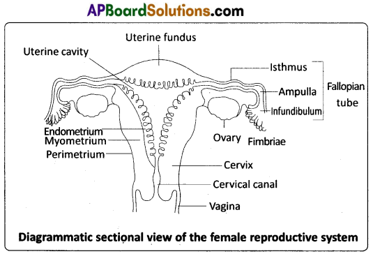
The wall of the uterus has three layers of tissue. The external thin membranous perimetrium, the middle thick layer of smooth muscle called myometrium and inner glandular lining layer called endometrium. The endometrium undergoes cyclic changes during menstrual cycle while the myometrium exhibits strong contractions during parturition.
Vagina : The vagina is a large, median, fibro – muscular tube that extends from the cervix to the vestibule (the space between the labia minora). It is lined by non – keratinised stratified squamous epithelium. It is highly vascular, and opens into the vestibule by the vaginal orifice.
Vulva : The term vulva (vulva = to wrap around) or pudendum refers to the external genitals of the female. The vestibule has two apertures – the upper external urethral orifice of the urethra and the lower vaginal orifice of vagina. Vaginal orifice is often covered partially by a membrane called hymen which is a mucous membrane. Vestibule is bound by two pairs of fleshy folds of tissue called labia minora (inner) and larger pair called labia majora (outer). Clitoris is a sensitive, erectile structure, which lies at the upper junction of the two labia minora above the urethral opening. The clitoris is homologous to the penis of a male as both are supported by corpora cavernosa internally. There is a cushion of fatty tissue covered by skin and public hair present above the labia majora. It is known as mons pubis.
Accessory reproductive glands of female : These glands include Bartholin’s glands, Skene’s glands and Mammary glands.
Bartholin’s glands : The Bartholin’s glands (Greater vestibular glands) are two glands located slightly posterior and to the left and right of the opening of the vagina. They secrete mucus to lubricate .the vagina and are homologous to the bulbourethral glands of the male reproductive system.
Skene’s glands : The Skene’s glands (Lesser vestibular glands) are located on the anterior wall of the vagina, around the lower end of the urethra. They secrete a lubricating fluid when stimulated. The Skene’s glands are homologous to the prostate glands, of the male reproductive system.
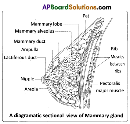
Mammary glands : A functional mammary gland is characteristic of all female mammals. The mammary glands are paired structures ( breasts) that contain glandular tissue and variable amount of fat. The glandular tissue of each breast is divided into 15 – 20 mammary lobes containing clusters of cells called alveoli. The cells of the alveoli secrete milk, which is stored in the cavities (lumens) of the alveoli. The alveoli open into mammary tubules. The tubules of each lobe join to form a mammary duct. Several mammary ducts join to form a wider mammary ampulla which is connected to lactiferous duct through which milk is sucked out by the baby.
![]()
Question 21.
What is Criss-cross inheritance ? Explain the inheritance of one sex linked recessive character in human beings.
Answer:
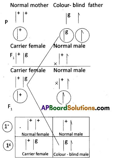
The crisscross pattern of inheritance (skip generation inheritance) is one in which a gene responsible for the sex linked recessive character is transmitted from a male parent.to a male grand child through a carrier female of the first generation. Colour blindness is the best example for criss – cross inheritance in human being.
Colour blindness : It is a sex-linked recessive disorder. Retina of the eye in man contains the cells sensitive to red and green colours. This phenotypic trait is genetically controlled. Its alleles are located on the X – chromosome. When a woman with normal vision (homozygous) marries a colour – blind man, all the sons and daughters are normal, but daughters are carriers (heterozygous). If a carrierjwoman marries a man with normal vision, all the daughters and half of the sons have normal vision and another half of sons are colour – blind. Colour – blind trait is inherited from a male parent to his grand sons through carrier daughter, which is an example of crisscross pattern of inheritance.
Haemophilia : Haemophilia A is recessive X – linked genetic disorder involving lack of the functional clotting Factor – VIII and represents 80% of haemophilia cases. Haemophilia B is also a recessive X – linked genetic disorder involving lack of the functional clotting Factor IX. When a person with hemophilia is injured, bleeding is prolonged because a firm clot is slow to form. Haemophilia follows the characteristic crisscross pattern of inheritance like that of colour – blindness.
Duchenne Muscular dystrophy : Duchenne muscular dystrophy (DMD) is a recessive X – linked form of muscular dystrophy, affecting around 1 in 3,600 boys. The disease is characterized by a progressive weakening of the muscles and loss of coordination. Affected individuals rarely live past their early 20s. The disorder is caused by a mutation in the dystrophin gene (the largest known gene in humans) located on the X – chromosome, which codes for the protein dystrophin, an important structural component within muscle tissue (connects sarcoiemma and the outer most
layer of muscle filaments and supports muscle fiber strength). If the mother is known to be a carrier of this gene, about half of her male children are expected to be affected. All female children born to a carrier mother are expected to be normal, since the possibility of their being homozygous for this sex – linked recessive gene is virtually non – existent.