Thoroughly analyzing AP Inter 2nd Year Zoology Model Papers Set 7 helps students identify their strengths and weaknesses.
AP Inter 2nd Year Zoology Model Paper Set 7 with Solutions
Time: 3 Hours
Maximum Marks: 60
General Instructions:
Note : Read the following instructions carefully.
- Answer all questions of Section – A. Answer ANY SIX questions in Section – B and answer ANY TWO questions in Section – C.
- In Section – A questions from SI. Nos. 1 to 10 are of Very Short Answer Type. Each question carries TWO marks. Every answer may be limited to 5 lines. Answer all these questions at one place in the same order.
- In Section – B, questions from SI. Nos. 11 to 18 are of Short Answer Type. Each question carries FOUR marks. Every answer may be limited to 20 lines.
- In Section – C, questions from SI. Nos. 19 to 21 are of Long Answer Type. Each question carries EIGHT marks. Every answer may be limited to 60 lines.
- Draw labelled diagrams wherever necessary in Sections – B and C.
Section – A (10 × 2 = 20)
Note : Answer ALL the questions.
Question 1.
Name the different types of salivary glands of man, and their locations in the human body.
Answer:
There are 3 pairs of salivary glands in man
- Parotid glands – present below the pinna and inner surface of cheeks.
- Sub maxillary glands – located at the angles of lower jaws.
- Sub lingual glands – present below the tongue.
Question 2.
Define vital capacity. What is its significance ?
Answer:
Vital Capacity (VC) : The maximum volume of air a person can breathe in after forced expiration. This includes ERV, TV and IRV or the maximum volume of air a person can breathe out after forced inspiration.
Question 3.
Write the differences between open and closed systems of circulation ?
Answer:
a) Open type : In this type blood flows from the heart into the arteries. The arteries open into large spaces called sinuses. From sinuses, blood is carried by the veins to the heart. These are no interconnecting vessels, capillaries between arteries and veins. It is found in leeches, arthropods, molluscs and echinoderms.
b) Closed type : In this type blood flows through blood vessels. Blood flows from arteries to the veins through capillaries. Closed type of blood vascular system is found in annelids, cephalopods among non-chordates and all vertebrates.
![]()
Question 4.
Write the difference between actin and myosin.
Answer:
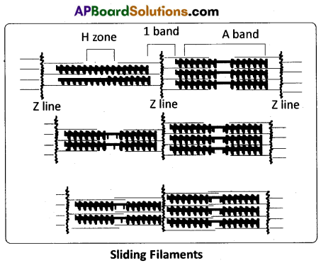
a) The light band in myofibril contains action and two regulatory proteins called troponin and tropomyosin. Actin filaments are thinner compared to myosin filaments.
b) The dark band in a myofibril contains (A band) myosin. Myosin filaments are thick and non contractile.
Question 5.
Name the type of joint between a) cranial bone b) Intertarsal joint.
Answer:
a) Joint between cranial bones is fibrous joint called sufure.
b) Intertarsal joint is Gliding joint.
Question 6.
What is insulin shock ?
Answer:
Hyper secretion of insulin leads to decreased level of glucose in the blood (hypoglycemia) resulting in insulin shock.
Question 7.
Mention various types of immunological disorders.
Answer:
- Acquired immuno deficiency syndrome (AIDS)
- Hypersensitivity disorders (allergies)
- Auto immune disorders (Grave’s disease, Rhematoidarthritis)
- Graft rejections.
Question 8.
Define spermiogenesis and spermiation.
Answer:
Development of spermatozoa from sperm mothers cells in male is called spermiogenesis. After spermiogenesis sperm heads become embedded in the sertoli cells and are finally released from the seminiferous tubules by the process called spermiation.
Question 9.
Mention any four fish by products.
Answer:
a) Shark and cod liver oils containing vit A and vit D
b) oil from sardine and salmon good source of omega – 3 – fattyacids (lowers blood cholesterol)
c) fish guano – fertiliser prepared from scrap fish
d) Shagreen.
![]()
Question 10.
What is popularly called Guardian Angel of Cell’s Genome.
Answer:
The protein P53 plays an important role with reference to the G1 check point in the regulation of cell division cycle. It guards the integrity of the DNA. Hence it is often called the Guardian Angel of Cell’s Genome.
Section – B (6 × 4 = 24)
Note : Answer ANY SIX questions.
Question 11.
What are the functions of liver ?
Answer:
Functions of the liver :
Liver performs a variety of functions such as synthesis, storage and secretion of various substances. There are as follows :
- Liver secretes bile juice. It does not contain enzymes, but it contains bile salts such as glycocholates and taurocholates of sodium and potassium and ‘bile pigments’ the bilirubin and biliverdin.
- Liver plays the ‘key role’ in carbohydrate metabolism (glycogenesis, glycogenolysis, gluconeogenesis and lipogenesis).
- Liver also plays a role in lipid metabolism (synthesis of cholesterol and production of triglycerides).
- Deamination of proteins (removal of NH2 group from the amino acids) and conversion of ammonia into to urea – via the ornithine cycle).
- The lactic acid formed during anaerobic muscle contraction is converted into glycogen (gluconeogenesis) in the liver by Coricycle.
- Liver is the chief organ of detoxification of toxic substances that enter the gut along with food.
- Liver acts as thermoregulatory organ (like skeletal muscle, liver too takes part in thermogenesis as it has high glucose at its disposal).
- Liver acts as a haemopoietic organ in the foetus and erythroclastic organ in the adult.
- The liver synthesizes the plasma proteins such as albumins, globulins, blood clotting factors such as fibrinogen, prothrombin, etc., and the anticoagulant, called heparin.
- Kupffer’s cells/ Kupffer cells are the large phagocytic cells which remove unwanted substances and microbes that attack the liver by phagocytosis. They are present in the sinusoids that lie in between hepatic cords and they are also called hepatic macrophages.
Question 12.
Describe the important steps in muscle contraction.
Answer:
Important steps in muscle contraction are
i) Excitation or stimulation of muscle : Muscle contraction is initiated by a signal sent by the central nervous system (CNS) via a motor neuron. A neural signal reaching the neuromuscular junction releases a neurotransmitter (acetylcholine) which generates an ‘action potential’ in the sarcolemma. When the action potential spreads to the triad system through the T – tubules, the cisternae of the sarcoplasmic reticulum release calcium ions into the sarcoplasm.
ii) Formation of Cross bridges : Increase in the Ca2+ level leads to the binding of calcium ions to the subunit Tn – C of the troponin of the thin filaments. This makes troponin and tropomyosin complex to move away from the active sites of action molecules. Now, the active sites are exposed to the heads of the myosin. Utilizing the energy released from hydrolysis of ATP, the myosin head now binds to the exposed ‘active sites’ on the actin molecules to form a cross bridge and P1 is released.
iii) Power Stroke: The cross bridge pulls the attached actin filaments towards the centre of the ‘A’ band. The ‘Z’ lines attached to these actin filaments are also pulled inwards from both the sides, thereby causing shortening of the sarcomere, i.e., contraction. During the shortening of the muscle, the T bands get reduced in size / length (Z membranes of the sarcomere are brought closer) whereas the’A’ bands retain their size/ length. It is important to note that myofilaments do not actually shorten. As the thin filaments are pulled deep in to the A bands making the H bands narrow, the muscle shows the effect contraction.
Question 13.
Compare a ‘pituitary dwarf and a ‘thyroid dwarf in respect of similarities and dissimilarities they possess.
Answer:
1. Pituitary dwarf : Hypo secretion of growth harmone (STH) during childhood retards growth, resulting in a pituitary dwarf / midget. The pituitary dwarf is sexually and intellectually a normal individual.
2. Thyroid dwarf : During pregnancy, due to hypothyroidism, defective development of the growing baby leads to a disorder called cretinism. Physical and mental growth get severely stunted and is called thyroid dwarf. This is due to untreated congenital hypothyroidism. Stunted growth, mental retardation, low intelligent quotient, abnormal skin, deafness and mutism are some of the characters of this disease.
![]()
Question 14.
Describe the surgical methods of contraception.
Answer:
Surgical procedure to prevent pregnancy is also known as sterilization. Sterilization procedure in the male is called vasectomy and that in the female tubectomy.
i) Vasectomy : A small part of the vas deferens on either side is removed or tied up through a small incision on the scrotum. Thus the sperms are prevented from reaching seminalvesicle and so the ‘semen’ in ‘vasectomised’ males do not contain sperms.
ii) Tubectomy : A small part of the fallopian tube on both sides is removed or tied up through a small incision made in the abdomen or through vagina. This will block the entry of ova into the fallopian tubes and thus pregnancy is prevented.
Question 15.
Describe male heterogamety.
Male Heterogamety: In this method of sex determination, the males (heterogametic) produce dissimilar gametes while females (homogametic) produce similar gametes. Male heterogamety is of two kinds, XX – XO type and XX – XY type.
i) XX – XO type : In some insects such as bugs, grasshoppers and cockroaches, females are with two X – chromosomes and males are with one X – chromosome in each somatic cell. Me Clung discovered this type in grasshoppers. The unpaired X – chromosome determines the male sex. The karyotype of the female (homogametic) is AAXX and that of the male (heterogametic) is AAXO. All the ova contain ‘AX’ complement of chromosomes and the sperms are of two types. One half of the sperms have ‘AX’ complement and the other half have ‘A’ complement of chromosomes. The sex of the offspring depends on the type of sperm that fertilizes the ovum.
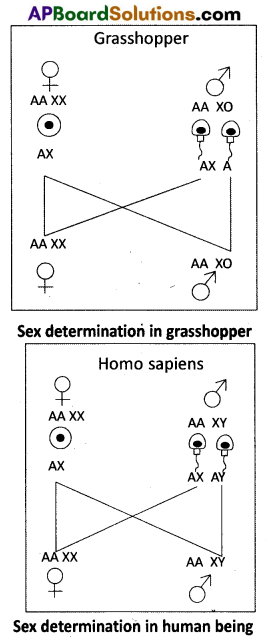
ii) XX – XY type : In human beings and some insects such as Drosophila, both females and males have the same number of chromosomes. The karyotype of the female is AAXX and that of the male is AAXY. Females are ‘homogametic’ with ‘XX’ chromosomes.
They produce similar ova having one X – chromosome each. Males are ‘heterogametic’ with X and Y – chromosomes. They produce two kinds of sperms; one half of them with X – chromosome and the other half with Y – chromosome. The sex of the offspring depends on the fertilizing sperm. The XX – XY type is also found in most other mammals.
Question 16.
Discuss the role of different patterns of selections in evolution.
Answer:
Selection is a process by which the organisms that are physically. Physiologically and behaviourally better adapted to the environment, survive and reproduce. Selection is an operative process. Selections are 3 types.
a) Stabilising or Centripetal selection : It is the selective elimination of phenotypically extreme individuals from the two ends of the phenotypic distribution and preserving those that are in the mean of the phenotypic distribution.
b) Directional selection : It operates in response to gradual changes in environmental Conditions. Directional selection works by constantly removing individuals from one end of the phenotypic distribution.
c) Disruptive or Centrifugal selection : It is a rarest form of selection and is very important in bringing about evolutionary change. As a result of increased competition, selection pressure acting within the population may push the phenotypes away from the population mean towards the ends of the population. This can split the population into two or more sub-population called species populations. Each population may give rise to a new species. It is also called as adaptive radiation.
![]()
Question 17.
Explain Darwin’s theory of Natural selection with industrial melanism as an experimental proof.
Answer:
Experimental verification of Natural Selection – Industrial melanism : An important practical proof for the operation of Natural Selection is the classical case of industrial melanism, exhibited by peppered moth – Biston betuiaria. These moths were available in two colours, grey and black. Prior to industrial revolution, the grey moths were abundant. During the industrial revolution, the black forms were more and the grey forms were less in the industrial cities like Birmingham. Biologists proposed that with the industrial revolution, more soot was released due to the burning of coal, which resulted in the darkening of the barks of trees.
Grey moths on the dark bark were easily identified and predated more by birds. Hence the number of grey moths decreased and that of the black moths increased in the population. It means Nature offered ‘positive selection’ pressure to the black (melanic) forms. Bernard Kettlewell, a British ecologist, tested this hypothesis experimentally. He collected both the grey and the black forms of Biston betuiaria for his experiment. He released them in two sets of equal numbers; one set in Birmingham, a polluted urban area, and the other set in Dorset, an unpolluted rural area. After a few days he recaptured them. Of those moths recaptured from Birmingham, there were more black forms. Among those recaptured from Dorset there were more grey forms.
The reason for such a difference is: the melanic forms could not be easily spotted by predator birds as their body colour merged with the dark colour of the bark of trees in Birmingham area. In the rural areas (Dorset) the grey forms had better survival chance as their body colour merged with the light coloured surroundings. This explains the differential survival of the moths due to Natural Selection. It will be interesting to know that there was a reversal in the selection process after the introduction of pollution check laws in the urban areas.
Question 18.
Write about the procedure involved in MRI.
Answer:
MRI scan (Magnetic Resonance Imaging) is a diagnostic Radiology Technique. MRI is a non invasive medical imaging technique that helps physicians diagnose certain anatomical abnormalities or pathological conditions.
MRI scanner and procedure: MRI scanner is a giant circular magnetic tube. The patient is placed on a movable bed that is inserted into the magnet. Human body is mainly composed of water molecules which contain two hydrogen nuclei /protons, each. The magnet creates a strong ‘magnetic field’ that makes these protons align with the direction of the magnetic field (protons are not aligned under normal conditions). A second radiofrequency electromagnetic field is then turned on for a ‘brief period’. The ‘protons’ absorb some energy from these ‘radio waves’. When this ‘second radio frequency emitting field’ is turned off, the protons release energy at a radiofrequency which can be detected by the MRI scanner (the protons return to their ‘equilibrium state’ from the ’energized state’ at different ‘relaxation’ rates).
Section – C (2 × 8 = 16)
Note : Answer ANY TWO of the following questions.
Question 19.
Explain the physiology of urine formation.
Answer:
Urine Formation : The formation of urine involves three main processes namely, glomerular filtration, selective reabsorption and tubular secretion.
a) Glomerular filtration : The first step in the formation of urine is the ‘filtration’ of the blood from the glomerulus into the lumen of the Bowman’s capsule and this ‘passive’ (non – energy consuming process) process is called glomerular filtration. The hydrostatic pressure of the blood while flowing in the glomerulus is 60 mm Hg. It is opposed by ‘glomerular colloidal osmotic pressure1 of 32 mm Hg (which is exerted by the non- filtered plasma proteins of the blood in the glomerular capillaries) and Bowman’s capsular hydrostatic pressure of 18mm Hg. The net filtration pressure is 10mm Hg (60 – 32 + 18 = 10).
This causes the filtration of blood through the 3 layered filtrate membrane formed by the endothelial cells of glomerular capillary together with the basement membrane and podocytes of the Bowman’s cup. Blood is filtered through the fine slit pores and fenestrations due to the NFP. Therefore, this process is called ‘ultrafiltration’. The filtrate contains almost all the constituents of the plasma, except the proteins. The filtrate thus formed is called ultra – filtrate or ‘glomerular filtrate’ or ‘primary urine’, which is hypotonic to the cortical fluid. It passes into the next part of the renal tubule.
b) Selective reabsorption and secretion: The tubular epithelial cells in different segments of a nephron reabsorb certain substances of the glomerular filtrate either by active or passive mechanisms. About 85% of the filtrate formed is reabsorbed in a constant, unregulated fashion by the PCT and descending limb of Henle’s loop (obligatory or mandatory reabsorption) and the reabsorption of the rest of the fluid is ‘regulated’. Based on the necessity of re-absorption, the substances of glomerular filtrate can be categorized into ‘high threshold substances’ (essential and are efficiently reabsorbed e.g. glucose, amino acids, vitamins, some salts etc.), ‘low threshold substances’ (absorbed in very little amounts e.g. urea, uric acid etc), or ‘athreshold substances’ (actual excretory products and are not reabsorbed at all e.g. creatinine).
During the formation of urine, the tubular cells secrete substances such as H+, K+ and NH3 into the filtrate. Tubular secretion is also an important step in the formation of urine as it helps in the maintenance of ionic and acid-base balance of the body fluids. Mechanism of selective reabsorption and secretion in different parts of a nephron takes place as follows.
i) In the proximal convoluted tubule : PCT is lined by simple cuboidal epithelium with ‘brush border’, which increases the surface area of absorption. Nearly all the essential nutrients and 70 – 80% of electrolytes and water are reabsorbed by this segment. Na+ is actively transported into the cortical interstitial fluid. This transfer of positive charge drives the passive transport of Cl–. Glucose, amino acids, and other essential substances are also ‘actively’ transported. Movement of water occurs by ‘osmosis’.
PCT also helps to maintain the pH and ionic balance of the body fluids by selective secretion of hydrogen ions, and ammonia into the filtrate and by the absorption of HCO3– from it.
ii) In the Henle’s loop : Reabsorption in this segment is minimum. However, this region plays a significant role in the maintenance of high osmolarity of the medullary interstitial fluid.
The descending limb of loop of Henle is permeable to water and almost impermeable to electrolytes, hence reabsorption of water continues as the filtrate moves along the descending limb (passive transport). As a result, the filtrate concentration gradually increases as it moves towards the inner medulla. The ascending limb has two specialized regions, a proximal thin segment, in which NaCl diffuses out into the interstitial fluid passively, and a distal thick segment, in which NaCl is actively pumped out. The ascending limb is impermeable to water. Thus the filtrate becomes progressively more dilute as it moves up to the cortex (towards the DCT).
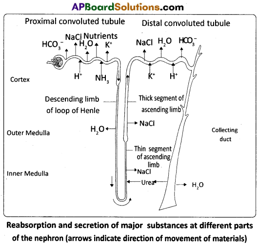
iii) In the distal convoluted tubule (DCT) : The cells here are shorter than those in the proximal tubule and lack ‘microvilli’, indicating that they are not involved much in reabsorption ‘conditional reabsorption’ /’facultative reabsorption’ of Na+ and water takes place in this segment. The reabsorption of water is variable depending on several conditions and is regulated by ADH. DCT is also capable of reabsorption of HCO3– and selective secretion of H+ and K+ ions and NH3 into the DCT from the peritubular network, to maintain the pH and sodium – potassium balance in the blood.
iv) In the collecting duct (CD) : This long duct carries the filtrate through the medulla to the renal pelvis. Considerable amount of water could be reabsorbed from the region to produce concentrated urine. This segment allows passage of small amount of urea to the medullary interstitum to keep up its osmolarity. It also plays a role in the maintenance of pH and ionic balance of blood by the selective secretion of H+ and K+ ions. The renal fluid after the process of facultative reabsorption in the CD, influenced by ADH, constitutes the ‘urine’, that is sent out. Urine in the CD is hypertonic to the plasma of blood.
![]()
Question 20.
Describe female reproductive system of a woman with the help of a labelled diagram.
Answer:
The female reproductive system consists of a pair of ovaries along with a pair of oviducts, uterus, vagina and the external genitalia located in the pelvic region. These parts of the system along with a pair of the mammary glands are integrated structurally and functionally to support the processes of ovulation, fertilization, pregnancy, birth and child care.
Ovaries : Ovaries are the primary female sex organs that produce the female gametes (ova) and several steroid hormones (ovarian hormones). A pair of ovaries is located one on each side of the lower abdomen. The double layered fold of peritoneum connecting the ovary with the wall of the abdominal cavity is known as the mesovarium.
The ovaries are covered on the outside by a layer of simple cuboidal epithelium called germinal (ovarian) epithelium. This is actually the visceral peritoneum that envelops the ovaries. Underneath this layer there is a dense connective tissue capsule, the tunica albuginea. The ovarian stroma is distinctly divided into an outer cortex and an inner medulla. The cortex appears more dense and granular due to the presence of numerous ovarian follicle in various stages of development. The medulla is a loose connective tissue with abundant blood vessels, lymphatic vessels, and nerve fibers.
Fallopian tubes (Oviducts) : Each fallopian tube extends from the periphery of each ovary to the uterus, and it bears a funnel shaped infundibulum. The edges of the infundibulum possess finger like projections called fimbriae, which help in collection of the ovum after ‘ovulation’. The infundibulum leads to a wider part of the oviduct called ampulla. The last part of the oviduct, isthmus has a narrow lumen and it joins the uterus. Fallopian tube is the site of fertilization. It conducts the ovum or zygote towards the uterus by peristalsis. The fallopian tube is attached to the abdominal wall by a peritoneal fold called mesosalpinx.
Uterus : The uterus is single and it is also called womb. It is a large, muscular, highly vascular and inverted pear shaped structure present in the pelvis between the bladder and the rectum. The uterus is connected to the abdominal wall by the peritoneal fold called mesometrium. The lower, narrow part through which the uterus opens into the vagina is called the cervix. The cavity of the cervix is called cervical canal which along with vagina forms the birth canal.
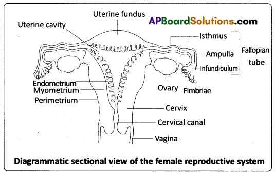
The wall of the uterus has three layers of tissue. The external thin membranous perimetrium, the middle thick layer of smooth muscle called myometrium and inner glandular lining layer called endometrium. The endometrium undergoes cyclic changes during menstrual cycle while the myometrium exhibits strong contractions during parturition.
Vagina : The vagina is a large, median, fibro – muscular tube that extends from the cervix to the vestibule (the space between the labia minora). It is lined by non – keratinised stratified squamous epithelium. It is highly vascular, and opens into the vestibule by the vaginal orifice.
Vulva : The term vulva (vulva = to wrap around) or pudendum refers to the external genitals of the female. The vestibule has two apertures – the upper external urethral orifice of the urethra and the lower vaginal orifice of vagina. Vaginal orifice is often covered partially by a membrane called hymen which is a mucous membrane. Vestibule is bound by two pairs of fleshy folds of tissue called labia minora (inner) and larger pair called labia majora (outer). Clitoris is a sensitive, erectile structure, which lies at the upper junction of the two labia minora above the urethral opening. The clitoris is homologous to the penis of a male as both are supported by corpora cavernosa internally. There is a cushion of fatty tissue covered by skin and public hair present above the labia majora. It is known as mons pubis.
Accessory reproductive glands of female : These glands include Bartholin’s glands, Skene’s glands and Mammary glands.
Bartholin’s glands : The Bartholin’s glands (Greater vestibular glands) are two glands located slightly posterior and to the left and right of the opening of the vagina. They secrete mucus to lubricate .the vagina and are homologous to the bulbourethral glands of the male reproductive system.
Skene’s glands : The Skene’s glands (Lesser vestibular glands) are located on the anterior wall of the vagina, around the lower end of the urethra. They secrete a lubricating fluid when stimulated. The Skene’s glands are homologous to the prostate glands, of the male reproductive system.
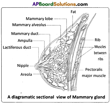
Mammary glands : A functio-nal mammary gland is characteristic of all female mammals. The mammary glands are paired structures ( breasts) that contain glandular tissue and variable amount of fat. The glandular tissue of each breast is divided into 15-20 mammary lobes containing clusters of cells called alveoli. The cells of the alveoli secrete milk, which is stored in the cavities (lumens) of the alveoli. The alveoli open into mammary tubules. The tubules of each lobe join to form a mammary duct. Several mammary ducts join to form a wider mammary ampulla which is connected to lactiferous duct through which milk is sucked out by the baby.
![]()
Question 21.
Why is the Human Genome Project called a mega project ?
Answer:
Human Genome Project (HGP) was called a mega project. It was an international effort formally begun in October, 1990. The HGP I was a 13 – year project coordinated by the U.S. Department of Energy and the National Institute of Health. During the early years of ; the HGP, the Wellcome Trust (U.K.) became a major partner, and ‘ additional contributions came from Japan, France, Germany, China and others. The project was almost completed in 2003. Knowledge about the effects of DNA variations among individuals can lead to revolutionary new ways to diagnose, treat and someday prevent the thousands of disorders that affect human beings. HGP was closely associated with the rapid development of a new area in biology called Bioinformatics.
Besides providing clues to under-standing human biology, learning about non – human organisms’ DNA sequences can lead to an understanding of their natural capabilities that can be applied toward solving challenges in health care, agriculture, energy production, environmental remediation. Genomes of many non – human model organisms, such as bacteria, yeast, Caenorhabditis elegans (a free living non – pathogenic nematode), Drosophila, plants (rice and Arabidopsis), etc. have also been sequenced. In a way they helped the progress of HGP.
Goals of HGP : Some of the important goals of HGP were as follows:
- Identify all the approximately 20,000 – 25,000 genes in human DNA.
- Determine the sequences of the 3 billion chemical base pairs that make up human DNA.
- Improve tools for data analysis.
- Address the ethical, legal, and social issues (ELSI) that may arise from the project.
Methodologies : The methods involved two major approaches. One approach focused on identifying all the genes that expressed as RNA (referred to as Expressed Sequence Tags (ESTs). The other took the blind approach of simply sequencing the whole set of genome that contained all the coding and non-coding sequence, and later assigning different regions in the sequence with functions (a term referred to as Sequence Annotation).
What is DNA sequencing ?
DNA sequencing, the process of determining the exact order of the 3 billion paired chemical building blocks (called ‘bases’ – A, T, C, and G) that make up the DNA of the 24 different human chromosomes (23 + Y in a male), was the greatest technical challenge in the Human Genome Project.
For sequencing, the total DNA from a cell is isolated and converted into random fragments of relatively smaller size and cloned in a suitable host using specialized vectors. The cloning results in the amplification of DNA fragments which are used for sequencing the bases. The commonly used hosts are bacteria and yeast, and the vectors are called BAC (bacterial artificial chromosomes), and YAC (yeast artificial chromosomes). The fragments were sequenced using automated DNA sequencers that worked on the principle of a method developed by Frederick Sanger. Alignment of these sequences was humanly not possible. Therefore, specialized computer based programs were developed. These sequences were subsequently annotated and were assigned to each chromosome. The latest method of sequencing even longer fragments, by a method called Shotgun sequencing using super computers, replaced the traditional sequencing methods.
Salient Features of Human Genome : Some of the salient observations drawn from human genome project are as follows :
- The human genome contains 3164.7 million nucleotide bases.
- The average gene consists of 3000 bases, but sizes vary greatly, with the largest known human gene being the one that codes for the protein called dystrophin.
- The total number of genes is estimated at 30,000. Almost all (99.9%) nucleotide bases are exactly the same in all people.
- The functions are unknown for over 50% of the genes discovered.
- Less than 2 per cent of the genome codes for proteins.
- Repeated sequences make up very large portion of the human genome.
- Repetitive sequences are stretches of DIMA sequences that are repeated many times. They are thought to have no direct coding functions, but they shed light on chromosome structure, dynamics and evolution.
- Chromosome 1 has the highest number of genes (2,968), and the Y – chromosome has the fewest genes (231).
- Scientists have identified about 1.4 million locations where single base DNA differences (SNPs – single nucleotide polymorphism, pronounced as snips) occur in humans. This information promises to revolutionise the processes of finding chromosomal locations for disease – associated sequences and tracing human history.
Advantages of HGP:
- In the area of health care, identification and mapping of the genes responsible for genetic diseases helps in diagnosis, treatment and prevention of these diseases.
- Detailed knowledge of the genomes of humans and other species will give a clearer picture of Gene expression, Cellular growth and differentiation and evolutionary biology.
- Earlier detection of genetic predispositions to disease, rational drug design, Gene therapy is going to be easy with more knowledge on human genome.
- A new era of Molecular Medicine, characterized by looking into the most fundamental causes of disease than treating the symptoms will be an important advantage.