Thoroughly analyzing AP Inter 2nd Year Zoology Model Papers Set 2 helps students identify their strengths and weaknesses.
AP Inter 2nd Year Zoology Model Paper Set 2 with Solutions
Time: 3 Hours
Maximum Marks: 60
Section – A (10 × 2 = 20)
Answer all the questions. (Very Short Answer Type)
Question 1.
What is chyme ?
Answer:
Semi fluid mass of partly digested acidic food formed in the stomach is called chyme.
Question 2.
What are the columns of Bertin ?
Answer:
Columns of Bertin are the medullary extensions of the renal cortex in between the renal pyramids.
Question 3.
What is corpus callosum ?
Answer:
Two cerebral hemispheres are internally connected by a transverse, wide and flat bundle of myelinated fibres beneath the cortex is called corpus callosum.
Question 4.
Define the terms immunity and immune system.
Answer:
Immunity: It is an ability of host or individual to fight against the disease causing organisms is called immunity.
Immune System : The network of organs, cells and proteins that protect the body from harmful, infectious agents such as bacteria, viruses, animal parasites, fungi etc., is called the immune system.
![]()
Question 5.
What are the functions of sertoli cells of the seminiferous tubules and the Leydig cells in man ?
Answer:
- Sertoli cells : Also known as ‘nourishing cells’ helps in the nourishment of spermatozoa and produce a hormone ‘inhibin’, which inhibits the secretion of FSH:
- Leydig cells : Produce Testosterone that controls the secondary sexual characters and spermatogenesis.
Question 6.
What is the genetic basis of blood types in ABO system in man ?
Answer:
Three alleles of gene I are responsible for ABO blood grouping. They are IA, IB and IO.
IA IA / IA IO – for A blood group
IB IB / IB IO – for B blood group
IA IA – for AB blood group
IO IO – for O blood group
Question 7.
Distinguish between allopatric and sympatric speciations.
Answer:
Allopatric speciation : Speciation occurring in which first geographical isolation occurs, then secondary reproductive isolation occurs.
Sympatric speciation : Reproduction isolation occurs without geographical isolation.
Question 8.
Name the keytone bone of the cranium. Where is it located.
Answer:
Sphenoid bone is the keystone bone of the cranium, because it articulates with all the other cranial bones. It is present at the middle part of the base of the skull.
Question 9.
What is common between Darwinism and Lamarckism ?
Answer:
Presence of variations is common to Darwinism and Lamarckism.
Question 10.
Distinguish between out-cross and cross-breed.
Answer:
Out cross : The offspring formed by mating of animals within the same breed, but having no ancestors on either side of pedigree for 4 – 6 generations.
A single out cross helps to overcome inbreeding depression. Cross breed : The offspring formed by a mating between superior males of one breed and superior females another breed.
Cross breed shows desirable qualities of two different breeds to be combined.
Section – B (6 × 4 = 24)
Answer any six questions. (Short Answer Type)
Question 11.
What are the functions of liver ?
Answer:
Liver performs a variety of functions such as synthesis, storage and secretion of various substances.
- Liver secretes bile juice, it contains bile salts such as sodium / potassium glycocholates and taurocholates, which helps in digestion and absorption of lipids.
- Liver plays the key role in carbohydrate metabolism.
a) Glycogenesis : formation of glycogen from glucose.
b) Glycogenolysis : breakdown of glycogen into glucose.
c) Gluconeogenesis : Synthesis of glucose from certain amino acids, lactate (or) glycerol. - Liver also plays an important role in synthesis of .cholesterol and production of triglycerides.
- Deamination of proteins occurs in the liver.
- Liver is the chief organ of detoxification of toxic substances that enter the gut along with food.
- Liver acts as thermoregulatory organ.
- Liver acts as a haemopoietic organ in the foetus and erythro- clastic organ in the adult.
- The liver synthesizes the plasma proteins such as albumin, globulins; blood clotting factors such as fibrinogen / prothrombin, etc., and the anticoagulant called heparin.
- The lactic acid formed during anaerobic muscle contraction is converted into glycogen (gluconeogenesis) in the liver by Cori cycle.
- Kupffer cells are the largest phagocytic cells which remove unwanted substances and microbes that attack the liver by phagocytosis.
![]()
Question 12.
Define the term breed. What are the objectives of animal breeding?
Answer:
Breed: A breed is a group of animals related by descent and similar in most characters such as general appearance, size, configuration and features with other members of the same species.
Jersery and Brown Swiss are example of foreign breeds of cattle. These two varieties of cattle have the ability to produce abundant quantities of milk. This milk is very nutritious with high protein content.
Objects of animal breeding :
- To produce disease resistant animals.
- Increase in the quality and quantity of milk, meat, wool etc.,
- Fast growth rate.
- Enhanced productive life by improving the genetic merit of livestock.
- Early maturity.
- Economy of feed.
Question 13.
Explain the mechonism of clotting of blood.
Answer:
When a blood vessel is injured a number of physiological mechanisms are activated that promote hemostasis, and stops bleeding. Blood clots within 3 – 6 minutes after damage of a blood vessel.
Mechanism of blood clotting: Blood clotting takes place in three essential steps.
i) Formation of prothrombin activator : It is formed by two pathways.
a) Intrinsic pathway : It occurs when the blood is exposed to collagen of injured wall of blood vessel. This activates factor XII, and in turn it activates another clotting factor, which activates yet another reaction, which results in the formation of prothrombin activator.
b) Extrinsic pathway : It occurs when the damaged vascular wall or extra vascular tissue comes into contact with blood. This activates the release of tissue thromboplastin, from the damaged tissue. It activates the factor VII. As a result of these cascade reactions, the final product formed is the prothrombin activator.
ii) Activation of prothrombin : The prothrombin activator, in the presence of sufficient amount of Ca2+, causes the convertion of inactive prothrombin to active thrombin.
iii) Convertion of soluble fibrinogen into fibrin : Thrombin converts the soluble protein fibrinogen into soluble, fibrin monomers, which are held together by weak hydrogen bonds. The factor XIII replaces hydrogen bonds with covalent bonds and cross links the fibers to form a meshwork and prevent the blood bleeding.
Question 14.
Describe the structure of synovial joint with the help of a neat labelled diagram.
Answer:
Synovial joints are characterised by the presence of a fluid filled synovial cavity between the articulating surfaces of the two bones.
Structure of synovial joint : Synovial joint is covered by a double layered synovial capsule. The outer layer consist of dense fibrous irregular connective tissue with more collagen fibers. This layer is continuous with the periosteum and resists stretching and prevents the dislocation of joints. Some fibres of these mem-branes are arranged in bundles called ligaments.
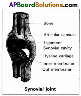
The inner layer of synovial capsule is formed of areolar tissue and elastic fibers. It secretes a viscous synovial fluid which contains hyaluronic acid, phagocytes etc., and acts as a lubricant for the free movement of the oints.
Question 15.
Describe the microscopic structure of ovary of woman.
Answer:
Ovaries are the primary female sex organs that produce the ‘female garnets’ or ‘ova’ and several steroid hormones (Ovarian hormones). A pair of ovaries is located one on each side of the lower abdomen. The double layered fold of peritoneum connecting the ovary with the wall of abdominal cavity is known as the ‘mesovarium’.
The ovaries are covered on the outside by a layer of simple cuboidal epithelium called ‘germinal (ovarian) epithelium. This is actually the visceral peritoneum that envelopes the ovaries. Under this layer there is a dense connective tissue capsule, the ’tunica albuginea’. The ovarian stroma is distinctly divided into an outer cortex and an inner medulla. The cortex is dense and granular due to the presence of numerous ovarian follicles in various stages of development. The medulla is a loose connective tissue with abundant blood vessels, lymphatic vessels and nerve fibers.
Question 16.
Describe the male and female sex harmones and their actions.
Answer:
The hormones, which are responsible for the development of secondary sexual characters and changes in different stages of life are called sex hormones.
Male sex hormones :
Androgens : Androgens are produced by the Leydig cells of the testes and to a minor extent by the adrenal glands in both sexes. Functions :
- Growth, development and maintenance of male reproductive organs.
- Sexual differentiation and secondary sexual characteristics.
- Spermatogenesis.
- Male pattern of aggressive behaviour.
- Increases the protein synthesis and increases the glycolysis.
Female sex hormones :
1) Estrogens : Synthesized by the follicles and corpus luteum of ovary.
Functions :
- Development and maintenance of female reproductive organs.
- Maintenance of menstrual cycle.
- Development of secondary sexual characters.
- Estrogen promotes the protein synthesis and calcification . and bone growth.
2) Progesterone : It is synthesized and secreted by corpus luteum and placenta.
Functions : required for implantation of fertilised ovum and maintenance of pregnancy.
3) Follicle stimulating and Lutenizing hormones : Both these hormones produced from anterior pituitary gland in both sexes.
Functions : Both these hormones play an important role in secondary sexual characters in both sexes.
Question 17.
What is meant by genetic drift ? Explain genetic drift citing the example of founder effect.
Answer:
The change in the frequency of a gene that occurs merely by chance and not by selection, in small proportion is called genetic drift.
Suppose, for a gene with two alleles, the frequency of a particular allele is 1% (e = 0.01) the probability of losing that allele by chance from small population is more. The end result is either fixation or loss of that allele.
Genetic drift tend to reduce the amount of genetic variation within the population mainly by removing the alleles with low frequencies. It can examplified by Founder and Bottleneck effect.
Founder effect: If a small group of individuals from a population starts a new colony in an isolated region, those individuals are called the founders of the new population. The allelic frequency of their descendants are similar those of the founders rather than to their ancestral parent population.
Eg: O+ve blood group is present in nearly 100% of the red Indians. It means the forefathers of the Red Indians tribe were predominantly 0+ve and they isolated themselves reproductively from other population.
![]()
Question 18.
Explain the role of animal husbandry in human welfare.
Answer:
Animal husbandary deals with the scientific management of livestock. It includes various aspects such as feeding, breeding and control diseases to raise the population of livestock. Animal husbandary usually includes buffaloes, cows, pigs, horses, cattle, sheep, camels, goats, poultry, fish etc which are useful for humans in various ways.
These animals are managed for production of commercially, important products such as milk, meat, wool, egg, honey, silk etc. The increase in human population has increased the demand of these products. Hence it is necessary to improve the management of livestock scientifically.
Section – C (2 × 8 = 16)
Answer any two questions. (Long Answer Type)
Question 19.
Describe the excretory system of man, giving the structure of a nephron.
Answer:
In humans, the excretory system consists of a pair of kidney, a pair of ureters, a urinary bladder and urethra.
Kidney : Kidneys are reddish brown, bean shaped structures, situated on either side of the vertebral column between the levels of last thoracic and third lumbar vertebrae in a retroperitoneal position. The right kidney is slightly lower than the left one due to the presence of liver.
The outer surface of the kidney is convex and the inner surface is concave, where it has a deep notch called hilum, the point at which the renal artery and nerves enter and renal vein and ureter leave. Each kidney is surrounded by a tough, fibrous tissue, called renal capsule.
Ureter : These are slender whitish tubes, which emerges from the pelvis of the kidney. The ureter rundown and open into the urinary bladder.
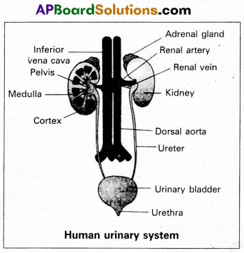
Urinary bladder : Urinary bladder is a pear shaped like muscular organ. It tempirarily stores the urine, situated in the lower abdominal cavity. The neck of the bladder leads into the urethra. Urethra opens near the vaginal orifice in the female and through the penis in the male.
Structure of nephron: Each kidney has nearly one mjllion nephrons. These are structural and functional units of kidney, embedded in the loose connective tissue of cortex and medulla. Nephron consist of malpighian body and renal tubule.
I) Malphigian body : It begins in the cortex of the kidney. It contains Bowman’s capsule and glomerulus.
a) Bowman’s capsule : It is a thin walled, double layered cup. The inner wall of the Bowman’s capsule has certain unique cells called podocytes.
b) Glomerulus : It is a dense network of capillaries in the cup of Bowman’s capsule. Afferent arteriole of renal artery enter the cavity of Bowman’s capsule and split into five branches. They unite and come out of the Bowman’s capsule as an afferent arteriole.
The podocytes of inner wall of Bowman’s capsule wrap around each capillary. The podocytes are arranged in an intricate manner so as to leave some minute spaces called filteration slits. The endothelium cells of capillaries have numerous pores called fenestrations.
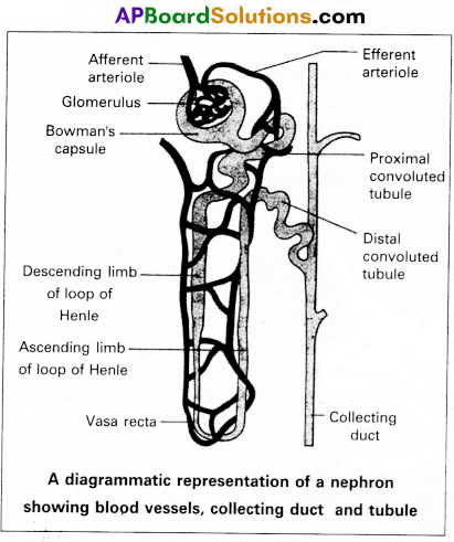
II) Renal tubule : It is narrow, delicate tubule arises from the posterior part of Bowman s capsule known as neck. It opens into along narrow convoluted tubule with three parts like proximal convoluted tubule, Loop of Henle and Distal convoluted tubule.
a) Proximal convoluted tubule : It is a lined by simple cuboidal epithelium with brush border to increase area of absorption.
b) Loop of Henle : It is a hairpin like tubule present in medulla region. It consist of a descending limb and an ascending limb. The proximal part of the ascending limb is thin and the distal part is thick. The thick ascending limb continuous into the distal convoluted tubule.
c) Distal convoluted tubule (DCT) : It is present in cortex. It is lined by simple cuboidal epithelium. The DCT continuous as the initial collecting duct in the cortex.
Collecting system : Some initial collecting ducts unite to form straight collecting duct, which passes through the medullary pyramid. In the medulla, the tubes of each pyramid join and form duct of Bellini, which finally opens into tip of the renal papilla.
Capillary network of nephron : The efferent arteriole emerging from the glomerulus forms a fine capillary network called the peritubular capillaries, around the renal tubule. The portion of the peritubular capillaries that surrounds the loop of Henle is called the vasa recta. The vasa recta is absent or highly reduced in the cortical nephrons. The juxtamedullary nephrons possess well developed vasa recta.
Question 20.
Write an essay on common genetic disorders.
Answer:
A number of disorders in human beings have been found to be associated with the inheritance of changed or altered genes of chromosomes.
Genetic disorders broadly grouped into two categories :
1) Mendalian disorders,
2) Chromosomal disorders
1) Mendelian disorders : These are genetic disorders showing Mendelian pattern of inheritance, caused by a single mutation in structure of DNA.
Most common and prevalent Mendelian disorders are : Haemophilia, Cystic fibrosis, sickle cell anaemia, colour blindness, phenyl ketonuria, thalassemia etc.
I. Haemophilia : It is also called as bleeder’s disease.
a) Haemophilia-A: This is sex linked recessive disorder, transmitted by females and affecting males. Haemophilia-A is the most common clotting abnormality and is due to the deficiency of clotting factor VIII.
Symptoms : The affected individuals have prolonged clotting time and suffer from internal bleeding.
b) Haemophilia – B : This is due to the deficiency of clotting factor IX.
Symptoms : Symptoms are similar to that found in haemophilia-A.
II. Sickle-cell anaemia : It is an autosomal recessive genetic dis-order, characterised by rigid, sickle-shaped red blood cells in hypoxia condition. It is due to point mutation in the β-globin gene causing replacement of glutamic acid in the sixth position by valine.
Symptoms: Haemolysis leads to sickle-cell anaemia sickle cells block. The capillaries resulting in poor blood supply to tissue leads to physical weakness, pain, organdamage, paralysis etc.
III. Phenylketonuria : This is an autosomal recessive metabolic genetic disorder caused by a mutation in the gene codes for phenylalanine hydroxylase. This enzyme catalyses the convertion of phenylalanine into tyrosine. Defect of this enzyme leads to accumulation of phenylalanine derivatives like phenylpyruvate, phenylacetate etc.
Symptoms : Mental retardation, failure to walk or talk, failure of growth etc.,
IV. Colour blindness : It is a sex linked disorder. It is the inability to differentiate between some colours. This phenotypic trait is duml mutation in certain genes located in X-chromosome.
Symptoms :
Protanopia – red colour blindness
Deuteranopia – green colour blindness
Tritanopia – blue colour blindness
V. Thalassemia : Thalassemia is an autosome linked recessive blood disorder. Thalassemias are characterised by a defect in the a or Globin chain, resulting in production of abnormal haemoglobin molecules leads to anaemia.
Symptoms : Anaemia
VI. Cystic fibrosis : It is an autosomal recessive genetic disorder. It is the result of mutation in the gene that influences salt and water movement across epithelial cell membrane.
Symptoms : The mucus builds up in organs such as lungs, pancreas, GI tracts etc., If they are not treated it may lead to death.
2. Chromosomal disorders : Chromosomal disorders are caused by errors in the number or structure of chromosomes.
Allosomal disorders :
I. Klinefelter’s syndrome : This genetic disorder due to the presence of additional X-chromosome along with the normal XY.
Symptoms : The resulting young sterile male shows feeble breast, small testicles, rounded hips etc.,
II. Turner’s syndrome : A female with 44 autosomes with one X- chromosome, such females are sterile.
Symptoms : Short structure, webbed neck, broad shield chest with widely spaced nipples, poorly developed ovaries etc.,
Autosomal disorders :
I. Down syndrome (Trisomy 21) : The cause of this genetic dis-order is the presence of an additional copy of chromosome numbered 21.
Symptoms: Small rounded head, furrowed tongue and partially open mouth mental retardent etc.,
II. Edwards syndrome (Trisomy 18) : This is due to presence of an extra copy of genetic material on the 18th chromosome, either in whole or a part.
Symptoms : Majority of people with the syndrome die during the foetal stage due to defect in heart and kidney.
III. Patau syndrome (Trisomy 13): Patau syndrome is due to presence of an addition copy of chromosome number 13.
Symptoms : Kidney and heart defects, intellectual disability etc.
![]()
Question 21.
Describe chromosomal theory of sex determination.
Answer:
Chromosomal sex determination : The chromosomes, which determine the somatic characters of an individual are known as auto somes. These chromosomes do not differ in morphology and number in male and female sex. Those chromosomes, which differ in morphology and number in male and female sex and contain genes responsible for the determination of sex are known as allosomes or sex chromosomes. There are two types of sex chromosomal mechanisms :
a) Heterogametic male and
b) Heterogametic female
a) Heterogametic male : In this mechanism, the female sex has. two ‘X’ chromosomes, while the male sex has only a single X’ chromosome. The heterogametic male may be of the following two types :
i) XX – XO
ii) XX – XY
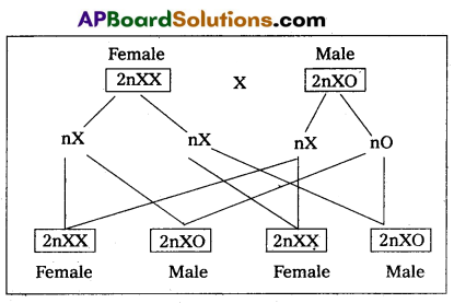
i) XX – XO type: In certain insects belonging to orders Hemiptera (true bugs), Orthoptera (grass hoppers) and Dictyoptera (cockroaches) female has two ‘X” chromosomes (XX) and are, thus homogametic, while male has only single ‘X” chromosome (XO). The male being heterogametic sex produces two types of sperms, half with X chromosome and half without X chromosome in equal proportions. The sex of the offspring depends upon the sperm that fertilises the egg, each of which carries a single X chromosome. Thus fertilisation between male and female gametes always produced zygotes with one ‘X’ chromosome from the female, but only 50% of the zygotes have an additional X Chromosome from the male. In this way, ‘XO’ and ‘XX’ types would be formed in equal proportions, the former being males and the latter being females.
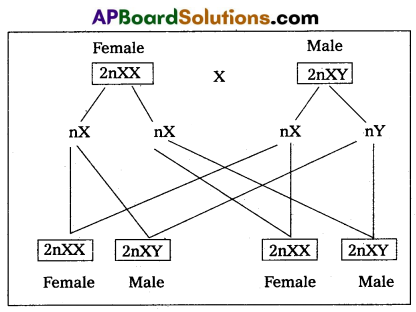
ii) XX – XY type: In man, other mammals, certain insects including Drosphila, the females possess two X chromosomes (XX) and are thus homogametic, produce one kind of eggs, each one with one X chromosome. While the males possess one X and one Y chromosome (XY) and are hence, heterogametic. They produce two kinds of sperms, half with X chromosome and half with Y chromosome. The sex of embryo depends on the kind of sperm. An egg fertilised by a X bearing sperm, produces a female, but if fertilised by a Y bearing sperm, a male is produced.
b) Heterogametic female : In this method of sex determination, the male produces similar type of gametes, while female produce dissimilar gametes. The heterogametic females may be of following two types.
i) ZO – ZZ
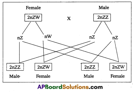
ii) ZW – ZZ.
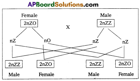
i) ZO – ZZ : This mechanism is found in certain moths and butterflies. In this case, female possesses one single ‘Z’ chromosome and hence is heterogametic, producing two kinds of eggs half with Z chromosome and another half without any Z chromosome. Male possesses two Z chromosomes and thus homogametic, producing single type of sperms, each carries single Z chromosome. The sex of the offspring depends on the kind of egg.
ii) ZW- ZZ : This system is found in certain insects (gypsy moth) and vertebrates such as fishes, reptiles anid birds. In this system, the female is heterogametic and produces two types of gametes, one with ‘Z’ Chromosome and the other with ‘W’ chromosome. On the other hand, male is homogametic and produces all sperms of same type carrying one Z’ chromosome. The sex of the offspring depends on the kind of egg being fertilised. The ‘Z’ chromosome bearing eggs produce males, but the ‘W’ chromosome bearing eggs produces females.