Thoroughly analyzing TS Inter 2nd Year Zoology Model Papers and TS Inter 2nd Year Zoology Question Paper May 2019 helps students identify their strengths and weaknesses.
TS Inter 2nd Year Zoology Question Paper May 2019
Time: 3 Hours
Maximum Marks: 60
Instructions:
Note : Read the following instructions carefully.
- Answer all questions of Section ‘A’. Answer ANY SIX questions in Section ‘B’ and answer ANY TWO questions in Section ‘C’.
- In Section ‘A’, questions from Sr. Nos. 1 to 10 are of ‘Very Short Answer Type’. Each question carries TWO marks. Every answer may be limited to 5 lines. Answer all these questions at the one place in the same order.
- In Section ‘B’, questions from Sr. Nos. 11 to 18 are of ‘Short Answer Type’. Each question carries FOUR marks. Every answer may be limited to 20 lines.
- In Section ‘C’, questions from Sr. Nos. 19 to 21 are of’ Long Answer Type’. Each question carries EIGHT marks. Every answer may be limited to 60 lines.
- Draw labelled diagrams, wherever necessary in Sections ‘B’ and ‘C’.
Section – A (10 × 2 = 20)
Note : Answer all the questions in 5 lines each:
Question 1.
Give the dental formula of adult human being.
Answer:
The arrangement of different types of teeth in each half of both the jaws in the order I, C, PM, M is represented by dental formula.
In adult it is \(\frac{2123}{2123}\) = 32.
Question 2.
What is haplo-diploidy ?
Answer:
It is a mechanism of sex determination. In this system the sex of the offspring is determined by the number of sets of chromosomes. Eg : Honeybees.
In honeybees fertilised eggs developed into female and un-fertilised eggs developed into male. This means male have half the number of chromosomes ie., haploid and the females have double the number i.e., diploid hence the name haplo-diploidy.
Question 3.
What is pleiotropy ?
Answer:
The ability of a gene to have multiple phenotypic effects because it influences a number of characters simultaneously is known as Pleiotropy. Eg : Phenylketonuria.
![]()
Question 4.
Define the terms layer and broiler.
Answer:
Layer : The birds which are raised exclusively for the production of eggs are called layers.
Broiler: The birds which are raised only for their meat are called broilers.
Question 5.
What is popularly called Guardian Angel of Cell’s Genome ?
Answer:
The protein p53 is a tumor suppressor protein, which plays an important role with reference to the “G1 check point”. In the regulation of cell division cycle. It guards the integrity of the DNA. So it is also called guardian angel of cell’s genome.
Question 6.
What in your view are the reasons for population explosion, especially in India ?
Answer:
The reasons for population explosion in India are :
a) Illiteracy among people
b) Decline in death rate
c) Increased health cafe facilities.
Question 7.
Distinguish between the enzymes renin and rennin.
Answer:
Renin: Renin is an enzyme produced by the JG cells. This enzyme catalyses the conversion of angiotensinogen into angio – tensin – I.
Rennin : Rennin is also an enzyme found in the gastric juice of infants. It acts on the milk protein casein in.the presence of calcium ions and convert it into calcium para caseinate and proteoses.
Question 8.
What is triad system ?
Answer:
In a skeletal muscle each transverse tubule (T-Tubule) is flanked on either side by several cisternae of the sarcoplasmic reticulum. T- tubule and the two terminal cisternae at its sides form the triad system.
Question 9.
What do you know about arbor vitae ?
Answer:
The white matter of cerebellum is branched, tree like appea-rance. Hence it is called arbor vitae and is surrounded by a sheath of grey matter.
Question 10.
What is amniocentesis ? Name any two disorders that can be detected by amniocentesis.
Answer:
Amniocentesis is a diagnostic procedure to detect genetic defects in the unborn baby, in which amniotic fluid is collected from foetus and diagnosed for abnormalities. Down’s syndrome, Turner’s syndrome and-Edward’s syndrome can be detected by amniocentesis.
Section – B (6 × 4 = 24)
Note : Answer any six questions in 20 lines each:
Question 11.
What are the functions of liver ?
Answer:
Liver performs a variety of functions such as synthesis, storage and secretion of various substances.
- Liver secretes bile juice, it contains bile salts such as sodium / potassium glycocholates and taurocholates, which helps in digestion and absorption of lipids.
- Liver plays the key role in carbohydrate metabolism.
a) Glycogenesis : formation of glycogen from glucose.
b) Glycogenolysis : breakdown of glycogen into glucose.
c) Gluconeogenesis : Synthesis of glucose from certain amino acids, lactate (or) glycerol. - Liver also plays an important role in synthesis of cholesterol and production of triglycerides.
- Deamination of proteins occurs in the liver.
- Liver is the chief organ of detoxification of toxic substances that enter the gut along with food.
- Liver acts as thermoregulatory organ.
- Liver acts as a haemopoietic organ in the foetus and erythro- clastic organ in the adult.
- The liver synthesizes the plasma proteins such as albumin, globulins, blood clotting factors such as fibrinogen / prothrombin, etc., and the anticoagulant called heparin.
- The lactic acid formed during anaerobic muscle contraction ‘ is converted into glycogen (gluconeogenesis) in the liver by Cori cycle.
- Kupffer cells are the largest phagocytic cells which remove unwanted substances and microbes that attack the liver by phagocytosis.
Question 12.
What are the major transport mechanisms for CO2 ? Explain.
Answer:
Carbondioxide is transported in three ways.
1. In dissolved state : 7% of CO2 is transported in dissolved state through plasma.
CO2 + H2O → H2CO3.
2. As Carbamino compounds : About 20 – 25% of CO2 com-bine directly with free amino group of haemoglobin and forms Carbamino haemoglobin in a reversible manner.
Hb – NH2 + CO2 → Hb – NHCOO– + H+.
pCO2 and pO2 could affect the binding of CO2 to haemoglobin.
- when pCO2 is high and pO2 is low as in the tissues, bind-ing of’more CO2 occurs.
- when pCO2 is low and pO2 is high as in the alveoli, dissociation of CO2 carbamino – haemoglobin takes place, (i.e., CO2 which is bound to haemoglobin from the tissues is delivered at the alveoli)
3. As Bicarbonates : About 70% of CO2 is transported as bicarbonate. RBCs contain a very high concentration of the enzyme, carbonic anhydrase and a minute quantity of the same is present in plasma too. This enzyme facilitates the following reaction in both the directions.
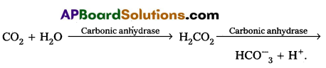
At the tissues where partial pressure of CO2 is high due to catabolism, CO2 diffuses into the blood and forms carbonic acid which dissociates into HCO–3 + H+
At the alveolar site where pCO2 is low, the reaction proceeds in the opposite direction leading to the formation of CO2 and water. Thus CO2 is mostly trapped as bicarbonate at the tissues and trans¬ported to the alveoli where it is dissociated out as CO2.
![]()
Every 100 ml of deoxygenated blood delivers approximately 4 ml of CO2 to the alveolar air.
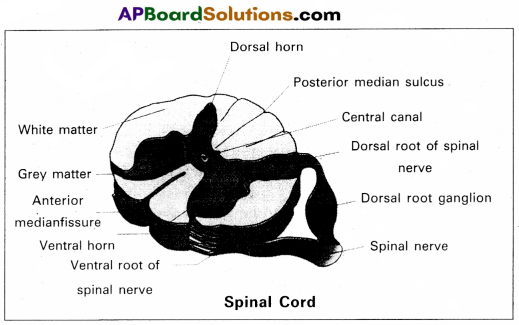
Question 13.
Draw a labelled diagram of the T.S. of the spinal cord of man.
Answer:
Question 14.
Give an account of the secretions of pituitary gland.
Answer:
The pituitary gland is also called hypophysis. Anatomically pituitary gland is divided into anterior and posterior pituitary.
I. Anterior Pituitary : It produces six important peptides. They are :
- Growth hormone (GH) Somatotropin : They promote growth of the entire body by increasing protein synthesis, cell division and cell differentiation.
- Prolactin : It causes enlargement of the mammary glands of the breast and initiate the maintenance of lactation in mammals. Prolactin also promote the growth of corpus luteum and stimulate the production of progesterone.
- Thyroid stimulating hormone (TSH) : It stimulates the production of thyroid hormones from thyroid gland.
- Adreno corticotropic hormone (ACTH) : Controls the production of steroid hormones called gluco corticoids, by the adrenal cortex.
- Follicle stimulating hormone (FSH) : It stimulates growth the development of the ovarian follicles in females. In males FSH along with the androgens, regulates spermatogenesis.
- Luteinizing hormone (LH) : In males. it stimulates production of androgens. In females it stimulates the ovaries to produce estrogens and progesterone and it maintains corpus luteum.
II. Posterior pituitary : It stores and releases two hormones called oxytocin and vasopressin.
Oxytocin : In females it stimulates contraction of pregnant uterus during child birth and ejection of milk from the mammary gland.
Vasopressin (ADH) : Affects the kidney and stimulates reabsorption of water and electrolytes by the DCT and collecting duct.
Question 15.
Write short notes on B-cells.
Answer:
The lymphocytes capable of producing antibodies and can capture circulating antigens are called B-cells. They are produced from the stem cells in the bone marrow, liver of foetus and bursa of fabricius in birds. Mature B-cells express or display Ig M and Ig D antibodies on their membrane surfaces. As these antibodies can take antigens, the mature B-cells are also called immuno-competent B-cells. In secondary lymphoid organs these immuno-competent B-cells develop into functional immune cells which later differentiate into long lived memory cells and effector plasma cells. The plasma cells produce antibodies specific to the antigen to which they are exposed. Memory cells store information about the specific antigens and show quick response, when the same type of antigen invades the body later.
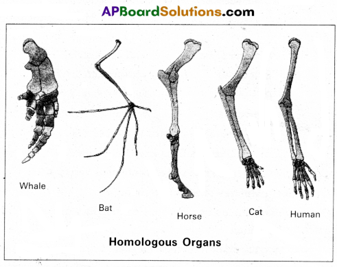
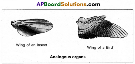
Question 16.
Distinguish between homologous and analogous organs.
Answer:
1. Homologous organs : The organs which have similar struc¬ture and origin but not necessarily the same function are called homologous organs. Eg : The appendages of vertebrates such as the flippers of whale, wings of bat, forelimbs of horse, paw of cat and hands of man have a common pattern in the arrangement of bones even though their external form and functions may vary to suit their mode of life.
2. Analogous organs : The organs which have dissimilar structure and origin but perform the same function are called the analogous organs. Eg : Wings of butterfly and wings of a bird.
Question 17.
Write a short note on the theory of mutations.
Answer:
Theory of mutation was proposed by Hugo de Varies, who coined the term mutation.
Mutations are sudden, random inheritable changes that occur in organisms. Hugo de Varies observed this phenomenon in the evening primrose plant Oenothera lamarckiana, which shows different forms like ;
- O. brevistylis – with small style.
- O. levifolia – with smooth leaves.
- O. gigas – with the giant form.
- O. nanella – with dwarf form.
These characters are inherited to the progeny.
- Darwin called mutations as sports of nature or saltations.
- Bateson called them as discontinuous variations
Salient features of mutation theory :
- Mutations occur from time to time in naturally breeding population.
- Mutants differ from their parents.
- Mutations are inheritable.
- Mutations occur in any direction i.e., they are random.
- They are discontinuous and not accumulated over generations.
- They are full-fledged and so there are no intermediate forms.
- They are subjected to natural selection.
Question 18.
Explain the different types of cancers.
Answer:
Based on the origin Cancers are classified into :
- Carcinomas: These are malignant tumor of epithelial cells. They are originating from the epithelial tissues of skin, lining of the respiratory, digestive, urinary and genetal systems or cells from various glands breast and nervous tissue etc. 85% of Cancers are Carcinomas.
- Sarcomas : These are malignant tumors of connective tissues or organs that originate from mesoderm. About 2% of tumors are Sarcomas.
- Leukemias : These are malignant tumors of stem cells of hematopoietic tissues, resulting in unrestrained production of WBC. They are liquid tumors. About 4% of Cancers are Leukemias.
- Lymphomas : These are malignant tumors of secondary lymphoid organs like spleen, and lymphnodes. About 4% of Cancers are Lymphomas.
![]()
Section – C (2 × 8 = 16)
Note : Answer any two questions in 60 lines each:
Question 19.
What is Criss-cross inheritance ? Explain the inheritance of one sex linked recessive character in human beings.
Answer:
The X-linked genes are represented twice in female (because female has two ‘X’-chromosomes) and once in males, (because male has one‘ X-chromosome). In male single X-linked recessive gene express it phenotypically, in contrast to female in which two ‘X’ linked recessive genes are necessary for the determination of a single phenotypic trait related to sex.
The recessive X-linked genes have chracteristic crisscross inheritance.
Crisscross inheritance : The inheritance of X-linked recessive trait (genes) to his grandson (F2) through his daughter (carrier) is called crisscross inheritance. Crisscross inheritance can be explained in humans by sex-linked recessive disorder, colour blindness.
Colourblindness : Colour blindness is a particular trait in human beings render them unable to differentiate between red and green colour. The gene for this colour blindness is located on X- chromosome. Colour blindness is recessive to normal vision so that if colour blind man marries a normal vision (homozygous) woman, all the sons and daughters are normal but daughter are heterozy-gous, which means that these daughters would be carrier for this trait. If such carrier woman marries a man with normal vision all the daughters and half of the sons have normal vision and half of sons are colour blind.
Colour blind trait is inhereted from a male parent to his grandson through carrier daughter-i.e., this trait shows crisscross inheritance.
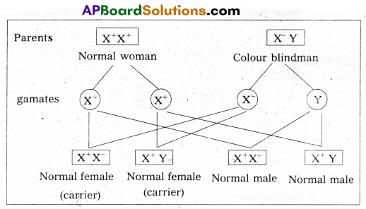
If carrier female is married to normal male
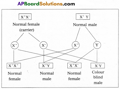
Characteristics of X-linked recessive traits :
- They never passed from father to son.
- Males are much more likely to be affected because they need only one copy of the mutant allele to express the phenotype.
- Affected males get the disease from their carrier another only.
- Sons of heterozygous female (i.e., carrier female) have 50% chance of receiving mutant alleles. These disorders are typically passed from an affected grandfather to 50% of his grandsons.
- The X-linked recessive traits shows Crisscross pattern of inhertance.
Eg : Colourblindness, Hemophilia, Muscular dystrophy etc.,
![]()
Question 20.
Describe male reproductive system of a man. Draw a labelled diagram of it.
Answer:
The male reproductive system or male genital system consists of a number of sex organs that are a part of the human reproductive process. The sex organs which are located in the pelvic region include a pair of testes, accessory ducts, glands and external genitalia.
1) Testes : The testes are a pair of oval pinkish male sex organs suspended in abdominal cavity within a pouch called scrotum. The scrotum helps in maintaining the low temperature of the testes (2 – 2.5°C) necessary for spermatogenesis. The cavity of scrotal sac is connected to the abdominal cavity through the ‘inguinal canal’. Testes is held in position in the scrotum of the ‘gubernaculum’, a fibrous cord that connects the testis with the bottom of scrotum and a ‘spermatic cord’, formed by the vas deferens, nerves, blood vessels and other tissues that run from abdomen down to each testicle,
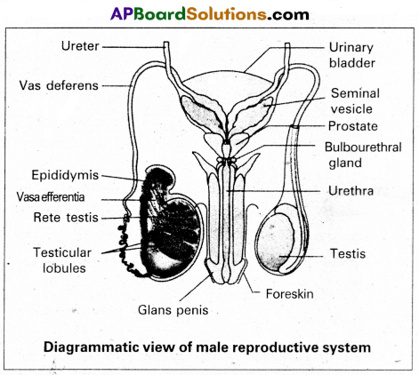
through inguinal canal. Each testis is enclosed in a fibrous envelope, ‘tunica albuginea’, which extends inwards into testis and divide it into lobules. Each lobule contains 1 to 3 highly coiled seminiferous tubules. A pouch of serous membrane ‘tunica vaginalis’ covers the testis.
Miniferous tubules : Each seminiferous tubule is lined by ‘germinal epithelium’ which consists of undifferentiated male gum cells called ‘spermatogonial mother cells’ and it also bears ‘nourishing cells’ called ‘sertoli cells’.
- Spermatogonial cells (or) primary spermatocytes undergo meiotic division, producing” spermatozoa or sperms by a process spermatogenesis.
- Sertoli cells provide nutrition to spermatozoa and produce a hormone inhibin’, which inhibits secretion of FSH.
The region outside the tubules, contain interstitial cells of Leydig cells’. They produce androgens, the most important in testosterone. It controls the development of secondary sexual characters and spermatogenesis. The seminiferous tubules open into vasa efferntia through the rete testis. Rete testis is a network of tubules is of the testis carrying spermatozoa from the seminiferous tubules to the vasa efferentia.
2) Epididymis : The vasa efferntia leave the testis and open into a narrow, tightly coiled tube called ‘epididymis’ located along the posterior surface of each testis. The epididymis provides a storage space for sperms and gives them time to nature.
It is differentiated into three regions,
a) Caput epididymis
b) Corpus epididymis
c) Cauda epididymis
The caput epididymis receives spermatozoa via the vasa efferntia of the mediastinum testis. It is mass of a connective tissue at the back of the testis that encloses the rete testis.
3) Vasa deferentia : The vas deferens or ductus deferens is a long, narrow mascular tube. The mucosa of the ductus deferens consists of a pseudo stratified columnar epithelium and lamina propia. It starts from the tail of epididymis, passes through the inguinal canal into the abdomen and loops over the urinary bladder. It receives a duct from seminal vesicle.
The vas deferens and the duct of the seminal vesicle units to form a ‘short ejaculatory duct’ or ‘ductus ejaculatorius’. The two ducts, carrying spermatozoa and the fluid secreted by the seminal vesicles, converge in the centre of prostate and open into urethra, which transports the sperms to outside.
4) Urethra : In male, Urethra is the shared terminal duct of the reproductive and urinary systems. The urethra originates from urinary bladder and extends through the penis to its external opening called ‘urethral meatus’. The urethra provides an exit for urine as well as semen during ejaculation.
5) Penis : Urethra opens into the major copulatory organ of male, the ‘penis’. The penis and scrotum constitute the male external genitalia. The penis serves as a urinal duct and intromittent organ the transfers spermatozoa to the vagina of a female.
The penis is made up of three columns of tissue : Two upper Corpora cavernosa on the dorsal aspect and one Corpus spongiosum on the ventral side. Skin and a subcutaneous layer encloses all three columns, which consists of special tissue that helps in erection of penis. The enlarged and bulbous end of penis is called glans penis’, which is covered by a loose fold of skin (foreskin) called prepuce.
Male accessory glands : Male accessory glands are :
a) Seminal vesicles
b) Prostate glands
c) Bulbourethral glands
a) Seminal vesicles : These are a pair of simple tubular glands present postero-inferior to the urinary bladder in the pelvis. Each seminal vesicle enters prostate gland through vas deferens. The vesicles produce seminal fluid rich is fructose, proteins, citric acid, in organic phosphorus, potassium and prosta- glandins. All these serve sperm cells.
b) Prostate gland : It is located directly beneath the urinary bladder. The gland surrounds the ‘Prostatic urethra’, and sends its secretions through prostatic ducts. The prostatic secretion activates spermatozoa and provides nutrition. In man, the prostate contributes 15-30% of the semen.
c) Bulbourethral glands : These are also called cowper’s glands located beneath the prostate gland at the beginning of the internal portion of the penis. They add an alkaline fluid to semen and the fluid secreted by them lubricates urethra. It acts as flushing agent washing out the acidic urinary residues that remain in the urethra, before the semen is ejaculated.
![]()
Question 21.
Describe the structure of the heart of man with the help of neat labelled diagram.
Answer:
Human heart is a hallow muscular, cone shaped, and pulsating organ situated between lungs. It is about the size of a closed fist.
The heart is covered by double walled pericardium, which consists of outer fibrous pericardium and inner serous pericardium. The serous pericardium is double layered, outer parietal layer and inner visceral layer. These two layers are separated by pericardial space, which is filled with pericardial fluid. This fluid reduces friction between the two membranes and allow free movement of the heart.
Human heart has four chambers with two smaller upper chambers called atria and two larger lower chambers called ventricles. Atria and ventricles are separated by a deep transverse groove called coronary sulcus.
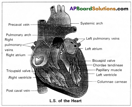
i) Atria :
- Atria are thin walled receiving chambers. The right one is larger than the left.
- The two atria are separated by thin inter-atrial septum. It has a small pore known as Foramen Ovale’ in fetal stage. Later it is closed and appears as depression (oval patch) known as ‘Fossa ovale’. If, the foramen ovale does not close properly it is called a patent foramen ovale.
- The right atrium receives deoxygenated blood from different ‘ parts of the body, through three caval veins like two precaval veins and one post caval vein.
- The right atrium also receives, blood from wall of the heart through coronary sinus, whose opening into the right atrium ‘ is guarded by the valve of Thebesius.
- Opening of the post caval vein is guarded by the Eustachian valve. It is functional in fetal stage and directs the blood from post caval vein into left atrium through foramen ovale. But it is non-functional in adult.
- The openings of the precaval veins into the right atrium have no valves.
- Left atrium receives oxygenated blood from lungs through a pair of pulmonary veins, which open into the left atrium through a common pore.
- Atrio-ventricular septum separates atria and ventricles. It has right and left atrio-venticular apertures.
- Tricuspid valve guards the right atrio-ventricular aper-ture. Bicuspid valve guards the left atrio-ventricular aperture.
ii) Ventricles :
These are the thick walled blood pumping chambers, separated by an interventricular septum. The wall of the left ventricle is thicker than that of the right ventricle as the left ventricle must force the blood to all the parts of the body.
The inner surface of the ventricles is raised into muscular ridges called columnae cameae. Some of them are large and conical and known as papillary muscles. Collagenous cords are known as chordae tendinae are present between atrio-ventricular valves and papillary muscles. They prevent the cusps of valves from bulging too far into atria during ventricular systole.
Nodal tissue : A specialized cardiac musculature called the nodal tissue is also distributed in the heart.
1) Sino-artrial node (SAN) – Present in the right upper comer of right atrium.
2) Atrio-ventricular node (AVN) – Present in the lower left comer of right atrium.
iii) Aortic arches : Human heart has two aortic arches.
1) Pulmonary arch : Arises from the left anterior angle of the right ventricle. It carries deoxygenated blood to lungs. It’s opening from right ventricle is guarded by pulmonary valve made with 3 semiluminar valves.
2) Left systemic arch : Arises from the left ventricle to distribute oxygenated blood to various parts in the body. Its opening is also guarded by aortic valve made with a set of 3 semilunar valves.
A fibrous strand, known as ligamentum arteriosm is present at the point of contact of the systemic and pulmonary arches. It is the remnant of the ductus arteriosus, which connects the systemic and pulmonary arches in the embryonic stage.