Thoroughly analyzing TS Inter 2nd Year Zoology Model Papers and TS Inter 2nd Year Zoology Question Paper March 2018 helps students identify their strengths and weaknesses.
TS Inter 2nd Year Zoology Question Paper March 2018
Time: 3 Hours
Maximum Marks: 60
Instructions:
Note : Read the following instructions carefully.
- Answer all the questions of Section ‘A’. Answer ANY SIX questions in Section ‘B’ and answer ANY TWO. questions in Section ‘C’,
- In Section ‘A’, questions from SI. Nos. 1 to 10 are of very short answer type. Each question carries TWO marks. Every answer may be limited to 5 lines. Answer all these questions at one place in the same order.
- In Section ‘B’, questions from SI. Nos. 11 to 18 are of short answer type. Each question carries FOUR marks. Every answer may be limited to 20 lines.
- In Section ‘C’, questions from SI. Nos. 19 to 21 are of long answer type. Each question carries EIGHT marks. Every answer may be limited to 60 lines.
- Draw labelled diagrams wherever necessary for questions in Section ‘B’ and ‘C’.
Section – A (10 × 2 = 20)
Note : Answer all the questions in 5 lines each:
Question 1.
Name different types of papillae present on the tongue of man.
Answer:
The upper surface of the tongue has small projections called papillae: In humans the tongue bears 3 (three) types of papillae namely
- fungi form
- filiform
- Circumvallate papillae.
Question 2.
Differentiate between the “Columns of Bertin” and “Duct of Bellini”.
Answer:
Columns of Bertin : Columns of Bertin are the medullary extensions of the renal cortex in between renal pyramids.
Ducts of Bellini: Some initial collecting ducts unite to form straight collecting duct, which passes through the medullary pyramid. In the medulla, the tubes of each pyramid join and form duct of Bellini.
![]()
Question 3.
Write any two differences between aerobic and anaerobic muscles.
Answer:
| Red muscle fiber (aerobic) | White muscle fiber (anaerobic) |
| 1. Red muscle fibers are thin and smaller in size. | 1. White muscle fibers are thick and larger in size. |
| 2. They are red in colour as they contain large amount of myoglobin. | 2. They are white in colour as they contain small amount of myoglobin. |
| 3. They contain numerous mitchondria. | 3. They contain less number of mitochondria. |
| 4. They carry out slow and sustained contractions for a long period. | 4. They carry out fast work for short duration. |
Question 4.
Name the keystone bone of the cranium. Where is it located ?
Answer:
Sphenoid bone is the keystone bone of the cranium, because it articulates with all the other cranial bones. It is present at the middle phrt of the base of the skull.
Question 5.
“Colostrum is very much essential for the newborn infants.” Justify.
Answer:
The colostrum secreted by the mother during the initial days of lactation has abdundant IgA antibodies to protect infant from initial sources of infection.
Question 6.
Define the terms immunity and immune system.
Answer:
Immunity: It is an ability of host or individual to fight against the disease causing organisms is called immunity.
Immune System: The network of organs, cells and proteins that protect the body from harmful, infections agents such as bacteria, viruses, animal parasites, fungi etc., is called the immune system.
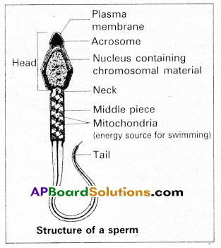
Question 7.
Define “Spermiation” and “Spermiogenesis”.
Answer:
Spermiogenesis : The process in which haploid spermatids are transformed into spermatozoa or sperms.
Spermiation : The process in which sperm head becomes embedded in the Sertoli cells and finally released from the semi-niferous tubules.
Question 8.
What is ‘Amniocentesis’ ? Name any two disorders that can be detected by “Amniocentesis”.
Answer:
Amniocentesis is a diagnostic procedure to detect genetic defects in the unborn baby, in which amniotic fluid is collected from foetus and diagnosed for abnormalities. Down’s syndrome, Turner’s syndrome and Edward’s syndrome can be detected by amniocentesis.
![]()
Question 9.
Differentiate between “Apiculture” and “Aquaculture”.
Answer:
| Apiculture | Aquaculture |
| Apiculture is the maintenance of hives of honeybees for the production of honey and wax. | Culturing of fishes and other aquatic organisms under regulated conditions to achieve better production. |
Question 10.
MRI scan is harmless. Justify.
Answer:
MRI does not use ionizing radiation, as involved in X-rays, and is generally safe and harmless procedure.
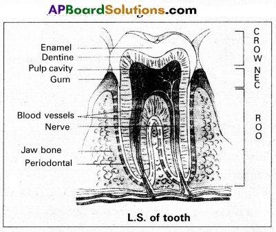
Question 11.
Draw a neat labelled diagram of L.S. of a tooth.
Answer:
Question 12.
Give an account of the disorders of Human respiratory system.
Answer:
Disorders of respiratory system.
1) Asthma : Asthma is a difficulty in breathing caused due to inflammation of bronchi and bronchioles. Symptoms include coughing, difficulty in breathing and wheezing.
2) Emphysema : It is a chronic disorder in which alveolar walls are damaged and their walls coalesce due to which respiratory surface area of exchange of gases is decreased. One of the major causes of this is smoking of tobacco.
3) Bronchitis : Bronchitis is the inflammation of the bronchi, resulting in the swelling of mucus lining of bronchi, increased mucus production and decrease in the diameter of bronchi. Symptoms include chronic cough with thick sputum.
4) Pneumonia : The infection of lungs caused by Stre-ptococcus pneumoniae and also by certain Virus, Fungi, Protozoans and Mycoplasmas. Symptoms include inflammation of lungs, accumulation of mucus in alveoli and impaired exchange of gases, leading to death if untreated.
Occupational dissorders : These are caused by exposure of the body to the harmful substances.
E.g. : i) Asbestosis: It occurs due to chronic exposure to asbestos dust in the people working in asbestos industry.
ii) Silicosis : It occurs because of long term exposure to silica dust.
iii) Siderosis : It occurs due to deposition of iron particles in tissues.
iv) Black lung disease : It develops from inhalation of coal dust.
Question 13.
Give an account of the retina of the human eye.
Answer:
Retina is the inner coat of the eye. It consist of a pigmented epithelium and a neural portion. The pigmented epithelium is a sheet of melanin containing epithelial cells. The neural portion has three layers namely photoreceptor layer, bipolar cell layer and ganglion cell layer.
Photoreceptor layer consist of rods and cones. Rods contain a protein called rhodopsin. Rods are concerned with dim light. Cones contain a visual pigment called iodopsin and they are important in daylight vision and colour vision. There are three types of cones and are response to red, green and blue colours.
The centre of the posterior portion of the retina is called yellow spot. A depression present in the yellow spot is called ‘Forea’ contractile and it contains only cones. Forea is responsible for sharp vision. The region of retina which is devoid of rods and cones is known as blind spot (or) optic disc, which form the optic nerve called 2nd cranial nerve.
Question 14.
Write a note on Addison’s disease and Cushing’s syndrome.
Answer:
Addison’s disease : It is caused due to hyposecretion of glucocorticoids by the adrenal cortex. This disease is characterised by loss of weight, muscle weakness, fatigue and reduced blood pressure. Sometimes darkening of the skin in both exposed and non-exposed parts of the body occurs in this disorder.
Cushing’s Syndrome : It results due to over production of glucocorticoids. This condition is characterised by breakdown of muscle proteins and redistribution of body fat resulting in spindly arms and legs, a round moon-face, buffalo hump on the back and pendulous abdomen is also observed. Wound healing is poor. The elevated level of cortisols causes hyperglycemia, over deposition of glycogen in liver and rapid gain of weight.
![]()
Question 15.
Describe the Genic Balance Theory of sex determination in Drosophila.
Answer:
Genic balance mechanism of determination of sex was first observed and studied by C.B.Bridges in 1921 in Drosophila. According to this mechanism, the sex of an individual in Drosophila melanogaster is determined by a balance between the genes for femaleness located in the X- chromosome and those for maleness located in autosomes. Hence, the sex of an individual is determined by the ratio of number of its X chromosomes and that of its autosomal sets, the ‘Y’ chromo-some being unimportant.

Individuals with sex index of 0.5 develop into normal males and those with sex index of 1 into normal females. If the sex index is between 0.5 and 1, the resulting individuals is called inter sex. Such individuals are sterile. Some flies have sex index of >1, such flies are called super females or metafemales. Super male flies have a sex index value of < (3.5 and are also weak, sterile and non-viable.
| Sex index = X/A | Phenotypes |
| 0.5 | Male |
| 1.0 | Female |
| Between 0.5 and 1 | Inter sex |
| Below 0.5 | Metamale |
| Above 1.0 | Metafemale |
Bridges drew, crossed a triploid females (3A + XXX) with normal diploid males (2A + XY). From such a cross he obtained normal diploid females, males, triploid females, intersexes, meta-males and metafemales.
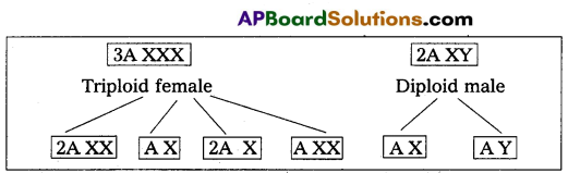
| A X | A Y | |
| 2A XX | 3A XXX Triploid female (1.0) |
3A XXY Triploid intersex (0.66) |
| A X | 2A XX Diploid female (1.0) |
2A XY Diploid male (0.5) |
| 2A X | 3A XX Triploid intersex (0.66) |
3A XY Metamales (0.33) |
| A XX | 2A X XX Metafemales (1.5) |
2A XXY Diploid female (1.0) |
Question 16.
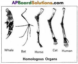
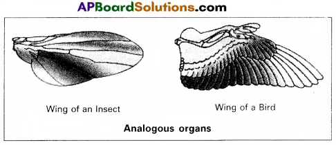
Distinguish between homologous and analogous organs with examples.
Answer:
1. Homologous organs : The organs which have similar structure and origin but not necessarily the same function are called homologous organs. Eg : The appendages of vertebrates such as the flippers of whale, wings of bat, forelimbs of horse, paw of cat and hands of man have a common pattern in the arrangement of bones even though their external form and functions may vary to suit their mode of life.
2. Analogous organs : The organs which have dissimilar structure and origin but perform the same function are called the analogous organs. Eg : Wings of butterfly and wings of a bird.
Question 17.
What is meant by genetic drift ? Explain genetic drift citing the example of Founder Effect.
Answer:
The change in the frequency of a gene that occurs merely by chance and not by selection, in small proportion is called genetic drift.
Suppose, for a gene with two alleles, the frequency of a particular allele is 1% (e = 0.01) the probability of losing that allele by chance from.small population is more. The end result is either fixation or loss of that allele.
Genetic drift tend to reduce the amount of genetic variation within the population mainly by removing the alleles with low frequencies. It can examplified by Founder and Bottleneck effect.
Founder effect : If a small group of individuals from a population starts a new colony in an isolated region, those individuals are called the founders of the new population. The allelic frequency of their descendants are similar those of the founders rather than to their ancestral parent population.
Eg : O+ve blood group is present in nearly 100% of the red Indians. It means the forefathers of the Red Indians tribe were predominantly O+ve and they isolated themselves reproductively from other population.
![]()
Question 18.
Discuss in brief about ‘Avian Flu’.
Answer:
Avian Flu or bird Flu is an important disease affecting poultry birds and man.
Causative organism : Avian Flu or bird Flu is caused by an “avian Flu virus” the H5NI. The virus that causes the bird infection infects humans too. It is a pandemic disease.
Mode of infection : Infection may be spread simply by touching contaminated surfaces. Birds infected by this type of influenza, continue to release the virus as in their faeces and saliva for as long as 10 days.
Symptoms: In humans it causes typical-flu-like symptoms, include cough, diarrhoea difficulty in breathing, fever, headache, malaise, muscle aches and sore throat.
Prevention :
- Avoiding consumption of under cooked chicken.
- People who work for poultry birds should use protective clothing and special breathing masks.
- Complete culling of infected flock by burying or burning them. ‘
Section – C (2 × 8 = 16)
Note : Answer any two questions in 60 lines each:
Question 19.
What are multiple alleles ? Describe multiple alleles with the help of ABO blood groups in man.
Answer:
Generally a gene has two alternative forms called allele. Sometimes a gene may have more than two alleles. These are referred to as multiple alleles. When more than two alleles exist in a population of a specific organism, the phenomenon is called multiple allelism. Multiple alleles cannot be observed in the genotype of a diploid individual, but can be observed in a population.
The number of genotypes that can occur for multiple alleles is given by the expression where ‘n’ = number of alleles.
ABO blood groups are the best example for multiple allelism in human beings.
The ABO blood group system was proposed by Karl Lands- steiner. The blood groups A, B, AB and O types are characterised by the presence or absence of antigens on the surface of RBC. Blood type ‘A’ person have antigen A on their RBCs and anti-B antibodies in the plasma. Blood type ‘B’ person have antigen B.
On their RBCs and anti-A antibodies in the plasma. Blood type ‘AB’ person have antigens A and B on the RBCs and no antibodies in the plasma. Blood type ‘O’ person have no antigens on their RBCs and both anti-A, and anti-B antibodies are present in the plasma.
| Blood group | Antigen on RBC | Antibodies in Plasma |
| A | A | b |
| B | B | a |
| AB | AB | — |
| O | — | a, b |
Bernstein discovered that these phenotypes were inherited by the interaction of three ‘autosomal alleles’ of the gene named ‘I’, located on chromosome 9. IA IB and IO are the three alleles of the gene I. The alleles IA and IB are responsible for the production of the respective antigens A and B. The allele IO does not produce any antigen. The alleles IA and IB are dominant to the allele IO but co-dominant to each other (IA = IB > IO).
A child receives one of the three alleles from each parent, giving rise to six possible genotypes and four possible blood types.
The genotypes are IA IA , IA IO , IB IB , IB IO , IA IB , IO IO. The phenotypic expressions of IA IA and IA IO , are A-type blood, the phenotypic expression of IBIB and IBIO are B-type blood, and that of IA IB , is AB blood type. The phenotype IO IO, is ‘O-type’ blood.
Question 20.
Describe male reproductive system of a man. Draw a labelled diagram of it.
Answer:
The male reproductive system or male genital system consists of a number of sex organs that are a part of the human reproductive process. The sex organs which are located in the pelvic region include a pair of testes, accessory ducts, glands and external genitalia.
1) Testes : The testes are a pair of oval pinkish male sex organs suspended in abdominal cavity within a pouch called scrotum. The scrotum helps in maintaining the low temperature of the testes (2 – 2.5°C) necessary for spermatogenesis. The cavity of scrotal sac is connected to the abdominal cavity through the ‘inguinal canal’. Testes is held in position in the scrotum of the ‘gubernaculum’, a fibrous cord that connects the testis with the bottom of scrotum and a ‘spermatic cord’, formed by the vas deferens, nerves, blood vessels and other tissues that run from abdomen down to each testicle, through inguinal canal. Each testis is enclosed in a fibrous envelope, ‘tunica albuginea’, which extends inwards into testis and divide it into lobules. Each lobule contains 1 to 3 highly coiled seminiferous tubules. A pouch of serous membrane ‘tunica vaginalis’ covers the testis.
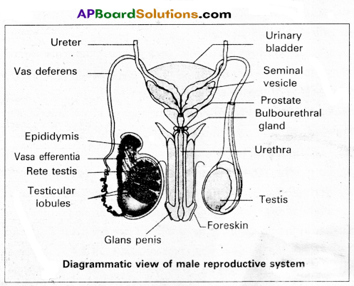
Miniferous tubules : Each seminiferous tubule is lined by ‘germinal epithelium’ which consists of undifferentiated male gum cells called ‘spermatogonial mother cells’ and it also bears ‘nourishing cells’ called ‘sertoli cells’.
- Spermatogonial cells (or) primary spermatocytes undergo meiotic division, producing spermatozoa or sperms by a process spermatogenesis.
- Sertoli cells provide nutrition to spermatozoa and produce a hormone ‘inhibin’, which inhibits secretion of FSH.
The region outside the tubules, contain interstitial cells of ‘Leydig cells’. They produce androgens, the most important in testosterone. It controls the development of secondary sexual characters and spermatogenesis. The seminiferous tubules open into vasa efferntia through the rete testis. Rete testis is a network of tubules is of the testis carrying spermatozoa from the seminiferous tubules to the vasa efferentia.
2) Epididymis: The vasa efferntia leave the testis and open into a narrow, tightly coiled tube called’epididymis’ located along the posterior surface of each testis. The epididymis provides a storage space for sperms and gives them time to nature.
It is differentiated into three regions.
a) Caput epididymis
b) Corpus epididymis
c) Cauda epididymis.
The caput epididymis receives spermatozoa via the vasa efferntia of the mediastinum testis. It is mass of a connective tissue at the back of the testis that encloses the rete testis.
3) Vasa deferentia : The vas deferens or ductus deferens is a long, narrow mascular tube. The mucosa of the ductus deferens consists of a pseudo stratified columnar epithelium and lamina propia. It starts from the tail of epididymis, passes through the inguinal canal into the abdomen and loops over the urinary bladder. It receives a duct from seminal vesicle.
The vas deferens and the duct of the seminal vesicle units to form a ‘short ejaculatory duct’ or’ductus ejaculatorius’. The two ducts, carrying spermatozoa and the fluid secreted by the seminal vesicles, converge in the centre of prostate and open into urethra, which transports the sperms to outside.
4) Urethra : In male, Urethra is the shared terminal duct of the reproductive and urinary systems. The urethra originates from urinary> bladder and extends through the penis to its external opening called ‘urethral meatus’. The urethra provides an exit for urine as well as semen during ejaculation.
5) Penis : Urethra opens into the major copulatory organ of male the ’penis’. The penis and scrotum constitute the male external genitalia. The penis serves as a urinal duct and intromittent organ the transfers spermatozoa to the vagina of a female.
The penis is made up of three columns of tissue : two upper Corpora cavernosa on the dorsal aspect and one Corpus spongiosum on the ventral side. Skin and a subcutaneous layer encloses all three columns, which consists of special tissue that helps in erection of penis. The enlarged and bulbous end of penis is called ‘glans penis’, which is covered by a loose fold of skin (foreskin) called prepuce.
Male accessory glands : Male accessory glands are :
(a) Seminal vesicles
(b) Prostate glands
(c) Bulbourethral glands.
a) Seminal vesicles : These are a pair of simple tubular glands present postero-inferior to the urinary bladder in the pelvis. Each seminal vesicle enters prostate gland through vas deferens. The vesicles produce seminal fluid rich is fructose, proteins, citric acid, in organic phosphorus, potassium and prostaglandins. All these serve sperm cells.
b) Prostate gland: It is located directly beneath the urinary bladder. The gland surrounds the ‘Prostatic urethra’, and sends its secretions through prostatic ducts. The prostatic secretion activates spermatozoa and provides nutrition. In man, the prostate contributes 15 – 30% of the semen.
c) Bulbourethral glands : These are also called cowper’s glands located beneath the prostate gland at the beginning of the internal portion of the penis. They add an alkaline fluid to semen and the fluid secreted by them lubricates urethra. It acts as flushing agent washing out the acidic urinary residues that remain in the urethra, before the semen is ejaculated.
![]()
Question 21.
Give an account of working of the Human heart.
Answer:
The human heart is an organ that provides a continuous blood circulation through the cardiac cycle.
Special conducting tissues of heart : Human heart is myogenic. It contains a specialized cardiac musculature called the nodal tissue. A patch of this tissue called Sino-Atrial Node (SAN), is present in the right upper corner of the right atrium. Another mass of this tissue, called the Atrio-Ventricular Tissue (AVN), is present in the lower left corner of the right atrium. A bundle of nodel fibers called AV bundles/His bundles continues from the AVN into the iriter-ventricular septum. It divides into right and left bundle branches. These branches give rise to minute fibers called purkinje fibers that extend throughout the ventricular musculature.
SAN has the ability to generate action potentials without any external stimuli, hence called pacemaker. AV node is a relay point that relay the action potentials received from the SA node to the ventricular musculature.
Cardiac cycle : Cardiac cycle consists of the sequence of the cardiac events that occur from the beginning of one heart beat to the beginning of next. At beginning of cardiac cycle all the four chambers of the heart are in relaxed state. Cardiac cycle is divided into three phases, namely.
1) Atrial systole
2) Ventricular systole
3) Cardiac diastole.
1) Atrial systole : It lasts about 0.1 seconds. SAN now stimulate an action potential which stimulates both the atria to contract simultaneously causing the atrial systole. This increases the flow of blood into the ventricles by about 30%, the remaining. blood flows into the ventricle before the atrial systole.
2) Ventricle systole : It lasts about 0.3 seconds. The action potential from the SAN reaches the AVN, from where they are conducted through the bundle of His, its branches and the Purkinje fibers to entire ventricular musculature. This causes the simul-taneous ventricular systole. The atria undergo relaxation coinciding with ventricular systole. Ventricular systole, increases the pressure causing closure of AV valves preventing the back flow of blood, results in the production of first heart beat sound ‘Lub’. When pressure in ventricles exceeds the pressure in aortic arches, semilunar valves open. It results in the flow of blood from ventricles into aortic arches.
3) Cardiac diastole: It lasts about 0.4 seconds. The ventricles now relax and ventricular pressure falls causing the closure of the semilunar valves which prevent the back flow of blood. This results in the production of second heart sound known as ‘Dup’. When pressure in ventricles falls below atrial pressure, AV valves open and ventricular filling begins. All the chambers are now again in’ relaxed state. Soon another cardiac cycle sets in.