Thoroughly analyzing AP Inter 2nd Year Zoology Model Papers and AP Inter 2nd Year Zoology Question Paper May 2016 helps students identify their strengths and weaknesses.
AP Inter 2nd Year Zoology Question Paper May 2016
Time: 3 Hours
Maximum Marks: 60
Instructions:
- Answer all questions of Section – A. Answer ANY SIX questions in Section – B and answer ANY TWO questions in Section – C.
- In Section – A questions from Sr. Nos. 1 to 10 are of Very Short Answer Type. Each question carries TWO marks. Every answer may be limited to 5 lines. Answer all these questions at one place in the same order.
- In Section – B, questions from Sr. Nos. 11 to 18 are of Short Answer Type. Each question carries FOUR marks. Every answer may be limited to 20 lines.
- In Section – C, questions from Sr. Nos. 19 to 21 are of Long Answer Type. Each question carries EIGHT marks. Every answer may be limited to 60 lines.
- Draw labelled diagrams wherever necessary in Sections – B and C.
Section – A
Note : Answer ALL the questions. (10 × 2 = 20)
Question 1.
What is meant by Chloride Shift ?
Answer:
The exchange of chloride and bicarbonate ions between RBC and plasma at the tissues is called chloride shift or Hamburger’s phenomenon or Hamburger’s shift.
Question 2.
Define Glomerular Filtration.
Answer:
Filtration of the blood from the glomerulas into the lumen of the Bowman’s Capsule and this passive process is called glomerular filtration.
Question 3.
Write the difference between actin and myosin.
Answer:
a) The light band in a myofibril contains actin and two regulatory proteins called troponin and tropomyosin. Actin filaments are thinner compared to myosin filaments.
b) The dark band in a myotfibril contains (A band) myosin. Myosin filaments are thick and non contractile.
![]()
Question 4.
How do rods and cones of human eye differ from each other chemically and functionally ?
Answer:
a) Rods contain a purplish red protein called rhodopsin or visual purple which contains a derivative of vitamin A and they are important in twilight.
b) Cones contain a visual pjgment iodopsin made of a protein called photopsin and they are important in day light and colour vision.
Question 5.
What is Insulin shock ?
Answer:
Hyper secretion of insulin leads to decreased level of glucose in the blood (hypoglycemia) resulting in insulin shock.
Question 6.
Write the names of any four mononuclear phagocytes.
Answer:
Mono nuclear phagocytes are
- Histocytes,
- Kupffer cells,
- Microglia,
- Osteoclasts and
- Synovial cells.
Question 7.
Name the yellow mass of cells accumulated in the empty follicle after ovulation. Name the hormone secreted by it. What is its function ?
Answer:
After ovulation the granulosa cells in the follicle proliferate and are transformed into a yellowish glandular mass called corpus luteum. It secretes progesterone harmone. This harmone is essential for maintenance of pregnancy in first few months.
Question 8.
What are the measures one has to take to prevent contracting STDS ?
Answer:
a) Avoiding sex with unknown partners/multiple partners.
b) Using condoms compulsorily during coitus.
c) Consulting qualified doctor for early detection of STDs and getting complete treatment in case of infections.
Question 9.
Distinguish between a drone and worker in a honeybee colony.
Answer:
a) Drones are robust, large winged small numbered, short lived and fed with bee bread by nurse workers. They are developed from unfertilized ova by arrhenotoky (male parthenogenesis).
b) Worker bees are multifaceted sterile females which develop from the fertilised eggs and perform diverse functions. They live for two or three months. They are very small in size.
Question 10.
MRI scan is harmless. – Justify.
Answer:
MRI (Magnetic Resonance Imaging) scan is harmless because MRI does not use ionizing radiation as involved in X-rays and is generally a very safe procedure.
![]()
Section – B (6 × 4 = 24)
Note : Answer ANY SIX questions.
Question 11.
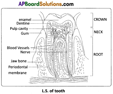
Draw a neat labelled diagram of L.S. of a tooth.
Answer:
Question 12.
Explain the process of inspiration and expiration under normal conditions.
Answer:
Inspiration : Intake of atmospheric air into the lungs is called inspiration. It is an active process, as it takes place by the contraction of the muscles of the diaphragm and the external inter – costal muscles, which extend in between the ribs. The contraction of the diaphragm (phrenic muscles) increases the volume of the thoracic chamber in the antero-posterior axis. The contraction of external inter – costal muscles lifts up the ribs and sternum causing an increase in the volume of the thoracic chamber in the dorso – ventral axis. The overall increase in the thoracic volume causes a similar increase in the ‘pulmonary volume’. An increase in the pulmonary volume decreases the intra – pulmonary pressure to less than that of the atmosphere, which forces the air from the outside to move into the lungs, i.e., inspiration.
Expiration : Release of alveolar air to the exterior is called expiration. It is a passive process. Relaxation of the diaphragm and the external inter – costal muscles returns the diaphragm and sternum to their normal positions, and reduces the thoracic volume and thereby the pulmonary volume. This leads to an increase in the intra – pulmonary pressure to slightly above that of the atmospheric pressure, causing the expulsion of air from the lungs, i.e., expiration.
Question 13.
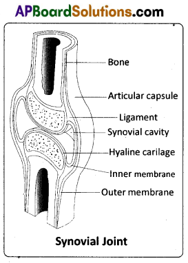
Describe the structure of synovial joint with the help of a neat labelled diagram.
Answer:
Synovial joint is covered by a double layered synovial capsule. The outer layer consists of dense fibrous irregular connective tissue with more collagen fibres. This layer is continuous with the periosteum and resists stretching and prevents the dislocation . of joints. Some fibres of these membranes are arranged in bundles called ligaments. The inner layer of synovial capsule is formed of areolar tissue and elastic fibers. It secretes a viscous synovial fluid which contains hyaluronic acid, phagocytes etc., and acts as a
‘lubricant’ for the free movement of the joints. Synovial joints include Ball and socket joint. Hinge joint, Pivot joint, Gliding joint, Condyloid joint, Saddle joint.
Question 14.
Compare a ‘pituitary dwarf and a thyroid dwarf’ in respect of similarities and dissimilarities they possess.
Answer:
1. Pituitary dwarf : Hypo secretion of growth harmone (STH) during childhood retards growth, resulting in a pituitary dwarf/ midget. The pituitary dwarf is sexually and intellectually a normal individual.
2. Thyroid dwarf : During pregnancy, due to hypothyroidism, defective development of the growing baby leads to a disorder called cretinism. Physical and mental growth get severely stunted and is called thyroid dwarf. This is due to untreated congenital hypothyroidism. Stunted growth, mental retardation, low intelligent quotient, abnormal skin, deafness and mutism are some of the characters of this disease.
Question 15.
Describe Erythroblastosis foetalis.
Answer:
Destruction of RBC of Rh positive foetus by anti Rh antibodies produced by Rh negative mother due to immunological incompatibility is called Erythroblastosis foetalis or Haemolytic disorder of newborn (HDNB). This is due to genetically incompatible marriage involving Rh positive father and Rh negative mother. At the time of birth the Rh positive foetal blood mixes with the Rh negative blood of mother, through the ruptured placenta. The Rh antigens sensitize the mother to produce anti Rh antibodies (IgG antibodies) and memory cells. This first Rh positive by is unaffected because it is delivered by the time mother is sensitized. During the next pregnancy bearing Rh positive foetus these antibodies increase in concentration due to memory cells and cross the placenta, enter the blood of baby and destroy the RBC. Haemolytic anaemia is the symptom in this disorder.
![]()
Question 16.
Distinguish between homologous and analogous organs.
Answer:
Homologous organs : The organs which have similar structure and origin but not necessarily the same function are called homologous organs. The evolutionary pattern that describes the occurrence of similarity in origin and internal structure is called homology. Such organs show adaptive radiation, hence ‘divergent evolution’, e.g. the appendages of vertebrates such as the flippers of whale, wings of bat, forelimbs of horse, paw of cat and hand of man, have a common pattern in arrangement of bones eventhough their external form and function may vary to suit their mode of life. It explains that all vertebrates might have had a common ancestor.
Analogous organs : The organs which have dissimilar structure and origin but perform the same function are called analogous organs. Analogous organs suggest ‘convergent evolution’, e.g. wings of a butterfly and wings of a bird.
Question 17.
Explain Darwin’s theory of Natural selection with industrial melanism as an experimental proof.
Answer:
Experimental verification of Natural Selection – Industrial melanism : An important practical proof for the operation of Natural Selection is the classical case of industrial melanism, exhibited by peppered moth – Bistort betularia. These moths were available in two colours, grey and black. Prior to industrial revolution, the grey moths were abundant. During the industrial revolution, the black forms were more and the grey forms were less in the industrial cities like Birmingham. Biologists proposed that with the industrial revolution, more soot was released due to the burning of coal, which resulted in the darkening of the barks of trees.
Grey moths on the dark bark were easily identified and predated more by birds. Hence the number of grey moths decreased and that of the black moths increased in the population. It means Nature offered ‘positive selection’ pressure to the black (melanic) forms. Bernard Kettlewell, a British ecologist, tested this hypothesis experimentally. He collected both the grey and the black forms of Bistort betularia for his experiment. He released them in two sets of equal numbers; one set in Birmingham, a polluted urban area, and the other set in Dorset, an unpolluted rural area. After a few days he recaptured them. Of those moths recaptured from Birmingham, there were more black forms. Among those recaptured from Dorset there were more grey forms.
The reason for such a difference is : the melanic forms could not be easily spotted by predator birds as their body colour merged with the dark colour of the bark of trees in Birmingham area. In the rural areas (Dorset) the grey forms had better survival chance as their body colour merged with the light coloured surroundings. This explains the differential survival of the moths due to Natural Selection. It will be interesting to know that there was a reversal in the selection process after the introduction of pollution check laws in the urban areas.
Question 18.
Explain the different types of cancers.
Answer:
Types of cancers : There are different types of cancers such as carcinomas (cancers of epithelial tissues / cells which are most common as epithelial cells divide more often), sarcomas (cancers of connective tissues), leukemias (cancers of bone marrow cells resulting in understrained production of WBC – a liquid tumor), lymphomas (cancers of the lymphatic system). Certain types of cancers are called ‘familial cancers’ (cancer that occurs in families; genetic based) and others ‘sporadic cancers’ (non-hereditary cancers occurring without any family history). Some types of cancers are caused by ‘tumor forming RNA viruses’ (oncoviruses), e.g. Rous sarcoma virus which causes ‘avian sarcoma’.
![]()
Section – C (2 × 8 = 16)
Note : Answer ANY TWO of the following questions.
Question 19.
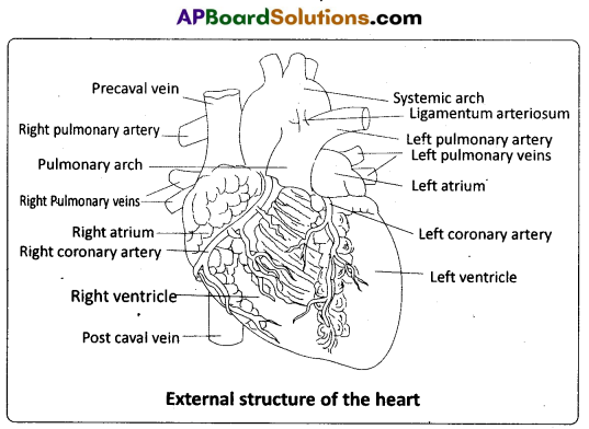
Describe the structure of the Heart of Man with the help of neat labelled diagram.
Answer:
The heart is mesodermal in origin. It is a thick walled, muscular and pulsating organ, situated in the mediastinum (the region in the thorax between the two lungs), and with its apex slightly turned to the left. It is the size of a clinched fist.
The heart is covered by a double walled pericardium which consists of the outer fibrous pericardium and the inner serous pericardiurh. The serous pericardium is double – layered, formed of an outer parietal layer and an inner visceral layer. The parietal layer is fused with the fibrous pericardium, whereas the visceral layer adheres to the surface Of the heart and forms its outer layer, the epicardium. The two layers are separated by a narrow pericardial space, which is filled with the pericardial fluid. This fluid reduces friction between the two membranes and allows free movement of the heart.
The wall of the heart consists of three layers. They are the outer epicardium, the middle myocardium (a thick layer of cardiac muscles), and the inner most endocardium (a thin layer of endothelium). The endothelium covers the heart valves also and is continuous with the endothelial lining of the large blood vessels connected to the heart.
External structure:
Human heart has four chambers, with two relatively smaller upper chambers, called atria and two larger lower chambers called ventricles. Atria and ventricles are separated by a deep transverse groove called coronary sulcus (atrio – ventricular groove). The muscular pouch like projection from each atrium is called auricular appendix (auricular appendage). The ventricles are separated by two inter ventricular grooves (anterior and posterior), in which the coronary arteries and their branches are lodged.
i) Atria : Atria are thin walled ‘receiving chambers’ (upper chambers). The right one is larger than the left. The two atria are separated by thin inter – atrial septum. In the fetal heart, the atrial septum has a small pore called foramen ovale. Normally the foramen ovale closes at birth, when lungs become functional. It is represented by a depression in the septum between the right and left atria, called fossa ovalis (that marks the position of the foramen ovale in the fetus). If, the foramen ovale does not close properly, it is called a patent foramen ovale.
The right atrium receives deoxygenated blood from different parts of the body (except the lungs) through three caval veins viz. the two precavals (right and left) and a post caval vein. It also receives blood from the myocardium (wall of the heart) through the coronary sinus, whose opening into the right atrium is guarded by the valve of Thebesius. Opening of the postcaval vein is guarded by the valve of the inferior vena cava or Eustachian valve. It directs the blood to the left atrium through the foramen ovale, in the foetal stage, but in the adult it becomes rudimentary and non – functional. The openings of the precaval veins into the right atrium have no valves. The left atrium receives blood from each lung through two pulmonary veins, which open into the left atrium. The two left pulmonary veins open by a common aperture in some.
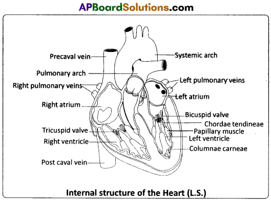
Atria and ventricles are separated by a membranous atrio – ventricular septum, which possesses left and right atrioventricular apertures. The left and right apertures are guarded by bicuspid (mitral valve) and tricuspid valves respectively.
ii) Ventricles : These are the thick walled blood pumping chambers (lower chambers), separated by an interventricular septum. The wall of the left ventricle is thicker than that of the right ventricle. The inner surface of the ventricles is raised into muscular ridges or columns called columnae carneae / trabeculae carneae projecting from the inner walls of the ventricles. Some of these ridges are large and conical, and are called papillary muscles, whose apices are connected to the chordae tendineae, or ‘heart strings’. They are cord – like collagenous processes that connect the papillary muscles to the tricuspid valve and the mitral valve in the heart. They prevent the cusps of the atrioventricular valves from bulging too far into atria during ventricular systole.
![]()
Question 20.
Describe the Male reproductive system of a man with the help of a labelled diagram.
Answer:
The Male Reproductive System : The male reproductive system (male genital system) consists of a number of sex organs that are a part of the human reproductive process. The sex organs which are located in the pelvic region include a pair of testes (sing : testis) along with accessory ducts, glands and the external genitalia.
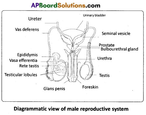
Testes : The testes (testicles) are a pair of oval pinkish male primary sex organs suspended outside the abdominal cavity with in a pouch called scrotum. The scrotum helps in maintaining the low temperature of the testes (2 – 2.5°C lower than the normal internal body temperature) necessary for spermatogenesis. The cavity of the scrotal sac is connected to the abdominal cavity through the inguinal canal.
Testis is held in position in the scrotum by the gubernaculum, a fibrous cord that connects the testis with the bottom of the scrotum and a spermatic cord, formed by the vas deferens, nerves, blood vessels and other tissues that run from the abdomen down to each testicle, through the inguinal canal. Each testis is enclosed in a fibrous envelope, the tunica albuginea, which extends inward to form septa that partition the testis into lobules. There are about 250 testicular lobules in each testis. Each lobule contains 1 to 3 highly coiled seminiferous tubules. A pouch of serous membrane (peritoneal layer) called tunica vaginalis covers the testis.
Each seminiferous tubule is lined by the germinal epithelium which consists of undifferentiated male germ cells called spermatogonial mother cells and it also bears ‘nourishing cells’ called Sertoli cells. The spermatogonia produce the primary spermatocytes which undergo meiotic division, finally leading to the formation of spermatozoa or sperms (spermatogenesis). Sertoli cells provide nutrition to the spermatozoa and also produce a hormone called inhibin, which inhibits the secretion of FSH.
The regions outside the seminiferous tubules, called interstitial spaces, contain interstitial cells of Leydig or Leydig cells. Leydig cells produce androgens, the most important of which is testosterone. Testosterone controls the development of secondary sexual characters and spermatogenesis. Other immunologically competent cells are also present. The seminiferous tubules open into the vasa efferentia through the rete testis (a network of tubules in of the testis carrying spermatozoa from the seminiferous tubules to the vasa efferentia).
Epididymis : The vasa efferentia leave the testis and open into a narrow, tightly coiled tube called epididymis located along the posterior surface of each testis. The epididymis provides a storage space for the sperms and gives the sperms time to mature. It is differentiated into three regions – caput epididymis, corpus epididymis and cauda epididymis. The caput epididymis receives spermatozoa via the vasa efferentia of the mediastinum testis (a mass of connective tissue at the back of the testis that encloses the rate testis).
Vasa deferentia : The vas deferens or ductus deferens is a long, narrow, muscular tube. The mucosa of the ductus deferens consists of pseudostratified columnar epithelium and lamina propria (areolar connective tissue). It starts from the tail of the epididymis, passes through the inguinal canal into the abdomen and loops over the urinary bladder. It receives a duct from the seminal vesicle. The vas deferens and the duct of the seminal vesicle unite to form a short ejaculatory duct / ductus ejaculatorius. The two ejaculatory ducts, carrying spermatozoa and the fluid secreted by the seminal vesicles, converge in the centre of the prostate and open into the urethra, which transports the sperms to outside.
Urethra : In males, the urethra is the shared terminal duct of the reproductive and urinary systems. The urethra originates from the urinary bladder and extends through the penis to its external opening called urethral meatus. The urethra provides an exit for urine as well as semen during ejaculation.
Penis : The penis and the scrotum constitute the male external genitalia. The penis serves as urinal duct and also intromittent organ that transfers spermatozoa to the vagina of a female. The human penis is made up of three columns of tissue; two upper corpora cavernosa on the dorsal aspect and one corpus spongiosum on the ventral side. Skin and a subcutaneous layer enclose all three columns, which consist of special tissue that helps in erection of the penis to facilitate insemination. The enlarged and bulbous end of penis called glans penis is covered by a loose fold of skin (foreskin) called prepuce. The urethra traverses the corpus spongiosum, and its opening lies at the tip of the glans penis (urethral meatus).
Male accessory genital glands : The male accessory glands include paired seminal vesicles, a prostate and bulbourethral glands. ’
Seminal vesicles : The seminal vesicles are a pair of simple tubular glands present postero- inferior to the urinary bladder in the pelvis. Each seminal vesicle opens into the corresponding vas deferens, as the vas deferens enters the prostate gland. The secretion of the seminal vesicles constitutes about 60 percent of the volume of seminal fluid. It is an alkaline, viscous fluid that contains fructose, proteins, citric acid, inorganic phosphorus, potassium, and prostaglandins. Once this fluid joins the sperm in the ejaculatory duct, fructose acts as the main energy source for the sperm outside the body. Prostaglandins are believed to aid fertilization by causing the mucous lining of the cervix to be more receptive to sperm as well as by aiding the movement of the sperm towards the ovum with peristaltic contractions of the uterus and fallopian tubes.
Prostate gland : Prostate gland is located directly beneath the urinary bladder. The gland surrounds the prostatic urethra, and sends its secretions through several prostatic ducts. In man, the prostate contributes 15 – 30 percent of the semen. The fluid from the prostate is clear and slightly acidic. The prostatic secretion ‘activates’ the spermatozoa and provides nutrition.
Bulbourethral Glands : Bulbourethral glands, also called Cowper’s glands, are located beneath the prostate gland at the beginning of the internal portion of the penis. They add an alkaline fluid to semen during the process of ejaculation. The fluid secreted by these glands lubricates the urethra. It is also thought to function as a ‘flushing agent’ that washes out the acidic urinary residues that remain in the urethra, before the semen is ejaculated.
![]()
Question 21.
What are multiple alleles ? Describe multiple alleles with the help of ABO blood groups in man.
Answer:
Multiple alleles and human blood groups : Generally a gene has two alternative forms / versions called alleles. They are present at the same locus in a pair of homologous chromosomes. Two alleles of a gene can form three genotypes in a diploid organism. Sometimes a gene may have more than two alleles. When more than two allelic forms occur at the same locus on the homologous chromosomes of an organism, they are called mutiple alleles when more than two alleles exist in a population of a specific organism, the phenomenon is called mutiple allelism.
As mentioned above ‘multiple alleles’ cannot be observed in the genotype of a diploid individual, but can be observed in a population. The number of genotypes that can occur for multiple alleles is given by the expression n (n + 1) /2 where, n = number of alleles. A well known example of multiple allelism in man is the expression of ABO blood types by three alleles of a single gene which can produce six genotypes.
ABO Blood Types : The ABO blood group system was proposed by Karl Landsteiner. He was awarded the Nobel Prize in Physiology or Medicine in 1930 for his work. The phenotypes (blood types) A, B, AB and O types are characterized by the presence or absence of ‘antigens’ on the plasma membrane of the RBCs. The A and B antigens are actually carbohydrate groups (sugar polymers) that are bound to lipid molecules (fatty acids) protruding from the membrane of the red blood cell.
They are also called isoagglutinogens because they cause blood cell agglutination in the case of incompatible blood transfusions. ‘Blood type A’ persons have antigen A on their RBCs and anti – B antibodies in the plasma. ‘Blood type B’ persons have antigen B on their RBCs and anti – A antibodies in the plasma. ‘Blood type AB’ person have antigens ‘A’ and ‘B’ on theRBCs and no antibodies in the plascna. ‘Blood type O’ persons have no antigens on their RBCs and both ‘anti – A, and ‘anti – B’ antibodies are present in the plasma.
Bernstein discovered that these phenotypes were inherited by the interaction of three ‘autosomal alleles’ of the gene named I, located on chromosome 9. IA, IB and i (or IO) are the three alleles of the gene I. The antibodies ‘anti – A’ and ‘anti – B’ are called isoagglutinins (also called isohaemagglutinins) which are usually IgM type. The isoagglutinins of an individual cause agglutination reactions with the antigens of another individual. The alleles IA and IB are responsible for the production of the respective antigens ‘A’ and ‘B’. The allele i does nto produce any antigen. The alleles lA and lB are dominant to the allele i, but co-dominant to each other (IA = IB > i). A child receives one of the three alleles from each parent, giving rise to six possible
Table : Genetic control of the human ABO blood groups
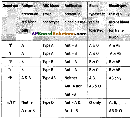
genotypes and four possible blood types (phenotypes). The genotypes are IAIA, IAi, IBIB, IBi, IAIB and ii. The phenotypic expressions of IAIA and IAi are ‘A’ – type blood, the phenotypic expressions of IBIB and IBi are ‘B’ – type blood, and that of IAIB is ‘AB’ – type blood. The phenotype of ii (IOIO) is ‘O’ – type blood.