Thoroughly analyzing AP Inter 2nd Year Zoology Model Papers and AP Inter 2nd Year Zoology Question Paper March 2016 helps students identify their strengths and weaknesses.
AP Inter 2nd Year Zoology Question Paper March 2016
Time: 3 Hours
Maximum Marks: 60
Instructions:
Note : Read the following instructions carefully.
- Answer all questions of Section – A. Answer ANY SIX questions in Section – B and answer ANY TWO questions in Section – C.
- In Section – A questions from Sr. Nos. 1 to 10 are of Very Short Answer Type. Each question carries TWO marks. Every answer may be limited to 5 lines. Answer all these questions at one place in the same order.
- In Section – B, questions from Sr. Nos. 11 to 18 are of Short Answer Type. Each question carries FOUR marks. Every answer may be limited to 20 lines.
- In Section – C, questions from Sr. Nos. 19 to 21 are of Long Answer Type. Each question carries EIGHT marks. Every answer may be limited to 60 lines.
- Draw labelled diagrams wherever necessary in Sections – B and C.
Section – A (10 × 2 = 20)
Note : Answer ALL the questions in 5 lines each.
Question 1.
What is meant by chloride shift ?
Answer:
The exchange of chloride and bicarbonate ions between RBCand plasma at the tissues is called chloride shift or Hamburger’s phenomenon or Hamburger’s shift.
Question 2.
Distinguish between the enzymes renin and rennin.
Answer:
a) A fall in glomerular blood flow / glomerular blood pressure / GFR can activate the JG cells to release an enzyme called renin into blood. This enzyme catalyses the conversion of angiotensinogen.
b) Rennin is a digestive enzyme produced by gastric glands that converts milk into curd in infants.
Question 3.
Name the ear ossicles and their evolutionary origin in human beings.
Answer:
Each middle ear contains 3 tiny bones – Malleus (modification of articular), Incus (modified quadrate) and stapes (modified hyomandibula) collectively called as ear ossicles.
Question 4.
Why the sympathetic division is called thoracolumbar division ?
Answer:
In the sympathetic division, pre ganglionic neurons arise from the thoracic and lumbar regions of the spinal cord, hence called ‘Thoraco – lumbar division’.
![]()
Question 5.
Distinguish between diabetes insipidus and diabetes mellitus.
Answer:
a) Diabetes insipidus: Deficiency of vasopressin causes diabetes insipidus in which the patient excretes large volumes of urine, resulting in dehydration and thirst.
b) Diabetes mellitus : A condition resulting from lack of insulin as a result of which the body cannot store or oxidise sugar efficiently (and sugar is lost through urine).
Question 6.
Differentiate between perforins and granzymes.
Answer:
Cytotoxic T-lymphocytes attach to the infected cells and release certain enzymes called perforins and granzymes.
Perforins form pores in the cell membrane of the infected cells. Then granzymes enter the infected cells through these perforations and activate certain proteins which help in the destruction of the infected cell.
Question 7.
Name the yellow mass of cells accumulated in the empty follicle after ovulation. Name the hormone secreted by it. What is its function ?
Answer:
After ovulation the granulosa cells in the follicle proliferate and are transformed into a yellowish glandular mass called corpus luteum. It secretes progesterone harmone. This harmone is essential for maintenance of pregnancy in first few months.
Question 8.
What is amniocentesis ? Name any two disorders that can be detected by amniocentesis.
Answer:
Amniocentesis is a diagnostic procedure to detect genetic defects . in the unborn baby. The most common abnormalities that can be detected by amniocentesis are Downsyndrome, Edward Syndrome and Turner’s syndrome.
Question 9.
Explain the term, hypophysation.
Answer:
Hypophysation or induced breeding is followed in artificial breeding. Pituitary extracts containing FSH and LH or ovaprim are injected into brood fish to induce release of spawn for seed production.
Question 10.
Define the term, vaccine.
Answer:
The term vaccine was coined by Edward Jenner. A vaccine is a biological preparation that improves immunity to a particular disease.
Section – B (6 × 4 = 24)
Note : Answer ANY SIX questions in 20 lines each.
Question 11.
Draw a neat labelled diagram of L.S. of a tooth.
Answer:
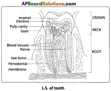
![]()
Question 12.
Describe the disorders of respiratory system.
Answer:
Disorders of the Respiratory System :
i) Asthma is a difficulty in breathing caused due to inflammation of bronchi and bronchioles. It is characterized by the spasm of smooth muscles present in the walls of the bronchi and bronchioles. Symptoms include coughing, difficulty in breathing and wheezing. In the case of asthma, the allergen causes release of histamine and other inflammatory substances which cause constriction of the bronchi.
ii) Emphysema is a chronic disorder in which alveolar walls are damaged and their walls coalesce due to which respiratory surface area of exchange of gases is decreased. The lung shows larger but fewer alveoli and more fibrous and less elastic. One of the major causes of this is ‘smoking’ of tobacco.
iii) Bronchitis is the inflammation of the bronchi, resulting in the swelling of mucous lining of bronchi, increased mucus production and decrease in the diameter of bronchi. Symptoms include chronic cough with thick mucus/ sputum (phlegm).
iv) Pneumonia is infection of lungs caused by bacteria such as Streptococcus pneumoniae and also by certain viruses, fungi, protozoans and mycoplasmas. Symptoms include inflammation of lungs, accumulation of mucus in alveoli, and impaired exchange of gases, leading to death if untreated.
v) Emphysema, chronic bronchitis and asthma come under Chronic Obstructive Pulmonary Diseases (COPDs).
Occupational Respiratory disorders : These are caused by exposure of the body to the harmful substances from certain industries, especially those involving grinding or stone breaking. Long term exposure of the body to such substances can give rise to inflammation of respiratory passage and lungs leading to several disorders.
- Asbestosis : It occurs due to chronic exposure to asbestos dust in the people working in asbestos industry.
- Silicosis : ft occurs because of long term exposure to ‘silica dust’ in the people working in mining industries, quarries etc.
- Siderosis : It occurs due to deposition of iron particles in tissues. It can cause different types of siderosis such as pneumoconiosis due to inhalation of iron particles, hyperferremia and hemosiderosis (which causes recurrent alveolar hemorrhage).
- Black – lung disease : It is a lung disease that develops from inhalation of coal dust. It is common in long time coal mine workers.
Question 13.
Give an account of synaptic transmission.
Answer:
The functional junction formed between two neurons is called synapse. In a chemical synapse, the presynaptic neuron synthesizes the neurotransmitter and stores in the synaptic vesicles of synaptic terminals.
When an action potential reaches a synaptic terminal, it depolarizes the membrane. By this voltage gated calcium channels open. The rise is the Ca2+ concentration leads to the release of neurotransmitters by exocytosis. The neuro transmitter diffuses across the synaptic cleft.
The postsynaptic membrane has ligandgated in channels. Binding of the neurotransmitter to a receptor of the channel opens the channel and allows specific ions to diffuse across the postsynaptic membrane. It results in a postsynoptic membrane potential. Excitatory neurotransmitters depolarize the postsynaptic membrane while inhibitory neurotransmitters cause hyperpolarization of post synaptic membrane.
Acetylcholine is the most common neurotransmitter. It may be excitatory or inhibitory. Gama aminobutyric acid (GABA) and glycine are the inhibitory neurotransmitters.
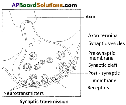
![]()
Question 14.
Explain how hypothyroidism and hyperthyroidism can affect the body.
Answer:
Over activity of the thyroid, cancer of the gland or development of nodules of thyroid lead to hyperthyroidism. In adults abnormal growth causes a disease called exophthalamic goiter, with characteristically protruded eyeballs. Hyperthyroidism also affects the physiology of the body (increased metabolic rate). Inadequate supply of iodine in the diet results in hypothyroidism and enlargement of the thyroid gland. This condition is called simple goiter.
During pregnancy, due to hypothyroidism, defective development of the growing baby leads to a disorder called cretinism. Physical and mental growth gets severely stunted (Thyroid dwarf) due to untreated ‘congenital hypothyroidism’ (deficiency of thyroid hormones by birth). Stunted growth, mental retardation, low intelligence quotient, abnormal skin, deafness and mutism are some of the characteristic features of the this disease. In adult women, hypothyroidism may cause irregular menstrual cycles. In adults the hypothyroidism results in a condition called myxedema, Lethargy, mental impairment, intolerance to cold, puffiness of face and dry skin and some of the symptoms of myxedema.
Question 15.
Describe erythroblastosis foetalis.
Answer:
Destruction of RBC of Rh positive foetus by anti Rh antibodies produced by Rh negative mother due to immunological incompatibility is called Erythroblastosis foetalis or Haemolytic disorder of newborn (HDNB). This is due to genetically incompatible marriage involving Rh positive father and Rh negative mother. At the time of birth the Rh positive foetal blood mixes with the Rh negative blood of mother, through the ruptured placenta. The Rh antigens sensitize the mother to produce anti Rh antibodies (IgG antibodies) and memory cells. This first Rh positive by is unaffected because it is delivered by the time mother is sensitized. During the next pregnancy bearing Rh positive foetus these antibodies increase in concentration due to memory cells and cross the placenta, enter the blood of baby and destroy the RBC. Haemolytic anaemia is the symptom in this disorder.
Question 16.
Explain Darwin’s theory of Natural selection with industrial melanism as an experimental proof.
Answer:
Experimental verification of Natural Selection – Industrial melanism : An important practical proof for the operation of Natural Selection is the classical case of industrial melanism, exhibited by peppered moth – Biston betuiaria. These moths were available in two colours, grey and Mack. Prior to industrial revolution, the grey moths were abundant. During the industrial revolution, the black forms were more and the grey forms were less in the industrial cities like Birmingham. Biologists proposed that with the industrial revolution, more soot was released due to the burning of coal, which resulted in the darkening of the barks of trees.
Grey moths on the dark bark were easily identified and predated more by birds. Hence the number of grey moths decreased and that of the black moths increased in the population. It means Nature offered ‘positive selection’ pressure to the black (melanic) forms. Bernard Kettlewell, a British ecologist, tested this hypothesis experimentally. He collected both the grey and the black forms of Biston betuiaria for his experiment. He released them in two sets of equal numbers; one set in Birmingham, a polluted urban area, and the other set in Dorset, an unpolluted rural area. After a few days he recaptured them. Of those moths recaptured from Birmingham, there were more black forms. Among those recaptured from Dorset there were more grey forms.
The reason for such a difference is: the melanic forms could not be easily spotted by predator birds as their body colour merged with the dark colour of the bark of trees in Birmingham area. In the rural areas (Dorset) the grey forms had better survival chance as their body colour merged with the light coloured surroundings. This explains the differential survival of the moths due to Natural Selection. It will be interesting to know that there was a reversal in the selection process after the introduction of pollution check laws in the urban areas.
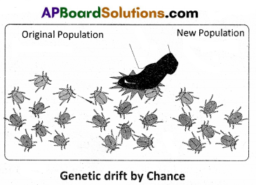
Question 17.
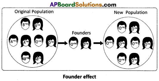
What is meant by genetic drift ? Explain the genetic drift citing the example of Founder Effect.
Answer:
Genetic Drift : The change in the frequency of a gene that occurs merely by chance and not by selection, in small populations, is called genetic drift or Sewail Wright effect. Suppose, for a gene with two alleles, the frequency of a particular allele is 1% (q = 0.01), the probability of losing that allele by chance from the small population is more. The end result is either Fixation (p or q = 1) or Loss (p or q = 0) of that allele. The probability of reaching the end point depends on the size of the population. Genetic drift tends to reduce the amount of genetic variation within the population mainly by removing the alleles with low frequencies, it can be exemplified by the Founder Effect and Bottleneck Effect.
Founder effect : If a small group of individuals from a population start a new colony in an isolated region, those individuals are called the founders of the new population. The allelic frequencies of their descendants are similar to those of the founders rather than to their ancestral parent population, e.g. presence of O+ve blood group in nearly 100% of the Red-lndians. It means the forefathers of the Red Indian tribe were predominantly O+ve and they isolated themselves reproductively from other populations.
![]()
Question 18.
“Honey bees are economically important”. Justify.
Answer:
Economic importance of Honey bees :
The bee products like
Honey, wax, propolis and bee venom are used in various ways.
- Honey is a rich source of fructose, water, glucose, minerals and vitamins.
- Bee’s wax is used in the preparation of cosmetics, polishes of various kinds and candles.
- Propolis is used in the treament of inflammation and superficial burns.
- Bee’s venom, which is extracted from the sting of worker bees, is used in the treatment of rheumatoid arthritis.
- Pollination : Bees are the pollinators of our crop plants such as sunflower, brassica, apple and pear.
Section – C (2 × 8 = 16)
Note: Answer ANY TWO questions in 60 lines each.
Question 19.
Describe the structure of the heart of man with the help of a neat labelled diagram.
Answer:
The heart is mesodermal in origin. It is a thick walled, muscular and pulsating organ, situated in the mediastinum (the region in the thorax between the two lungs), and with its apex slightly turned to the left. It is the size of a clinched fist.
The heart is covered by a double walled pericardium which consists of the outer fibrous pericardium and the inner serous pericardiurh. The serous pericardium is double – layered, formed of an outer parietal layer and an inner visceral layer. The parietal layer is fused with the fibrous pericardium, whereas the visceral layer adheres to the surface Of the heart and forms its outer layer, the epicardium. The two layers are separated by a narrow pericardial space, which is filled with the pericardial fluid. This fluid reduces friction between the two membranes and allows free movement of the heart.
The wall of the heart consists of three layers. They are the outer epicardium, the middle myocardium (a thick layer of cardiac muscles), and the inner most endocardium (a thin layer of endothelium). The endothelium covers the heart valves also and is continuous with the endothelial lining of the large blood vessels connected to the heart.
External structure :
Human heart has four chambers, with two relatively smaller upper chambers, called atria and two larger lower chambers called ventricles. Atria and ventricles are separated by a deep transverse groove called coronary sulcus (atrio – ventricular groove). The muscular pouch like projection from each atrium is called auricular appendix (auricular appendage). The ventricles are separated by two inter ventricular grooves (anterior and posterior), in which the coronary arteries and their branches are lodged.
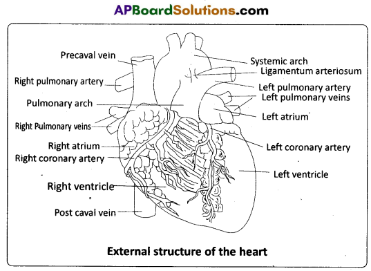
Internal structure :
i) Atria : Atria are thin walled ‘receiving chambers’ (upper chambers). The right one is larger than the left. The two atria are separated by thin inter – atrial septum. In the fetal heart, the atrial septum has a small pore called foramen ovale. Normally the foramen ovale closes at birth, when lungs become functional. It is represented by a depression in the septum between the right and left atria, called fossa ovalis (that marks the position of the foramen ovale in the fetus). If, the foramen ovale does not close properly, it is called a patent foramen ovale.
The right atrium receives deoxygenated blood from different parts of the body (except the lungs) through three caval veins viz. the two precavals (right and left) and a post caval vein. It also receives blood from the myocardium (wall of the heart) through the coronary sinus, whose opening into the right atrium is guarded by the valve of Thebesius. Opening of the postcaval vein is guarded by the valve of the inferior vena cava or Eustachian valve. It directs the blood to the left atrium through the foramen ovale, in the foetal stage, but in the adult it becomes rudimentary and non – functional. The openings of the precaval veins into the right atrium have no valves. The left atrium receives blood from each lung through two pulmonary veins, which open into the left atrium. The two left pulmonary veins open by a common aperture in some.
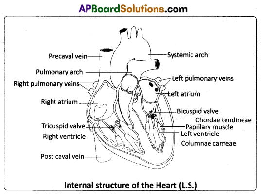
Atria and ventricles are separated by a membranous atrio – ventricular septum, which possesses left and right atrioventricular apertures. The left and right apertures are guarded by bicuspid (mitral valve) and tricuspid valves respectively.
ii) Ventricles : These are the thick walled blood pumping chambers (lower chambers), separated by an interventricular septum. The wall of the left ventricle is thicker than that of the right ventricle. The inner surface of the ventricles is raised into muscular ridges or columns called columnae carneae / trabeculae carneae projecting from the inner walls of the ventricles. Some of these ridges are large and conical, and are called papillary muscles, whose apices are connected to the chordae tendineae, or ‘heart strings’. They are cord – like collagenous processes that connect the papillary muscles to the tricuspid valve and the mitral valve in the heart. They prevent the cusps of the atrioventricular valves from bulging too far into atria during ventricular systole.
![]()
Question 20.
Describe male reproductive system of a man. Draw a labelled diagram of it.
Answer:
The Male Reproductive System : The male reproductive system (male genital system) consists of a number of sex organs that are a part of the human reproductive process. The sex organs which are located in the pelvic region include a pair of testes (sing: testis) along with accessory ducts, glands and the external genitalia.
Testes : The testes (testicles) are a pair of oval pinkish male primary sex organs suspended outside the abdominal cavity with in a pouch called scrotum. The scrotum helps in maintaining the low temperature of the testes (2 – 2.5°C lower than the normal internal body temperature) necessary for spermatogenesis. The cavity of the scrotal sac is connected to the abdominal cavity through the inguinal canal.
Testis is held in position in the scrotum by the gubernaculum, a fibrous cord that connects the testis with the bottom of the scrotum and a spermatic cord, formed by the vas deferens, nerves, blood vessels and other tissues that run from the abdomen down to each testicle, through the inguinal canal. Each testis is enclosed in a fibrous envelope, the tunica albuginea, which extends inward to form septa that partition the testis into lobules. There are about 250 testicular lobules in each testis. Each lobule contains 1 to 3 highly coiled seminiferous tubules. A pouch of serous membrane (peritoneal layer) called tunica vaginalis covers the testis.
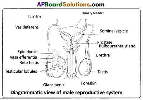
Each seminiferous tubule is lined by the germinal epithelium which consists of undifferentiated male germ cells called spermatogonial mother cells and it also bears ‘nourishing cells’ called Sertoli cells. The spermatogonia produce the primary spermatocytes which undergo meiotic division, finally leading to the formation of spermatozoa or sperms (spermatogenesis). Sertoli cells provide nutrition to the spermatozoa and also produce a hormone called inhibin, which inhibits the secretion of FSH.
The regions outside the seminiferous tubules, called interstitial spaces, contain interstitial cells of Leydig or Leydig ceils. Leydig cells produce androgens, the most important of which is testosterone. Testosterone controls the development of secondary sexual characters and spermatogenesis. Other immunologically competent cells are also present. The seminiferous tubules open into the vasa efferentia through the rete testis (a network of tubules in of the testis carrying spermatozoa from the seminiferous tubules to the vasa efferentia).
Epididymis : The vasa efferentia leave the testis and open into a narrow, tightly coiled tube called epididymis located along the posterior surface of each testis. The epididymis provides a storage space for the sperms and gives the sperms time to mature. It is differentiated into three regions – caput epididymis, corpus epididymis and cauda epididymis. The caput epididymis receives spermatozoa via the vasa efferentia of the mediastinum testis (a mass of connective tissue at the back of the testis that encloses the rate testis).
Vasa deferentia : The vas deferens or ductus deferens is a long, narrow, muscular tube. The mucosa of the ductus deferens consists of pseudostratified columnar epithelium and lamina propria (areolar connective tissue). It starts from the tail of the epididymis, passes through the inguinal canal into the abdomen and loops over the urinary bladder. It receives a duct from the seminal vesicle. The vas deferens and the duct of the seminal vesicle unite to form a short ejaculatory duct / ductus ejaculatorius. The two ejaculatory ducts, carrying spermatozoa and the fluid secreted by the seminal vesicles, converge in the centre of the prostate and open into the urethra, which transports the sperms to outside.
Urethra : In males, the urethra is the shared terminal duct of the reproductive and urinary systems. The urethra originates from the urinary bladder and extends through the penis to its external opening called urethral meatus. The urethra provides an exit for urine as well as semen during ejaculation.
Penis: The penis and the scrotum constitute the male external genitalia. The penis serves as urinal duct and also intromittent organ that transfers spermatozoa to the vagina of a female. The human penis is made up of three columns of tissue; two upper corpora cavernosa on the dorsal aspect and one corpus spongiosum on the ventral side. Skin and a subcutaneous layer enclose all three columns, which consist of special tissue that helps in erection of the penis to facilitate insemination. The enlarged and bulbous end of penis called glans penis is covered by a loose fold of skin (foreskin) called prepuce. The urethra traverses the corpus spongiosum, and its opening lies at the tip of the glans penis (urethral meatus).
Male accessory genital glands : The male accessory glands include paired seminal vesicles, a prostate and bulbourethral glands.
Seminal vesicles : The seminal vesicles are a pair of simple tubular glands present postero- inferior to the urinary bladder in the pelvis. Each seminal vesicle opens into the corresponding vas deferens, as the vas deferens enters the prostate gland. The secretion of the seminal vesicles constitutes about 60 percent of the volume of seminal fluid.
It is an alkaline, viscous fluid that contains fructose, proteins, citric acid, inorganic phosphorus, potassium, and prostaglandins. Once this fluid joins the sperm in the ejaculatory duct, fructose acts as the main energy source for the sperm outside the body. Prosta glandins are believed to aid fertilization by causing the mucous lining of the cervix to be more receptive to sperm as well as by aiding the movement of the sperm towards the ovum with peristaltic contractions of the uterus and fallopian tubes.
Prostate gland : Prostate gland is located directly beneath the urinary bladder. The gland surrounds the prostatic urethra, and sends its secretions through several prostatic ducts. In man, the prostate contributes 15 – 30 percent of the semen. The fluid from the prostate is clear and slightly acidic. The prostatic secretion ‘activates’ the spermatozoa and provides nutrition.
Bulbourethral Glands : Bulbourethral glands, also called Cowper’s glands, are located beneath the prostate gland at the beginning’of the internal portion of the penis. They add an alkaline fluid to semen during the process of ejaculation. The fluid secreted by these glands lubricates the urethra. It is also thought to function as a ‘flushing agent’ that washes out the acidic urinary residues that remain in the urethra, before the semen is ejaculated.
![]()
Question 21.
What are multiple alleles ? Describe multiple alleles with the help of ABO blood groups in man.
Answer:
Multiple alleles and human blood groups : Generally a gene has two alternative forms / versions called alleles. They are present at the same locus in a pair of homologous chromosomes. Two alleles of a gene can form three genotypes in a diploid organism. Sometimes a gene may have more than two alleles. When more than two allelic forms occur at the same locus on the homologous chromosomes of an organism, they are called mutiple alleles when more than two alleles exist in a population of a specific organism, the phenomenon is called mutiple allelism.
As mentioned above ‘multiple alleles’ cannot be observed in the genotype of a diploid individual, but can be observed in a population. The number of genotypes that can occur for multiple alleles is given by the expression n (n + 1) /2 where, n = number of alleles. A well known example of multiple allelism in man is the expression of ABO blood types by three alleles of a single gene which can produce six genotypes.
ABO Blood Types : The ABO blood group system was proposed by Karl Landsteiner. He was awarded the Nobel Prize in Physiology or Medicine in 1930 for his work. The phenotypes (blood types) A, B, AB and O types are characterized by the presence or absence of ‘antigens’ on the plasma membrane of the RBCs. The A and B antigens are actually carbohydrate groups (sugar polymers) that are bound to lipid molecules (fatty acids) protruding from the membrane of the red blood cell.
They are also called isoagglutinogens because they cause blood cell agglutination in the case of incompatible blood transfusions. ‘Blood type A’ persons have antigen A on their RBCs and anti – B antibodies in the plasma. ‘Blood type B’ persons have antigen B on their RBCs and anti – A antibodies in the plasma. ‘Blood type AB’ person have antigens ‘A’ and ‘B’ on theRBCs and no antibodies in the plascna. ‘Blood type O’ persons have no antigens on their RBCs and both ‘anti – A, and ‘anti – B’ antibodies are present in the plasma.
Bernstein discovered that these phenotypes were inherited by the interaction of three ‘autosomal alleles’ of the gene named I, located on chromosome 9. IA, IB and i (or IO) are the three alleles of the gene I. The antibodies ‘anti – A’ and ‘anti – B’ are called isoagglutinins (also called isohaemagglutinins) which are usually IgM type. The isoagglutinins of an individual cause agglutination reactions with the antigens of another individual. The alleles IA and IB are responsible for the production of the respective antigens ‘A’ and ‘B’. The allele i does nto produce any antigen. The alleles lA and lB are dominant to the allele i, but co-dominant to each other (IA = IB > i). A child receives one of the three alleles from each parent, giving rise to six possible
Table : Genetic control of the human ABO blood groups
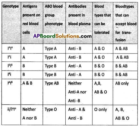
genotypes and four possible blood types (phenotypes). The genotypes are IAIA, IAi, IBIB, IBi, IAIB and ii. The phenotypic expressions of IAIA and IAi are ‘A’ – type blood, the phenotypic expressions of IBIB and IBi are ‘B’ – type blood, and that of IAIB is ‘AB’ – type blood. The phenotype of ii (IOIO) is ‘O’ – type blood.