Thoroughly analyzing AP Inter 2nd Year Zoology Model Papers Set 9 helps students identify their strengths and weaknesses.
AP Inter 2nd Year Zoology Model Paper Set 9 with Solutions
Time: 3 Hours
Maximum Marks: 60
General Instructions:
Note : Read the following instructions carefully.
- Answer all questions of Section – A. Answer ANY SIX questions in Section – B and answer ANY TWO questions in Section – C.
- In Section – A questions from SI. Nos. 1 to 10 are of Very Short Answer Type. Each question carries TWO marks. Every answer may be limited to 5 lines. Answer all these questions at one place in the same order.
- In Section – B, questions from SI. Nos. 11 to 18 are of Short Answer Type. Each question carries FOUR marks. Every answer may be limited to 20 lines.
- In Section – C, questions from SI. Nos. 19 to 21 are of Long Answer Type. Each question carries EIGHT marks. Every answer may be limited to 60 lines.
- Draw labelled diagrams wherever necessary in Sections – B and C.
Section – A (10 × 2 = 20)
Note : Answer ALL the questions.
Question 1.
Name the heart sounds. When are they produced ?
Answer:
Heart sounds are named as LUB and DUB.
a) The first heart sound LUB is produced when atrio – ventricular valves (AV valves) close preventing the back flow of blood.
b) The second heart sound DUB is produced when the semilunar valves close preventing the back flow of blood.
Question 2.
Name the keystone bone of the cranium. Where is it located ?
Answer:
Sphenoid bone is called keystone bone. It is present at the middle part of the base of the skull. It is named keystone bone because it articulates with all other cranial bones.
Question 3.
Distinguish between red muscle fibres and white muscle fibres.
Answer:
a) Red muscle fibres : Myoglobin of these fibres is high which give a reddish appearance. Such muscle fibres are called red fibres. They also contain plenty of mitochondria.
b) White muscle fibres: Some of the muscle fibres possess very less quantity of myoglobin in their muscle fibres and therefore appear pale or whitish. Hence called white muscle fibres.
![]()
Question 4.
What are islets of langerhans ?
Answer:
The Endocrine portion of pancreas is Just 1 to 2% and consists of 1 to 2 millions of islets of langerhans which are having α – cells and β – cells, α – cells produce hormone glucagon, β cells produce insulin.
Question 5.
How can the graft rejections be avoided in patients ?
Answer:
Graft reactions can be avoided in patients by tissue matching and blood group matching. Even after this, the patient has to take immuno – suppressant drugs throughout their life.
Question 6.
Define probiotic soup. Who coined this term ?
Answer:
The molecules of ammonia, hydrocarbons and water underwent condensation, oxidation, reduction and polymerisation due to energy sources to produce complex molecules like sugars, amino acids, fatty acids, purines, pyramidines and later nucleosides and nucieotides. All these reactions occurred in the ocean which was described as the hot dilute soup or probiotic soup by Haldane.
Question 7.
Distinguish between allopatric and sympatric speciations.
Answer:
a) If speciation takes place due to geographical isolation, it is called allopatric speciation.
b) If speciation takes place in the organisms which live in the same habitat, capable of interbreeding, but do not interbreed due to some isolation mechanisms is called sympatric speciation.
Question 8.
Define biogenetic law by giving an example.
Answer:
Biogenetic law was proposed by Haeckel. It states that Ontogeny repeats Phylogeny which means the developmental history of an organism repeats the evolutionary history of its ancestor, eg : Tadpole larva of frog resembles fish both externally and internally. It possesses a tail, gills and 2 chambered heart like that offish. Later it metamorphoses into adult frog.
Question 9.
Define the term transgenic animal.
Answer:
Animals that have their own genome and had their DNA manipulated to possess and express an extra gene are known as transgenic animals.
Question 10.
Explain the term hypophysation.
Answer:
Hypophysation or induced breeding is followed in artificial breeding. Pituitary extracts contains FSH and LH or Ovaprin are injected into brood fish to induce release of spawn for seed production.
Section – B (6 × 4 = 24)
Note : Answer ANY SIX questions.
Question 11.
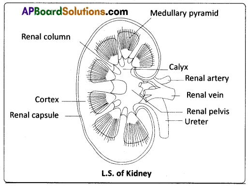
Draw neat labelled diagram of the V.S of Kidney.
Answer:
![]()
Question 12.
Explain the mechanism of clotting of blood.
Answer:
Mechanism of blood clotting : Clotting takes place in three essential steps.
i) Step – 1: It involves the formation of a complex of activated substances collectively called, prothrombin activator. It is formed by a complex cascade of chemical reactions that occur in the blood by the involvement of clotting factors in two pathways.
a) Intrinsic pathway : It occurs when the blood is exposed to collagen of injured wall of blood vessel. This activates Factor XII, and in turn it activates another clotting factor, which activates yet another reaction (cascade fashion) which results in the formation of the prothrombin activator.
b) Extrinsic pathway : It occurs when the damaged vascular wall or extra vascular tissue comes into contact with blood. This activates the release of tissue thromboplastin, from the dampged tissue. It activates the Factor VII. As a result of these cascade reactions, the final product formed is the prothrombin activator.
ii) Step – 2 : The prothrombin activator, in the presence of sufficient amounts of ionic Ca++, causes the conversion of inactive prothrombin to active thrombin (activation of prothrombin).
iii) Step – 3 : Thrombin converts the soluble protein fibrinogen into soluble fibrin monomers, which are held together by weak hydrogen bonds. The fibrin stabilizing factor(Factor XIII, released from platelets) replaces hydrogen bonds with covalent bonds and cross links the fibres to form a ‘mesh work’. The insoluble mesh work of fibrin fibers spreading in all directions adhere to the damaged surfaces and trap the blood cells and platelets.
Question 13.
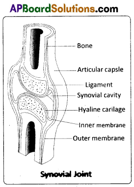
Describe the structure of synovial joint with the help of a neat labelled diagram.
Answer:
Synovial joint is covered by a double layered synovial capsule. The outer layer consists of dense fibrous
irregular connective tissue with more collagen fibres. This layer is continuous with the periosteum and resists stretching and prevents the dislocation of joints. Some fibres of these membranes are arranged in bundles called ligaments.
The inner layer of synovial capsule is formed of areolar tissue and elastic fibers. It secretes a viscous synovial fluid which contains hyaluronic acid, phagocytes, etc. and acts as a ‘lubricant’ for the free movement of the joints. Synovial joints include Bad and Socket joint, Hinge joint, Pivot joint, Gliding joint, Condyloid joint, Saddle joint.
Question 14.
Write short notes on immunoglobulins.
Answer:
Antibodies (Immunoglobulins): Whenever pathogens enter our body, the B – lymphocytes produce an army of proteins called antibodies to fight with them. They are highly specialized for binding with specific antigens. The part of an antibody that recognizes an antigen is called the paratope (antigen binding site). Based on their mobility, antibodies are of two types, namely circulating or free antibodies and surface antibodies. The circulating or free antibodies are present in the body fluids whereas the surface antibodies are present on the surface of the mature B – cells as well as the memory cells.
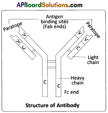
Structure : The basic structure of an antibody was proposed by Rodney Porter. It is a Y shaped molecule with four polypeptide chains of which two are long, identical heavy chains (H) and two are small, identical light chains (L). Hence, an antibody is represented as H2L2. The two chains are linked by disulphide bonds. One end of the antibody molecule is called Fab end (Fragment antigen binding) and the other end is called Fc end (Fragment – crystallizable or Fragment – cell binding). Based on the structure, the antibodies are of five types, namely IgD, IgE, IgG, IgA and IgM.
![]()
Question 15.
What is oogenesis ? Give a brief account of oogenesis in a woman.
Answer:
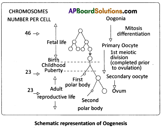
Oogenesis : The process of formation of a mature female gamete is called oogenesis. Oogenesis is initiated during the embryonic development stage when a couple of million gamete mother cells (oogonia) are formed within each foetal ovary and do not multiply thereafter. These cells start division and stop the process at prophase – 1 of the meiosis – 1 At this stage these are called primary oocytes.
Question 16.
Explain the inheritance of sex linked recessive character in human being.
Answer:
Colour Blindness : It is a sex -linked recessive disorder. Retina of the eye in man contains the cells sensitive to red and green colours.
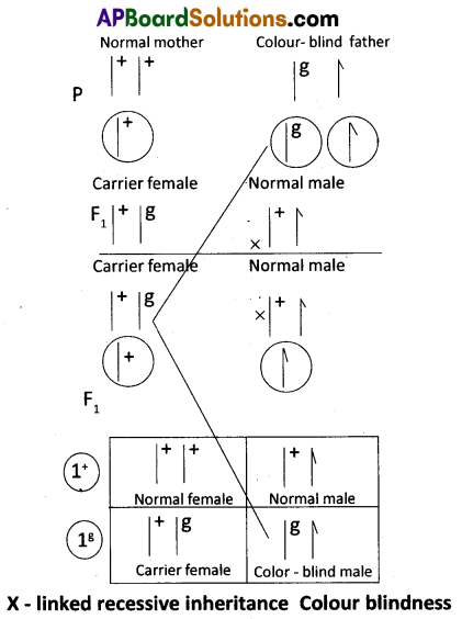
This phenotypic trait is genetically controlled. Its alleles are located on the X – chromosome. When a woman with normal vision (homozygous) marries a colour-blind man, all the sons and daughters are normal, but daughters are carriers (heterozygous). If a carrier woman marries a man with normal vision, all the daughters and half of the sons have normal vision and another half of sons are colour – blind. Colour – blind trait is inherited from a male parent to his grand sons through carrier daughter, which is an example of crisscross pattern of inheritance.
Question 17.
Distinguish between homologous and analogous organs.
Answer:
Homologous organs : The organs which have similar structure and origin but not necessarily the same function are called homologous organs. The evolutionary pattern that describes the occurrence of similarity in origin and internal structure is called homology. Such organs show adaptive radiation, hence ‘divergent evolution’, e.g. the appendages of vertebrates such as the flippers of whale, wings of bat, forelimbs of horse, paw of cat and hand of man, have a common pattern in arrangement of bones even though their external form and function may vary to suit their mode of life. It explains that all vertebrates might have had a common ancestor.
Analogous organs : The organs which have dissimilar structure and origin but perform the same function are called analogous organs. Analogous organs suggest ‘convergent evolution’, e.g. wings of a butterfly and wings of a bird.
Question 18.
Discuss briefly the process of indirect ELISA.
Answer:
Indirect ELISA : It is used to detect antibodies. The blood of the person undergoing the ‘assay’ (for example the HIV test) is allowed to clot and the cells are centrifuged out to obtain the clear serum with antibodies (called primary antibodies).
Protocol:
- It is used to detect ‘antibodies’.
- A known ‘antigen’ is added to the ‘well’ (adsorbed).
- Patient’s antiserum (serum with specific antibodies) is added.
- The ‘antibodies’ in the patient’s ‘antiserum’ (primary/complementary antibodies) bind to the antigens coated on the surface of the ‘well’.
- Enzyme linked antihuman serum globulins (anti HISGs) are added. They bind to the antibody which is already bound to the antigen.
- Enzyme’s substrate is added and the reaction produces a visible colour change which can be measured by a spectrophotometer.
Section – C (2 × 8 = 16)
Note : Answer ANY TWO of the following questions.
Question 19.
Write an essay on the transport of oxygen and carbondioxide by blood.
Answer:
Transport of gases: Blood is the medium of transport for O2 and CO2.
I. Transport of Oxygen : Oxygen is transported from the lungs to the tissues through the plasma and RBC of the blood. 100 ml of oxygenated blood can deliver 5 ml of O2 to the tissues under normal conditions.
i) Transport of oxygen through plasma : About 3% of O2 is carried through the blood plasma in a dissolved state.
ii) Transport of oxygen by RBC: About 97% of O2 is transported by the RBCs in the blood. Haemoglobin is a red coloured iron containing pigment present in the RBCs. Each haemoglobin molecule can carry a maximum of four molecules of oxygen. Binding of oxygen with haemoglobin is primarily related to the partial pressure of O2. At lungs, where the partial pressure of O2(oxygen tension) is high, oxygen binds to haemoglobin (purplish – bluish-red in colour) in a reversible manner to form oxyhaemoglobin (bright red in colour). This is called oxygenation of haemoglobin.
At the tissues, where the partial pressure of O2 is low, oxyhaemoglobin dissociates into haemoglobin and oxygen. The other factors that influence binding of oxygen with haemoglobin are the partial pressure of CO2, the hydrogen ion concentration (pH) and the temperature.
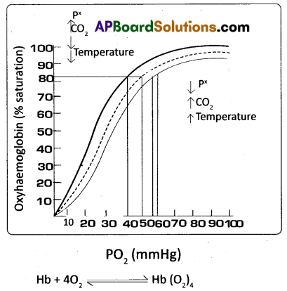
iii) Oxygen – haemoglobin dissociation curve : It explains the relation between percentage saturation of haemoglobin and partial pressure of oxygen. A sigmoid curve is obtained when percentage saturation of haemoglobin with O2 is plotted against the pO2. This curve is called ‘oxyhaemoglobin dissociation curve’ and is highly useful in studying the effect of factors such as pCO2, H+ concentration, temperature, etc., on the binding of O2 with haemoglobin. In the alveoli, where there is a high pO2, low pCO2, lesser H+ concentration (high pH) and lower temperature, the factors are all favourable for the formation of oxyhaemoglobin.
In the tissues where low pO2, high pCO2, high H+ concentration (low pH) and higher temperature exist, the conditions are favourable for dissociation of oxygen from oxyhaemoglobin. Under these conditions, oxygen dissociation curve shifts away from the Y – axis (to the right). The effect of pCO2 and H+ concentration on the oxygen affinity of haemoglobin is called Bohr Effect (increase of carbondioxide in the blood and decrease in pH results in the reduction of the affinity of haemoglobin for oxygen).
II. Transport of Carbon Dioxide : CO2 is transported in three ways.
i) In dissolved state : 7 percent of CO2 is carried in a dissolved state (physical solution) through plasma.
CO2 + H2O → H2 CO3
ii) As carbamino compounds : About 20 – 25 percent of CO2 combines directly with free amino group of the haemoglobin and forms carbamino-haemoglobin in a reversible manner.
Hb – NH2 + CO2 → Hb – NHCOO– + H+
This binding of CO2 is related to the partial pressure of CO2. pO2 is a major factor which could affect this binding. When pCO2 is high and pO2 is low as in the tissues, binding of more carbon dioxide occurs. When pCO2 is low and pO2 is high as in the alveoli, dissociation of CO2 from carbamino – haemoglobin takes place, i.e., CO2 which is bound to haemoglobin from the tissues is delivered at the alveoli. Carbamino compounds are also formed by the union of CO2 with plasma proteins.
iii) As Bicarbonates : About 70 percent of CO2 is transported as bicarbonate. RBCs contain a very high concentration of the enzyme, carbonic anhydrase and a minute quantity of the same is present in the plasma too. This enzyme facilitates the following reaction in both the directions.

At the tissues where partial pressure of CO2 is high due to catabolism, CO2 diffuses into the blood (RBC and Plasma) and forms carbonic acid which dissociates into HCO3– and H+. At the alveolar site where pCO2 is low, the reaction proceeds in the opposite direction leading to the formation of CO2 and water. Thus CO2 is mostly trapped as bicarbonate at the tissues and transported to the alveoli where it is released out as CO2. Every 100 mL of deoxygenated blood delivers approximately 4mL of CO2 to the alveolar air.
![]()
Question 20.
Describe the male reproductive system of a man. Draw a labelled diagram.
Answer:
The Male Reproductive System : The male reproductive system (male genital system) consists of a number of sex organs that are a part of the human reproductive process. The sex organs which are located in the pelvic region include a pair of testes (sing : testis) along with accessory ducts, glands and the external genitalia.

Testst : The tester (testicles) are pair of oval pinkish male primary sex organs suspended outside the abdominal cavity with in a pouch called scrotum. The scrotum helps in maintaining the low temperature of the testes (2 – 2.5°C lower than the normal internal body temperature) necessary for spermatogenesis. The cavity of the scrotal sac is connected to the abdominal cavity through the inguinal canal.
Testis is held in position in the scrotum by the gubernaculum, a fibrous cord that connects the testis with the bottom of the scrotum and a spermatic cord, formed by the vas deferens, nerves, blood vessels and other tissues that run from the abdomen down to each testicle, through the inguinal canal. Each testis is enclosed in a fibrous envelope, the tunica albuginea, which extends inward to form septa that partition the testis into .lobules. There are about 250 testicular lobules in each testis. Each lobule contains 1 to 3 highly coiled seminiferous tubules. A pouch of serous membrane (peritoneal layer) called tunica vaginalis covers the testis.
Each seminiferous tubule is lined by the germinal epithelium which consists of undifferentiated male germ cells called spermatogonial mother ceils and it also bears ‘nourishing cells’ called Sertoli cells. The spermatogonia produce the primary spermatocytes which undergo meiotic division, finally leading to the formation of spermatozoa or sperms (spermatogenesis). Sertoli cells provide nutrition to the spermatozoa and also produce a hormone called inhibin, which inhibits the secretion of FSH.
The regions outside the seminiferous tubules, called interstitial spaces, contain interstitial cells of Leydig or Leydig cells. Leydig cells produce androgens, the most important of which is testosterone. Testosterone controls the development of secondary sexual characters and spermatogenesis. Other immunologically competent cells are also present. The seminiferous tubules open into the vasa efferentia through the rete testis (a network of tubules in of the testis carrying spermatozoa from the seminiferous tubules to the vasa efferentia).
Epididymis : The vasa efferentia leave the testis and open into a narrow, tightly coiled tube called epididymis located along the posterior surface of each testis. The epididymis provides a storage space for the sperms and gives the sperms time to mature. It is differentiated into three regions – caput epididymis, corpus epididymis and cauda epididymis. The caput epididymis receives spermatozoa via the vasa efferentia of the mediastinum testis (a mass of connective tissue at the back of the testis that encloses the rate testis).
Vasa deferentia : The vas deferens or ductus deferens is a long, narrow, muscular tube. The mucosa of the ductus deferens consists of pseudostratified columnar epithelium and lamina propria (areolar connective tissue). It starts from the tail of the epididymis, passes through the inguinal canal into the abdomen and loops over the urinary bladder. It receives a duct from the seminal vesicle. The vas deferens and the duct of the seminal vesicle unite to form a short ejaculatory duct / ductus ejaculatorius. The two ejaculatory ducts, carrying spermatozoa and the fluid secreted by the seminal vesicles, converge in the centre of the prostate and open into the urethra, which transports the sperms to outside.
Urethra : In males, the urethra is the shared terminal duct of the reproductive and urinary systems. The urethra originates from the urinary bladder and extends through the penis to its external opening called urethral meatus. The urethra provides an exit for urine as well as semen during ejaculation.
Penis : The penis and the scrotum constitute the male external genitalia. The penis serves as urinal duct and also intromittent organ that transfers spermatozoa to the vagina of a female. The human penis is made up of three columns of tissue; two upper corpora cavernosa on the dorsal aspect and one corpus spongiosum on the ventral side. Skin and a subcutaneous layer enclose all three columns, which consist of special tissue that helps in erection of the penis to facilitate insemination. The enlarged and bulbous end of penis called glans penis is covered by a loose fold of skin (foreskin) called prepuce. The urethra traverses the corpus spongiosum, and its opening lies at the tip of the glans penis (urethral meatus).
Male accessory genital glands : The male accessory glands – include paired seminal vesicles, a prostate and bulbourethral glands.
Seminal vesicles : The seminal vesicles are a pair of simple tubular glands present postero- inferior to the urinary bladder in the pelvis. Each seminal vesicle opens into the corresponding vas deferens, as the vas deferens enters the prostate gland. The secretion of the seminal vesicles constitutes about 60 percent of the volume of seminal fluid. It is an alkaline, viscous fluid that contains fructose, proteins, citric acid, inorganic phosphorus, potassium, and prostaglandins. Once this fluid joins the sperm in the ejaculatory duct, fructose acts as the main energy source for the sperm outside the body Prostaglandins are believed to aid fertilization by causing the mucous lining of the cervix to be more receptive to sperm as well as by aiding the movement of the sperm towards the ovum with peristaltic contractions of the uterus and fallopian tubes.
Prostate gland : Prostate gland is located directly beneath the urinary bladder. The gland surrounds the prostatic urethra, and sends its secretions through several prostatic ducts. In man, the prostate contributes 15 – 30 percent of the semen. The fluid from the prostate is clear and slightly acidic. The prostatic secretion ‘activates’ the spermatozoa and provides nutrition.
Bulbourethral Glands : Bulbourethral glands, also called Cowper’s glands, are located beneath the prostate gland at the beginning of the internal portion of the penis. They add an alkaline fluid to semen during the process of ejaculation. The fluid secreted by these glands lubricates the urethra. It is also thought to function as a ‘flushing agent’ that washes out the acidic urinary residues that remain in the urethra, before the semen is ejaculated.
![]()
Question 21.
What is DNA finger printing ? Mention its applications.
Answer:
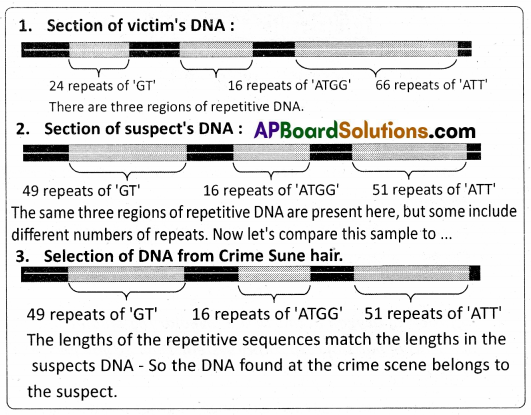
DNA Finger Printing : Over 99% of the 3 billion nucleotide pairs in human DNA are identical among all individuals. No two people (other than identical twins) have exactly the same sequence of bases in their DNA. Restriction Fragment Length Polymorphisms (RFLPs – pronounced riflips) are characteristic to every person’s DNA. They are called Variable Number Tandem Repeats (VNTRs) and are useful as Genetic markers. The VNTRs of two persons generally show variations. DNA fingerprinting involves identifying differences in some specific regions in DNA sequence called repetitive DNA, because in these sequences, a small stretch of DNA is repeated many times.
These sequences show high degree of polymorphism and form the basis of DNA finger printing. They are bits of chromosomes that can be cut by restriction endonucleases. The ‘fundamental technique’ involved in DNA Finger Printing was pioneered and perfected by Jeffrys of Great Britain. He observed that the gene pertaining to myoglobin of muscles contains many segments that vary in size and composition, from one person to another. For example in the following hypothetical example nucleotide base sequence, there are 6 Tandem Repeats of 16 bases each (count the first 16 and note how they are repeated).
5’GACTGCCTGCTAAGATGACTGCCtGCTAAGATGACTGCCTGCTAAGATGA CTGCCTGCTA AGATGACTGCCTGCTAAGATGACTGCCTGCTAAGAT3′
Such clusters of 10 – 100 nucleotides are called mini satellites. Such tandem repeats are characteristic of every person’s DNA. The VNTRs of two persons differ in the number of tandem repeats or the sequence of bases. Such changes are caused due to mutations and gene recombinations. For example, a child might inherit a chromosome with 6 tandem repeats from the mother and the same tandem repeated 4 times from the father in a homologous chromosome. It means half of the VNTR alleles of the child resemble those of the mother and the other half those of the father. This is a ‘heterozygous condition with reference to VNTR alleles’. These tandem repeats serve as basis of a technique called DNA finger printing.
DNA Finger printing – Protocol :
1. Obtaining DNA (Isolation /Extraction) : The first step is to obtain a sample of DNA from blood, saliva, hair roots, semen etc. If needed many copies of the DNA can be produced by PCR (cloning / DNA amplification).
2. Fragmenting DNA (Restriction Digestion): Treating DNA with Restriction Enzymes (Restriction endonucleases) which cut the DNA into smaller fragments by cutting it at specific sites.
3. Separation of DNA fragments by electrophoresis : DNA fragments are applied at one end of agarose gel plate. When an electric current is applied to the gel, the DNA fragments (which are slightly negatively charged) travel across the gel (smaller and more mobile pieces travel farther). This technique of separation of DNA fragments into individual bands is called Gel Electrophoresis.
4. Denaturing DNA : The DNA on the gel is ‘denatured’ using alkaline chemicals or by heating, (denaturing means separation / splitting of the double helix into ‘single strands’ by breaking hydrogen bonds between the two strands.)
5. Blotting:Athinnylonmembrane is placed overthe’size fractionated DNA strands’ and covered by paper towels. As the towels draw moisture the DNA strands are transferred on to the nylon membrane by capillary action. This process is called ‘Blotting’ – more precisely Southern blotting, after the name of its inventor E.M. Southern.
6. Using probes to identify specific DNA : A radioactive probe (DNA is labeled with a radioactive substance) is added to the DNA bands. The Probe is a single stranded DNA molecule that is ‘complementary’ to the gene of interest in the sample under study. The probe attaches by base pairing to those restriction fragments that are complementary to its sequence. The probes can be prepared by using either ‘fluorescent substances’ or ‘radioactive isotopes’.
7. Hybridisation with probe: After the probe hybridises and the excess prob washed off, a photographic film is placed on the membrane containing ‘DNA hybrids’.
8. Exposure on film to make a Genetic / DNA Finger Print: The radioactive label exposes the film to form an image (image of bands) corresponding to specific DNA bands. The thick and thin dark bands form a pattern of bars which constitute a Genetic fingerprint.
A given person can never have a VNTR which his parents do not have. Obtaining hybrid with radioactive probe and matching DNAs of different members of a family with biological children and adopted children, gives us an idea of how DNA Finger Prints help identification of paternity/ maternity, by studying the ‘DNA Finger Prints’ of members of a Family – Biological and non-biological relationships.
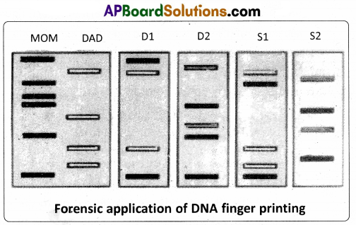
The illustrations given below are the VNTR patterns for, Mrs. Rose (blue), Mr. Rao (yellow), and their four children : D1 (Mr. Rao’s biological daughter), D2 (Mr. Rao’s step -daughter, child of Mrs. Rose and her former husband (red), SI (Mr. Rao’s biological son), and S2 (Mr. Rao’s adopted son not biologically related, his parents’ DNA marked in light and dark green bands).
Applications of DNA Finger Printing :
- Conservation of wild life – protection of endangered species. By maintaining their DNA records for identification of tissues of the dead endangered organisms.
- Taxonomical applications – study of phylogeny.
- Pedigree analysis – inheritance pattern of gene through generations.
- Anthropological studies – charting of origin and migration of human population.
- Medico – legal cases – establishing paternity and / or maternity more accurately.
- Forensic analysis – positive Identification of a suspect in a crime.