Thoroughly analyzing AP Inter 2nd Year Zoology Model Papers Set 5 helps students identify their strengths and weaknesses.
AP Inter 2nd Year Zoology Model Paper Set 5 with Solutions
Time: 3 Hours
Maximum Marks: 60
General Instructions:
Note : Read the following instructions carefully.
- Answer all questions of Section – A. Answer ANY SIX questions in Section – B and answer ANY TWO questions in Section – C.
- In Section – A questions from SI. Nos. 1 to 10 are of Very Short Answer Type. Each question carries TWO marks. Every answer may be limited to 5 lines. Answer all these questions at one place in the same order.
- In Section – B, questions from SI. Nos. 11 to 18 are of Short Answer Type. Each question carries FOUR marks. Every answer may be limited to 20 lines.
- In Section – C, questions from SI. Nos. 19 to 21 are of Long Answer Type. Each question carries EIGHT marks. Every answer may be limited to 60 lines.
- Draw labelled diagrams wherever necessary in Sections – B and C.
Section – A (10 × 2 = 20)
Note : Answer ALL the questions.
Question 1.
Bile juice contains no digestive enzymes yet it is important for digestion. How ?
Answer:
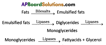
Bile salts of the bile help in the emulsification of fats (breakdown of fats into very small micelles). Bile also activates lipases of pancreatic juice (steapsin) and intestinal lipases.
Question 2.
What happens to the respiratory process in man going up a hill?
Answer:
At a height of about 6000 m the PO2 becomes almost half of what it is at the mean sea level, hence the mountain sickness in people ascending mountains.
Question 3.
What is Juxta glomerular apparatus ?
Answer:
The Juxta glomerular apparatus plays a complex regulating role. The JGA is the region in each nephron where the afferent arteriole comes into contact with DCT. Macula densa together with JG cells form the JGA. (Juxta Glomerulor Apparatus)
![]()
Question 4.
What is a ‘motor unit’ with reference to a muscle and nerve ?
Answer:
A motor neuron and the set of muscle fibres innervated by all the telodendrites constitute a motor unit.
Question 5.
Name the type of joint between a) Atlas and axis b) Carpal and metacarpal of the human thumb.
Answer:
a) Type of joint between atlas and axis is pivot joint.
b) Type of joint between carpal/ metacarpal is condyloid joint.
Question 6.
Distinguish between diabetes insipidus and diabetes mellitus,
Answer:
a) Diabetes insipidus : Deficiency of vasopressin causes diabetes insipidus in which the patient excretes large volumes of urine resulting in dehydration and thirst.
b) Diabetes mellitus : A condition resulting from lack of insulin as a result of which the body connot store or oxidise sugar efficiently (and sugar is lost through urine).
Question 7.
Colostrum is very much essential for the new born infants’. Justify.
Answer:
The colostrum secreted by the mother during the initial days of lactation has abundant IgA antibodies, to protect the infant. It is called natural passive acquired immunity.
Question 8.
Define gestation period. What is the duration of gestation period in the human beings ?
Answer:
Intra uterine development of the embryo or foetus is called gestation period. In humanbeing gestation period is 266 days or 38 weeks.
Question 9.
Distinguish between out cross and cross breed.
Answer:
Out crossing is the crossing of unrelated pure breeding animals of different traits with in the same breed whereas cross breeding is the mating of animals of different breeds.
Question 10.
Define the term vaccine.
Answer:
The term vaccine was coined by Edward Jenner. A vaccine is a biological preparation that improves immunity to a particular disease.
Section – B (6 × 4 = 24)
Note : Answer ANY SIX questions.
Question 11.
if, you take butter in your food, how does it get digested and absorbed in the body ? Explain.
Answer:
- Butter is a content of macromolecules and is fat or lipid.
- Bile salts of the bile help in breaking down of large fat molecules into very small micelles. This process is called Emulsification. They move into intestinal mucosal cells.
- These micelles are reformed into very small protein coated fat globules called chylomicrons which are transported into the lacteals in the villi by exocytosis.
- Bile also activates lipases of pancreatic juice (steapsin) and intestinal lipases. These lipases act on emulsified fats and convert them into fatty acids and glycerols.
![]()
Question 12.
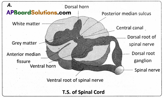
Draw a labelled diagram of the T.S. of the spina I cord of man.
Answer:
Question 13.
Give an account of the secretions of pituitary gland.
Answer:
Previously, pituitary gland was called the “master” endocrine gland, because it controls several endocrine glands. Release of hormones by adenohypophysis is stimulated by releasing hormones and suppressed by inhibiting hormones of the hypothalamus.
Growth Hormone or Somatotropin : In response to human growth hormone, cells in the liver, skeletal muscle, cartilage, bone, and other tissues secrete insulin – like growth factors that cause cells to grow and multiply. These factors accelerate protein synthesis and decrease catabolism of proteins.
Thyroid – Stimulating Hormone : It stimulates the synthesis and secretion of thyroid hormones by the thyroid gland.
Adrenocorticotropic Hormone (ACTH) : It controls the secretion of glucocorticoids by the adrenal cortex.
Follicle – Stimulating Hormone : In females FSH initiates the development of ovarian follicles. In males FSH stimulates spermatogenesis.
Luteinizing Hormone : In females, LH stimulates ovulation, formation of the corpus luteum andthe secretion of progesterone by the corpus luteum. In males, this hormone is called interstitial cell stimulating hormone. It stimulates leydig cells in the testis to secrete testosterone. FSH and LH are termed gonadotropins because their target organs are gonads.
Prolactin : Prolactin, together with other hormones, initiates and maintains milk secretion by the mammary glands. The function of prolactin is not known in males.
Melanocyte – Stimulating Hormone (MSH) : MSH increases skin pigmentation in lower vertebrates by stimulating the dispersion of melanin granules in melanocytes.
Neurohypophysis : It does not synthesize hormones. It stores and releases oxytocin and vasopressin.
Oxytocin : During delivery, oxytein enhances contraction of smooth muscle cells in the wall of uterus. After delivery, it stimulates milk ejection.
Vasopressin or Antidiuritic Hormone : ADH causes the kidneys to absorb more water into the blood. In the absence of ADH, urine output increases from the normal 1 to 2 litres about 20 litres a day. ADH causes constriction of arterioles, which increases blood pressure. The amount of ADH secreted is regulated.
Question 14.
Briefly describe the common sexually transmitted diseases in human beings.
Answer:
Sexually Transmitted Diseases (STDs) : Diseases or infections which are transmitted through sexual contact (intercourse) are collectively called sexually transmitted diseases (STDs) or venereal diseases (VDs) or reproductive tract infections (RTI). Most common STDs and their causative organisms are shown in the table below.
| S.No. | Name of the Disease | Causative organism |
| 1. | Gonorrhea | Neisseria gonorrhoeae (bacteria) |
| 2. | Syphilis | Treponema pallidum (spirochete bacterium) |
| 3. | Genital herpes | Herpes simplex virus (HSV) |
| 4. | Genital warts, cervical cancer | Human Papilloma virus (HPV) |
| 5. | Trichomoniasis | Trichomonas vaginalis (a protozoan parasite) |
| 6. | Chlamydiasis | Chlamydia trachomatis (bacteria) |
| 7. | Hepatitis – B | HBV |
| 8. | HIV infection / AIDS | HIV (Human immuno deficiency virus) |
Question 15.
How is sex determined in human beings ?
Answer:
Sex determination in Humans : It has already been mentioned that the sex determining mechanism in case of humans is XX – XY type. Out of 23 pairs of chromosomes present, 22 pairs are exactly same in both males and females; these are the autosomes. A pair of X- chromosomes is present in the female, where as the presence of an X and Y – chromosome are determinant of the male characteristic. During spermatogenesis among males, two types of gametes are produced.
50 percent of the total sperm produced carry the X – chromosome and the rest 50 percent has Y – chromosome besides the autosomes. Females, however, produce only one type of ovum with an X chromosome. There is an equal probability of fertilisation of the ovum by the sperm carrying either X or Y chromosome. In case the ovum is fertilised by a sperm carrying X – chromosome, the zygote develops into a female and the fertilisation of ovum with Y – chromosome carrying sperm results into a male offspring. Thus, it is evident that it is the genetic makeup of the sperm that determines the sex of the child. It is also evident that in each pregnancy there is . always 50 percent probability of either a male or a female child.
![]()
Question 16.
Distinguish between homologous and analogous organs.
Answer:
Homologous organs : The organs which have similar structure and origin but not necessarily the same function are called homologous organs. The evolutionary pattern that describes the occurrence of similarity in origin and internal structure is called homology. Such organs show adaptive radiation, hence ‘divergent evolution’, e.g. the appendages of vertebrates such as the flippers of whale, wings of bat, forelimbs of horse, paw of cat and hand of man, have a common pattern in arrangement of bones eventhough their external form and function may vary to suit their mode of life. It explains that all vertebrates might have had a common ancestor.
Analogous organs : The organs which have dissimilar structure and origin but perform the same function are called analogous organs. Analogous organs suggest ‘convergent evolution’, e.g. wings of a butterfly and wings of a bird.
Question 17.
Write a short note on Neo Darwinism.
Answer:
Modern synthetic theory of Evolution or Neo-Darwinism : Weismann’s germplasm theory, de Vries’ mutation theory and Mendel’s laws of inheritance helped a lot in understanding the origin and inheritance of variations. The scientists such as Huxley, Haeckel, Simpson, etc., supported Darwinism. Later Fisher, Sewall Wright, Mayr explained Natural Selection in the light of post- Darwinian discoveries (Synthetic theory / Genetical theory / Neo-Darwinism). According to this theory, five basic factors are involved in the process of organic evolution.
They are
(i) Gene mutations,
(ii) Chromosomal mutations,
(iii) Genetic recombinations,
(iv) Natural Selection and
(v) Reproductive isolation.
i) Gene mutations : Changes in the structure of a gene (DNA molecule) are called gene mutations or point mutations. They alter the phenotypic characters of the individuals. Thus, gene mutations tend to produce ‘variations’ in the offspring.
ii) Chromosomal mutations : Changes in the structure of chromosomes (due to deletion, addition, duplication, inversion or translocation) are called chromosomal mutations. They also bring about variations in the phenotype of organisms which lead to the occurrence of variations in the offspring.
iii) Genetic recombinations : Recombinations of genes due to crossing over during meiosis are also responsible for bringing about genetic variability among the individuals of the same species, thus, contributing to the occurrence of heritable variations.
iv) Natural Selection : Natural selection does not produce any genetic changes but once genetic changes occurred, it favours some genetic changes while rejecting others. Hence it is considered the driving force of evolution.
v) Reproductive isolation : The absence of gene exchange between populatioris is called the reproductive isolation. It plays a great role in giving rise to new species and preserving the species integrity.
Question18.
Explain the different types of Cancer.
Answer:
Types of cancers : There are different types of cancers such as carcinomas (cancers of epithelial tissues / cells which are most common as epithelial cells divide more often), sarcomas (cancers of connective tissues), leukemias (cancers of bone marrow cells resulting in understrained production of WBC – a liquid tumor), lymphomas (cancers of the lymphatic system). Certain types of cancers are called ‘familial cancers’ (cancer that occurs in families; genetic based) and others ‘sporadic cancers’ (non-hereditary cancers occurring without any family history). Some types of cancers are caused by ‘tumor forming RNA viruses’ (oncoviruses), e.g. Rous sarcoma virus which causes ‘avian sarcoma’.
Section – C (2 × 8 = 16)
Note : Answer ANY TWO of the following questions.
Question 19.
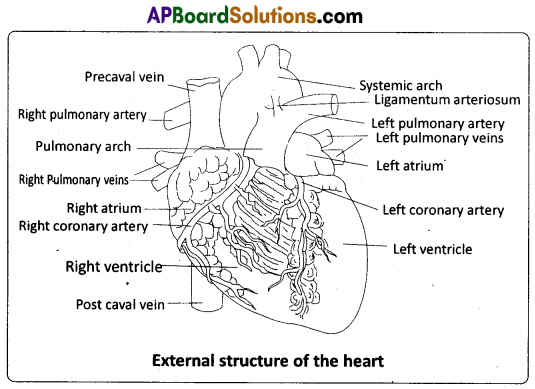
Describe the structure of the heart of man with the help of neat labelled diagram.
Answer:
The heart is mesodermal in origin. It is a thick walled, muscular and pulsating organ, situated in the mediastinum (the region in the thorax between the two lungs), and with its apex slightly turned to the left. It is the size of a clinched fist.
The heart is covered by a double walled pericardium which consists of the outer fibrous pericardium and the inner serous pericardiurh. The serous pericardium is double – layered, formed of an outer parietal layer and an inner visceral layer. The parietal layer is fused with the fibrous pericardium, whereas the visceral layer adheres to the surface Of the heart and forms its outer layer, the epicardium. The two layers are separated by a narrow pericardial space, which is filled with the pericardial fluid. This fluid reduces friction between the two membranes and allows free movement of the heart.
The wall of the heart consists of three layers. They are the outer epicardium, the middle myocardium (a thick layer of cardiac muscles), and the inner most endocardium (a thin layer of endothelium). The endothelium covers the heart valves also and is continuous with the endothelial lining of the large blood vessels connected to the heart.
External structure:
Human heart has four chambers, with two relatively smaller upper chambers, called atria and two larger lower chambers called ventricles. Atria and ventricles are separated by a deep transverse groove called coronary sulcus (atrio – ventricular groove). The muscular pouch like projection from each atrium is called auricular appendix (auricular appendage). The ventricles are separated by two inter ventricular grooves (anterior and posterior), in which the coronary arteries and their branches are lodged.
Internal structure :
i) Atria : Atria are thin walled ‘receiving chambers’ (upper chambers). The right one is larger than the left. The two atria are separated by thin inter – atrial septum. In the fetal heart, the atrial septum has a small pore called foramen ovale. Normally the foramen ovale closes at birth, when lungs become functional. It is represented by a depression in the septum between the right and left atria, called fossa ovalis (that marks the position of the foramen ovale in the fetus). If, the foramen ovale does not close properly, it is called a patent foramen ovale.
The right atrium receives deoxygenated blood from different parts of the body (except the lungs) through three caval veins viz. the two precavals (right and left) and a post caval vein. It also receives blood from the myocardium (wall of the heart) through the coronary sinus, whose opening into the right atrium is guarded by the valve of Thebesius. Opening of the postcaval vein is guarded by the valve of the inferior vena cava or Eustachian valve. It directs the blood to the left atrium through the foramen ovale, in the foetal stage, but in the adult it becomes rudimentary and non – functional. The openings of the precaval veins into the right atrium have no valves. The left atrium receives blood from each lung through two pulmonary veins, which open into the left atrium. The two left pulmonary veins open by a common aperture in some.
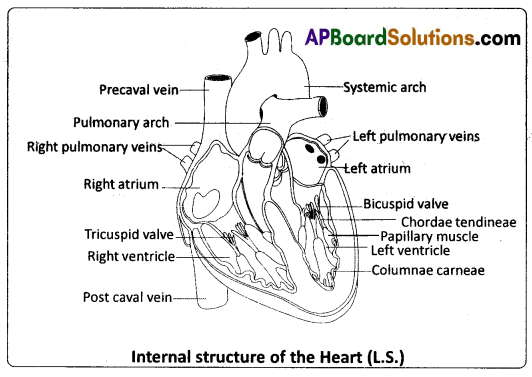
Atria and ventricles are separated by a membranous atrio – ventricular septum, which possesses left and right atrioventricular apertures. The left and right apertures are guarded by bicuspid (mitral valve) and tricuspid valves respectively.
ii) Ventricles : These are the thick walled blood pumping chambers (lower chambers), separated by an interventricular septum. The wall of the left ventricle is thicker than that of the right ventricle. The inner surface of the ventricles is raised into muscular ridges or columns called columnae carneae / trabeculae carneae projecting from the inner walls of the ventricles. Some of these ridges are large and conical, and are called papillary muscles, whose apices are connected to the chordae tendineae, or ‘heart strings’. They are cord – like collagenous processes that connect the papillary muscles to the tricuspid valve and the mitral valve in the heart. They prevent the cusps of the atrioventricular valves from bulging too far into atria during ventricular systole.
![]()
Question 20.
Describe the male reproductive system of a man. Draw a labelled diagram of it.
Answer:
The Male Reproductive System : The male reproductive system (male genital system) consists of a number of sex organs that are a part of the human reproductive process. The sex organs which are located in the pelvic region include a pair of testes (sing : testis) along with accessory ducts, glands and the external genitalia.
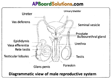
Testst : The tester (testicles) are pair of oval pinkish male primary sex organs suspended outside the abdominal cavity with in a pouch called scrotum. The scrotum helps in maintaining the low temperature of the testes (2 – 2.5°C lower than the normal internal body temperature) necessary for spermatogenesis. The cavity of the scrotal sac is connected to the abdominal cavity through the inguinal canal. Testis is held in position in the scrotum by the gubernaculum, a fibrous cord that connects the testis with the bottom of the scrotum and a spermatic cord, formed by the vas deferens, nerves, blood vessels and other tissues that run from the abdomen down to each testicle, through the inguinal canal. Each testis is enclosed in a fibrous envelope, the tunica albuginea, which extends inward to form septa that partition the testis into .lobules. There are about 250 testicular lobules in each testis. Each lobule contains 1 to 3 highly coiled seminiferous tubules. A pouch of serous membrane (peritoneal layer) called tunica vaginalis covers the testis.
Each seminiferous tubule is lined by the germinal epithelium which consists of undifferentiated male germ cells called spermatogonial mother ceils and it also bears ‘nourishing cells’ called Sertoli cells. The spermatogonia produce the primary spermatocytes which undergo meiotic division, finally leading to the formation of spermatozoa or sperms (spermatogenesis). Sertoli cells provide nutrition to the spermatozoa and also produce a hormone called inhibin, which inhibits the secretion of FSH. The regions outside the seminiferous tubules, called interstitial spaces, contain interstitial cells of Leydig or Leydig cells. Leydig cells produce androgens, the most important of which is testosterone. Testosterone controls the development of secondary sexual characters and spermatogenesis. Other immunologically competent cells are also present. The seminiferous tubules open into the vasa efferentia through the rete testis (a network of tubules in of the testis carrying spermatozoa from the seminiferous tubules to the vasa efferentia).
Epididymis : The vasa efferentia leave the testis and open into a narrow, tightly coiled tube called epididymis located along the posterior surface of each testis. The epididymis provides a storage space for the sperms and gives the sperms time to mature. It is differentiated into three regions – caput epididymis, corpus epididymis and cauda epididymis. The caput epididymis receives spermatozoa via the vasa efferentia of the mediastinum testis (a mass of connective tissue at the back of the testis that encloses the rate testis).
Vasa deferentia : The vas deferens or ductus deferens is a long, narrow, muscular tube. The mucosa of the ductus deferens consists of pseudostratified columnar epithelium and lamina propria (areolar connective tissue). It starts from the tail of the epididymis, passes through the inguinal canal into the abdomen and loops over the urinary bladder. It receives a duct from the seminal vesicle. The vas deferens and the duct of the seminal vesicle unite to form a short ejaculatory duct / ductus ejaculatorius. The two ejaculatory ducts, carrying spermatozoa and the fluid secreted by the seminal vesicles, converge in the centre of the prostate and open into the urethra, which transports the sperms to outside.
Urethra : In males, the urethra is the shared terminal duct of the reproductive and urinary systems. The urethra originates from the urinary bladder and extends through the penis to its external opening called urethral meatus. The urethra provides an exit for urine as well as semen during ejaculation.
Penis : The penis and the scrotum constitute the male external genitalia. The penis serves as urinal duct and also intromittent organ that transfers spermatozoa to the vagina of a female. The human penis is made up of three columns of tissue; two upper corpora cavernosa on the dorsal aspect and one corpus spongiosum on the ventral side. Skin and a subcutaneous layer enclose all three columns, which consist of special tissue that helps in erection of the penis to facilitate insemination. The enlarged and bulbous end of penis called glans penis is covered by a loose fold of skin (foreskin) called prepuce. The urethra traverses the corpus spongiosum, and its opening lies at the tip of the glans penis (urethral meatus).
Male accessory genital glands : The male accessory glands – include paired seminal vesicles, a prostate and bulbourethral glands.
Seminal vesicles : The seminal vesicles are a pair of simple tubular glands present postero- inferior to the urinary bladder in the pelvis. Each seminal vesicle opens into the corresponding vas deferens, as the vas deferens enters the prostate gland. The secretion of the seminal vesicles constitutes about 60 percent of the volume of seminal fluid. It is an alkaline, viscous fluid that contains fructose, proteins, citric acid, inorganic phosphorus, potassium, and prostaglandins. Once this fluid joins the sperm in the ejaculatory duct, fructose acts as the main energy source for the sperm outside the body. Prostaglandins are believed to aid fertilization by causing the mucous lining of the cervix to be more receptive to sperm as well as by aiding the movement of the sperm towards the ovum with peristaltic contractions of the uterus and fallopian tubes.
Prostate gland : Prostate gland is located directly beneath the urinary bladder. The gland surrounds the prostatic urethra, and sends its secretions through several prostatic ducts. In man, the prostate contributes 15 – 30 percent of the semen. The fluid from the prostate is clear and slightly acidic. The prostatic secretion ‘activates’ the spermatozoa and provides nutrition.
Bulbourethral Glands : Bulbourethral glands, also called Cowper’s glands, are located beneath the prostate gland at the beginning of the internal portion of the penis. They add an alkaline fluid to semen during the process of ejaculation. The fluid secreted by these glands lubricates the urethra. It is also thought to function as a ‘flushing agent’ that washes out the acidic urinary residues that remain in the urethra, before the semen is ejaculated.
![]()
Question 21.
What are multiple alleles ? Describe multiple alleles with the help of ABO blood groups in man.
Answer:
Multiple alleles and human blood groups : Generally a gene has two alternative forms / versions called alleles. They are present at the same locus in a pair of homologous chromosomes. Two alleles of a gene can form three genotypes in a diploid organism. Sometimes a gene may have more than two alleles. When more than two allelic forms occur at the same locus on the homologous chromosomes of an organism, they are called mutiple alleles when more than two alleles exist in a population of a specific organism, the phenomenon is called mutiple allelism.
As mentioned above ‘multiple alleles’ cannot be observed in the genotype of a diploid individual, but can be observed in a population. The number of genotypes that can occur for multiple alleles is given by the expression n (n + 1) /2 where, n = number of alleles. A well known example of multiple allelism in man is the expression of ABO blood types by three alleles of a single gene which can produce six genotypes.
ABO Blood Types : The ABO blood group system was proposed by Karl Landsteiner. He was awarded the Nobel Prize in Physiology or Medicine in 1930 for his work. The phenotypes (blood types) A, B, AB and O types are characterized by the presence or absence of ‘antigens’ on the plasma membrane of the RBCs. The A and B antigens are actually carbohydrate groups (sugar polymers) that are bound to lipid molecules (fatty acids) protruding from the membrane of the red blood cell.
They are also called isoagglutinogens because they cause blood cell agglutination in the case of incompatible blood transfusions. ‘Blood type A’ persons have antigen A on their RBCs and anti – B antibodies in the plasma. ‘Blood type B’ persons have antigen B on their RBCs and anti – A antibodies in the plasma. ‘Blood type AB’ person have antigens ‘A’ and ‘B’ on theRBCs and no antibodies in the plascna. ‘Blood type O’ persons have no antigens on their RBCs and both ‘anti – A, and ‘anti – B’ antibodies are present in the plasma.
Bernstein discovered that these phenotypes were inherited by the interaction of three ‘autosomal alleles’ of the gene named I, located on chromosome 9. IA, IB and i (or IO) are the three alleles of the gene I. The antibodies ‘anti – A’ and ‘anti – B’ are called isoagglutinins (also called isohaemagglutinins) which are usually IgM type. The isoagglutinins of an individual cause agglutination reactions with the antigens of another individual. The alleles IA and IB are responsible for the production of the respective antigens ‘A’ and ‘B’. The allele i does nto produce any antigen. The alleles lA and lB are dominant to the allele i, but co-dominant to each other (IA = IB > i). A child receives one of the three alleles from each parent, giving rise to six possible
Table : Genetic control of the human ABO blood groups
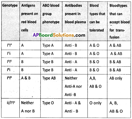
genotypes and four possible blood types (phenotypes). The genotypes are IAIA, IAi, IBIB, IBi, IAIB and ii. The phenotypic expressions of IAIA and IAi are ‘A’ – type blood, the phenotypic expressions of IBIB and IBi are ‘B’ – type blood, and that of IAIB is ‘AB’ – type blood. The phenotype of ii (IOIO) is ‘O’ – type blood.