Thoroughly analyzing AP Inter 2nd Year Zoology Model Papers Set 4 helps students identify their strengths and weaknesses.
AP Inter 2nd Year Zoology Model Paper Set 4 with Solutions
Time: 3 Hours
Maximum Marks: 60
General Instructions:
Note : Read the following instructions carefully.
- Answer all questions of Section – A. Answer ANY SIX questions in Section – B and answer ANY TWO questions in Section – C.
- In Section – A questions from SI. Nos. 1 to 10 are of Very Short Answer Type. Each question carries TWO marks. Every answer may be limited to 5 lines. Answer all these questions at one place in the same order.
- In Section – B, questions from SI. Nos. 11 to 18 are of Short Answer Type. Each question carries FOUR marks. Every answer may be limited to 20 lines.
- In Section – C, questions from SI. Nos. 19 to 21 are of Long Answer Type. Each question carries EIGHT marks. Every answer may be limited to 60 lines.
- Draw labelled diagrams wherever necessary in Sections – B and C.
Section – A (10 × 2 = 20)
Note : Answer ALL the questions.
Question 1.
Define chyme.
Answer:
The food is mixed thoroughly with the acidic gastric juice of the stomach by the churning movements of its muscular wall and the product is called Chyme. (The semi digested paste like acidic food of stomach is called chyme).
Question 2.
Name the muscles that help in breathing movement of man.
Answer:
Normal breathing movements are aided by
- Phrenic muscles of diaphragm,
- External and internal costal muscles of ribs.
Question 3.
How many pace makers are present in the human heart ? What are they ?
Answer:
In Human heart there are two pacemakers.
- Sinauricular node (SAN)
- Auriculo ventricular node (AVN)
Question 4.
What is a sarcomere ?
Answer:
The portion of the myofibril between two successive ‘Z’ lines is called Sarco/nere. It is the functional unit of contraction.
![]()
Question 5.
Name the meninges of the mammalian nervous system from the outer most to the inner most.
Answer:
The cranial meninges covering the brain of man are duramater (outer) arachnoidmater (middle) and piamater (inner most).
Question 6.
Write the functions of Leydig cells and Sertoli cells.
Answer:
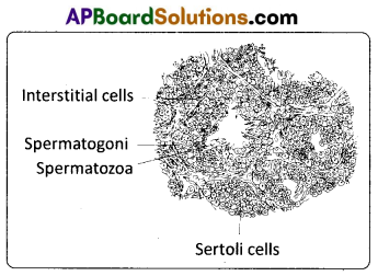
a) Sertoli cells : Nourishes the growing sperms and also produce a harmone called inhibin.
b) Leidig cells : Present in seminiferous tubules produce androgens, the most important of which is testosterone.
Question 7.
Distinguish between ‘Humoral immunity’ and ‘Cell mediated immunity’.
Answer:
a) The immunity mediated by the antibodies that are released into the fluids of the body (humors) such as plasma, lymph etc., is called humoral immunity.
b) Cell mediated Immunity : The immunity mediated by the activated T – cells, natural killer cells etc., is known as cell mediated immunity.
Question 8.
How is acromegaly caused ?
Answer:
Hyper secretion of growth stimulating hormone in adult results in an abnormality called acromegaly. It is characterised by enlargement of bones of Jaw, hand and feet, thickned nose, lips and eyelids and gorilla like appearance of the person affected.
Question 9.
Write any four important advantages of poultry farming.
Answer:
Advantage of poultry farming:
- Maximum returns with minimum investments.
- Eggs are nutritious and of good demand as food.
- Broiler rearing yields benefits in short span of time.
- Meat is with less cholestorol.
Question 10.
What is the principle involved in X – ray radiography ?
Answer:
A beam of X – rays is produced by an X – ray generator and is projected on the body parts. X – rays that pass through the body parts are recorded on a photographic film or observed on a fluorescent screen. Photographs develop using X – rays are known as radiographs or skiagraphs.
Section – B (6 × 4 = 24)
Note : Answer ANY SIX questions.
Question 11.
Explain the process of digestion in the stomach.
Answer:
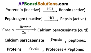
The gastric glands of stomach secrete acidic gastric juice. Gastric juice contains HCl, prorenin, pepsinogen and bicarbonates.
Proenzymes pepsinogen and prorennin, on exposure to HCI are converted into the active enzymes, pepsin and rennin respectively.
Pepsin converts proteins into proteoses and peptones. Rennin is found in gastric juice of infants. It acts on milk protein casein in the presence of calcium ions and converts it into calcium paracaseinate (curd) and proteoses. Pepsin again acts on calcium para- caseinate and converts it into peptones. Certain other cells in the wall of stomach produce bicarbonate, a base to buffer the acidic contents of the stomach. Bicarbonates and mucus produced by stomach wall forms a physical barrier to prevent HCl from damaging the wall of stomach.
![]()
Question 12.
What are the bio-chemical changes that occur in a muscle during contraction ?
Answer:
For the contraction of muscle, continuous supply of energy is needed. ATP is the immediate source of energy for muscle contraction. As the ATP content is very low, it is actively replenished, continuously, by an energy rich muscle phosphagen.
ATP → ADP + Pi
The high energy phosphates of muscles that donate energy phosphate group to ADP are known as phosphagen. In vertebrate muscles creatine phsophate (CP) is the phsophagen, which is an immediate backup source. In invertebrate muscles, it is in the arginine phosphate. This reaction is catalysed by creatine kinase (CK). Creatine Phosphate is the immediate additional source of energy in the muscle.
Creatine phosphate + ADP → Creatine
When creatine phosphate gets exhausted, the next source of reserve energy is utilised, which includes the oxidation of glucose and fatty acids. The energy liberated in this process is transferred to ADP and creatine. Thus ATP and creatine phosphate are formed and which in turn supply energy for muscle contraction.
During rapid activity of a muscle, the respiratory system is unable to supply sufficient oxygen needed by it, which leads to oxygen debt. It is defined as the amount of extra oxygen required by a muscle during recovery from vigorous exercise. Thus the pyruvic acid produced by glycolysis is transformed into lactic acid in the absence of oxygen. Accumulation of lactic acid in the muscle leads to muscle fatigue.
Lactic acid, formed in the anaerobic degradation in the muscle, reaches the liver through blood circulation. In liver, during rest, 80% of lactic acid is utilised in the resynthesis of glycogen, which is transported back to muscle. This is known as Cori cycle. 20% of lactic acid is oxidised as C02 and H20.
Question 13.
Write a brief note on Morphological Evidences in favour of organic evolution.
Answer:
Evidences from comparative anatomy : When we compare the anatomy of different animals, we find some similarities among them. For example, the fore limbs of different vertebrates are similar in origin and internal structure. All these indicate that there is a relationship among the organisms. These relationships can be studied under
i) Homologous organs,
ii) Analogous organs,
iii) Vestigial organs,
iv) Atavistic organs and
v) Connecting links.
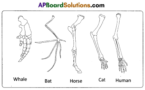
i) Homologous organs: The organs which have similar structure and origin but not necessarily the same function are called homologous organs. The evolutionary pattern that describes the occurrence of similarity in origin and internal structure is called homology. Such organs show adaptive radiation, hence ‘divergent evolution’, e.g. the appendages of vertebrates such as the flippers of whale, wings of bat, forelimbs of horse, paw of cat and hand of man have a common pattern in the arrangement of bones even though their external form and functions may vary to suit their mode of life. It explains that all the vertebrates might have had a common ancestor.
ii) Analogous organs: The organs which have dissimilar structure and origin but perform the same function are called the analogous organs. Analogous organs suggest ‘convergent evolution’, e.g. wings of a butterfly and wings of a bird.
iii) Vestigial organs : The organs which were functional in the ancestors but non-functional and reduced in the descendants are called vestigial organs. Presence of vestigial organs is the most convincing evidence in favour of organic evolution and also supports the concept of disuse proposed by Lamarck, e.g. Hind limbs in python, hind limbs and pelvic girdle in whale, wings of flightless birds, vermiform appendix, coccyx, plica semilunaris (vestigial nictitating membrane), auricular muscles that move the pinna, hair on the body, mammary glands in males, etc., in human beings.
iv) Atavistic organs: Sudden appearance of some vestigial organs in a better developed condition as in the case of the tailed human baby is called atavism. Such organs are called atavistic organs.They strongly support the concept of organic evolution.
v) Connecting links: The organisms which possess the characters of two different groups between which they are transitional are called connecting links. They clearly explain the path of evolution, e.g. Peripatus between annelida and arthropoda, prototherians between reptilia and mammalia, etc.
![]()
Question 14.
Draw a diagram of internal structure of testis.
Answer:
Question 15.
Write a brief note on different types of immunities.
Answer:
Types of immunity: Based on the nature of response, immunity is mainly of two types, namely
(i) Innate immunity and
(ii) Acquired immunity.
i) Innate Immunity (Innate – inborn or present at the time of birth) : The inborn resistance to diseases, possessed by all the living organisms is called innate immunity. It is a non – specific type of defence and does not depend on prior contact with the micro – organisms. This is executed by providing different types of barriers like :
a) Physical barriers : Skin and mucous membranes are the main physical barriers. Skin prevents the entry of micro – organisms whereas the mucus membranes help in trapping the microbes entering our body.
b) Physiological barriers : Secretions of the body like HCl in the stomach, saliva in the mouth, tears from the eyes are the main physiological barriers against microbes.
c) Cellular barriers : Certain types of cells like polymorpho – nuclear leukocytes (PMN – neutrophils), monocytes and natural killer cells in the blood as well a macrophages in the tissues are the main cellular barriers. They phagocytose and destroy the microbes.
d) Cytokine barriers: The cytokines secreted by the immune cells are involved in differentiation of the cells of immune system and protect the non – infected cells from further infection.
ii) Acquired Immunity or Adaptive immunity: The immunological resistance developed by an individual throughout the life after the birth is known as acquired immunity or adaptive immunity. It is pathogen specific and depends on prior contact with the infections micro – organisms. Hence it is characterised by immunological memory. It varies from person to person. It is again of two types namely (A) Active acquired immunity and (B) Passive acquired immunity.
A) Active acquired immunity : The immunological resistance developed by the organisms through the production of antibodies in their body, is called active immunity. It is a lifetime – immunity (long lasting immunity). But it is slow and takes time to show its fully effective response. It is again of two types, namely
(a) Natural active acquired immunity and
(b) Artificial active acquired immunity.
a) Natural active immunity: The resistance developed by an individual in response to natural infection, from which a person recovers is called natural active acquired immunity, e.g. the lifetime immunity acquired by an individual after recovering from infections such as smallpox, chickenpox, etc.
b) Artificial active immunity : The immunity developed by an individual due to the inoculation of weakened antigens into the body is called artificial active acquired immunity. e.g. immunity that develops due to vaccination.
B) Passive acquired immunity : The immunological resistance developed by an organism due to the transfer of ready – made (preformed) antibodies is called passive acquired immunity. It is again of two types, namely (a) Natural passive immunity and (b) Artificial passive immunity.
i) Natural passive Immunity : If the preformed antibodies are transferred from mother to child, it is called natural passive acquired immunity e.g. Transfer of antibodies from mother to foetus across the placenta or from mother to child through colostrum.
ii) Artificial passive immunity: If the pre-formed antibodies are transferred from an immunised donor to a non – immunised individual, it is called artificial passive acquired immunity, e.g. Injection of anti – tetanus serum (ATS), anti- rabies serum and serum containing antivenin against the venom of a snake, etc. These antibodies are generally produced in the body of an immunised horse or sheep.
Based on the types of responses evoked, immunity is of two types, namely
(i) Humoral immunity and
(ii) Cell mediated immunity.
i) Humoral Immunity (HI): The immunity mediated by the antibodies that are released into the fluids of the body (humors) such as plasma, lymph, etc, is called humoral immunity. It is due to the interaction of B – cells with free antigens.
ii) Cell Mediated Immunity (CMI): The immunity mediated by the activated T – cells, natural killer cells, etc., is known as cell mediated immunity. It is effective against both exogenous and endogenous antigens. It does not involve antibodies.
![]()
Question 16.
Explain the following phenomena.
a) Turner’s Syndrome.
b) Down Syndrome.
Answer:
a) Turner’s Syndrome: The karyotype is 45, X is due to monosomy 23rd pair where one X – chromosome is last. A turner female does not show Barr bodies in her somatic cells. The symptoms are short stature, gonadal dysgenesis, webbed neck and broad shield like chest with widely spaced nipples.
b) Down syndrome: Down syndrome is a genetic condition that causes delays in physical and intellectual development. The cause ; of this genetic disorder is the presence of an additional copy ; of the chromoso me numbered 21 (Trisomy of 21st set). The Karyotype is designated as TRISOMY 21 (47, XX, + 21). Characters are short statured, round head, furrowed tongue and partially opened mouth. Mental development and physical development is retarded.
Question 17.
How does Hardy Weinberg principle explain equilibrium of allelic frequency ?
Answer:
Hardy-Weinberg equilibrium was explained independently by Hardy of U.K. and Weinberg of Germany. It is a principle stating that the allelic frequencies in a population will remain constant from generation to generation under certain conditions.
They are:
- Size of the population should be large.
- Mating should be random (panmictic).
- There should no evolutionary forces like Natural Selection or mutations or large scale migrations.
- There should be no differential reproductive success, among the organisms of a population.
- All the members of a population should be homogenous in age.
Significance of Hardy – Weinberg equilibrium : Any deviation from one or more of these conditions will disturb the equilibrium by changing allelic frequency or genotypic frequency or both. These changes in frequencies are significant in producing variations, which are the raw materials for evolution.
Hardy – Weinberg Equation : It is a mathematical model which explains the genetic equilibrium in a population. In a stable population, for a gene with two alleles ‘A’ (dominant) and ‘a’ (recessive), three genotypes, namely ‘AA’ (homozygous dominant), ‘Aa’ (heterozygous) and ‘aa’ (recessive)’ are possible. If the frequency of ‘A’ is ‘p’ and that of ‘a’ is ‘q’, then the genotypic frequencies of AA, Aa and aa’ can be expressed by the equation p2 + 2pq + q2 = 1 or (p + q)2 = 1.
In mathematical terms, if (p + q)2 = 1, then (p + q) = 1. Where
p = frequency of dominant allele, q = frequency of the recessive allele,
p2 = frequency of the homozygous dominant genotype,
2pq = frequency of the heterozygous dominant genotype and
q2 = frequency of the recessive genotype.
Question 18.
What is the importance of MRI in diagnostic imaging ?
Answer:
An MRI (Magnetic Resonance Imaging) scan is a’Diagnostic Radiology Technique1 that uses magnetism, radio waves and a computer to produce images of body components’. It is important to note that MRI does not use ionizing radiation, as involved in X -rays, and is generally a very safe procedure. MRI is a non – invasive medical ‘imaging technique’ that help physicians diagnose certain anatomical abnormalities or pathological conditions. This technique uses nuclear magnetic resonance of protons to generate’proton density images of body parts’. Magnetic resonance Imaging uses a
powerful magnetic field, radio frequency pulses and a computer to produce detailed pictures of organs, soft tissues, bones and virtually all other internal body structures.
Section – C (2 × 8 = 16)
Note : Answer ANY TWO of the following questions.
Question 19.
How does human heart function to pump blood to the body parts?
Answer:
The cardiac events that occur from the beginning of one heart beat to the beginning of the next constitute a cardiac cycle. This cardiac cycle consists of three phases, namely atrial systole, ventricular systole and cardiac diastole.
To begin with, all the four chambers of the heart are in a relaxed state / joint diastole stage. Blood from the pulmonary veins and venae cavae flows into the respective atria. As the A – V valves are in open condition, blood flows into the left and right ventricles, through the left and right atrioventricular apertures. The semilunar valves of the pulmonary and aortic arches are closed at this stage.
Atrial systole : The SAN now generates an action potential which stimulates both the atria to contract simultaneously causing the ‘atrial systole’. It lasts about 0.1 sec. This increases the flow of blood into the ventricles by about 30%. It means atrial systole accounts for about 30% of the filling of the ventricles, the remaining blood flows into the ventricles before the atrial systole.
Ventricular systole : The action potentials from the SAN reach the AVN from where they are conducted through the bundle of His, its branches and the Purkinje fibres to the entire ventricular musculature. This causes the simultaneous ventricular systole. It lasts for about 0.3 sec. The atria undergo relaxation coinciding with the ventricular systole. Ventricular systole increases the pressure causing the closure of the AV valves preventing the ‘backflow’ of blood. It results in the production of the first heart sound known as ‘Lub’ . As the ventricular pressure increases further, the semilunar valves guarding the pulmonary artery and the aorta are forced open. This allows the blood in the ventricles to flow into the aortic arches and enter the circulatory pathway.
Cardiac diastole : The ventricles now relax and the ventricular pressure falls causing the closure of the semilunar valves which prevents the back flow of blood. This result in the production of the second heart sound known as ‘Dup’. As the ventricular pressure declines further, the AV valves are pushed open by the pressure in the atria exerted by the blood, which flowed into them through the larger veins. The blood now once again flows freely into the ventricles. All the heart chambers are now again in a relaxed state (joint diastolic phase). Soon, another cardiac cycle sets in.
Cardiac output: The volume of blood pumped out by each ventricle, for each heart beat, is known as the stroke volume. The volume of blood pumped out by the heart from each ventricle per minute is termed cardiac output.
Cardiac output = Stroke volume × No. of beats per minute = 70 ml / beat × 72 beats / minute = 5040 ml/ mtn. or approximately 5 litres.
![]()
Question 20.
Describe the female reproductive system with the help of a labelled diagram.
Answer:
The female reproductive system consists of a pair of ovaries along with a pair of oviducts, uterus, vagina and the external genita¬lia located in the pelvic region. These parts of the system along with a pair of the mammary glands are integrated structurally and functionally to support the processes of ovulation, fertilization, pregnancy, birth and child care.
Ovaries : Ovaries are the primary female sex organs that produce the female gametes (ova) and several steroid hormones (ovarian hormones). A pair of ovaries is located one on each side of the lower abdomen. The double layered fold of peritoneum connecting the ovary with the wall of the abdominal cavity is known as the mesovarium.
The ovaries are covered on the outside by a layer of simple cuboidal epithelium called germinal (ovarian) epithelium. This is actually the visceral peritoneum that envelops the ovaries. Underneath this layer there is a dense connective tissue capsule, the tunica albuginea. The ovarian stroma is distinctly divided into an outer cortex and an inner medulla. The cortex appears more dense and granular due to the presence of numerous ovarian follicle in various stages of development. The medulla is a loose connective tissue with abundant blood vessels, lymphatic vessels, and nerve fibers.
Fallopian tubes (Oviducts): Each fallopian tube extends from the periphery of each ovary to the uterus, and it bears a funnel shaped infundibulum. The edges of the infundibulum possess finger like projections called fimbriae, which help in collection of the ovum after ‘ovulation’. The infundibulum leads to a wider part of the oviduct called ampulla. The last part of the oviduct, isthmus has a narrow lumen and it joins the uterus. Fallopian tube is the site of fertilization. It conducts the ovum or zygote towards the uterus by peristalsis. The fallopian tube is attached to the abdominal wall by a peritoneal fold called mesosalpinx.
Uterus : The uterus is single and it is also called womb. It is a large, muscular, highly vascular and inverted pear shaped structure present in the pelvis between the bladder and the rectum. The uterus is connected to the abdominal wall by the peritoneal fold called mesometrium. The lower, narrow part through which the uterus opens into the vagina is called the cervix. The cavity of the cervix is called cervical canal which along with vagina forms the birth canal.
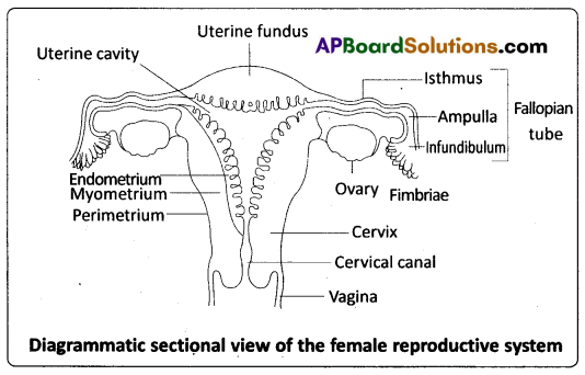
The wall of the uterus has three layers of tissue. The external thin membranous perimetrium, the middle thick layer of smooth muscle called myometrium and inner glandular lining layer called endometrium. The endometrium undergoes cyclic changes during menstrual cycle while the myometrium exhibits strong contractions during parturition.
Vagina : The vagina is a large, median, fibro – muscular tube that extends from the cervix to the vestibule (the space between the labia minora). It is lined by non – keratinised stratified squamous epithelium. It is highly vascular, and opens into the vestibule by the vaginal orifice.
Vulva: The term vulva (vulva = to wrap around) or pudendum refers to the external genitals of the female. The vestibule has two apertures – the upper external urethral orifice of the urethra and the lower vaginal orifice of vagina. Vaginal orifice is often covered partially by a membrane called hymen which is a mucous membrane. Vestibule is bound by two pairs of fleshy folds of tissue called labia minora (inner) and larger pair called labia majora (outer). Clitoris is a sensitive, erectile structure, which lies at the upper junction of the two labia minora above the urethral opening. The clitoris is homologous to the penis of a male as both are supported by corpora cavernosa internally. There is a cushion of fatty tissue covered by skin and public hair present above the labia majora. It is known as mons pubis.
Accessory reproductive glands of female : These glands include Bartholin’s glands, Skene’s glands and Mammary glands.
Bartholin’s glands : The Bartholin’s glands (Greater vestibular glands) are two glands located slightly posterior and to the left and right of the opening of the vagina. They secrete mucus to lubricate .the vagina and are homologous to the bulbourethral glands of the male reproductive system.
Skene’s glands: The Skene’s glands (Lesser vestibular glands) are located on the anterior wall of the vagina, around the lower end of the urethra. They secrete a lubricating fluid when stimulated. The Skene’s glands are homologous to the prostate glands, of the male reproductive system.
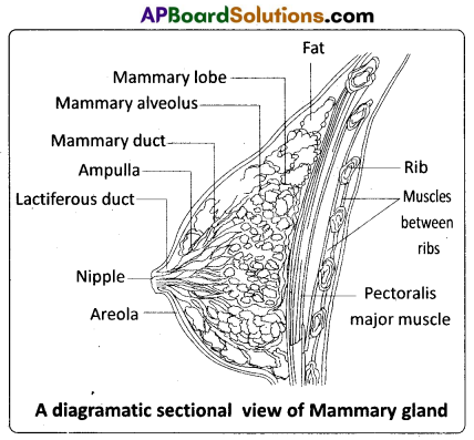
Mammary glands: A functio-nal mammary gland is characteristic of all female mammals. The mammary glands are paired structures ( breasts) that contain glandular tissue and variable amount of fat. The glandular tissue of each breast is divided into 15 – 20 mammary lobes containing clusters of cells called alveoli. The cells of the alveoli secrete milk, which is stored in the cavities (lumens) of the alveoli. The alveoli open into mammary tubules. The tubules of each lobe join to form a mammary duct. Several mammary ducts join to form a wider mammary ampulla which is connected to lactiferous duct through which milk is sucked out by the baby.
![]()
Question 21.
Describe the determination of sex by Genetic Balance theory of Bridges in Drosophila.
Answer:
Genetic Balance Theory: By studying the Chromosomal theory of sex determination, it may appear at first glance that some genes carried on the sex chromosomes, for example, X and Y are entirely responsible for determining the sex of the offspring. But this is not always true.
The Genetic Balance Theory of determination of sex was proposed by Bridges while working on Drosophila. This theory was devised to explain the mechanics of sex determination in Drosophila melanogaster According to his concept, the sex of an individual is determined by the ‘balance’ between the genes for femaleness located on the X – chromosome and those for maleness located on the ‘Autosomes’. Hence, the sex of an individual is determined by the ratio of number of its X chromosomes and that of its autosomal sets, the ‘Y’ chromosome taking no part in the determination of the sex. The ratio is termed as sex index arid is expressed as follows.
Sex index = Number of X chromosomes/ Number of sets of autosomes (X/A).
Bridges studied the offspring resulting from the non – disjunction of X chromosomes during meiosis in females Non – disjunction (not coming apart) is the failure of paired chromosomes to segregate or separate during the anaphase stage of the first or second meiotic divisions. The result is the production of aberrant gametes with abnormal numbers of chromosomes. There were various types of gametes such as the ones which cotnained one extra X – chromosome (AXX) and some others contained one chromosome less (AO).
Syngamy of such aberrant gametes and normal gametes produces zygotes with aneupioid karyotypes such as 2n + 1, 2n -1 etc. AAXO male is produced when an unusual ovum (AO) in the female is fertilized by a sperm with AX chromosome complement. An ‘AAXXY female’ is produced when an unusual ovum with AXX and a sperm with AY fuse. Bridges found that the ‘AAXXY flies’ were ‘fertile females’ and ‘AAXO flies’ were ‘sterile males’. It is important to note that the presence of Y – chromosome in the ‘AAXXY flies’ did not cause maleness, and its abnsence in the ‘AAXO flies’ did not produce femaleness.
| Sex Chromosome Complement | Haploid Sets of Autosomes | X : A Ratio | Sexual Phenotype |
| XX | AA | 1.0 | Female |
| XY | AA | 0.5 | Male |
| XO | AA | 0.5 | Male |
| XXY | AA | 1.0 | Female |
| XXX | AA | 1.5 | Matafemale |
| XXXY | AA | 1.5 | Metafemale |
| XX | AAA | 0.67 | Intersex |
| XO | AAA | 0.33 | Metamale |
| XXXX | AAA | 1.3 | Metafemale |
When Bridges crossed triploid females (AAAXXX) with normal diploid males (AAXY), he obtained normal diploid females, males, triploid females, intersexes, super males and super females. The occurrence of triploid intersexes from such a cross clearly established that autosomes also carry genesfor sex determinaton.
Bridges realized that the critical factor in determining sex is the ratio of X chromosomes to the number of haploid sets of auto-somes (A) present. He concluded that Y – chromosome in Drosophila lacks male determining factor, TDF (encoded by the SRY gene -Sex – determining Region Y). However, the Y – chromosome of Drosophila is required for male fertility. In ‘XO males’, sperms develop but are non – motile.