Varied difficulty levels in AP Inter 1st Year Zoology Model Papers and AP Inter 1st Year Zoology Question Paper March 2017 cater to students with diverse academic strengths and challenges.
AP Inter 1st Year Zoology Question Paper March 2017 with Solutions
Time: 3 Hours
Max. Marks: 60
Note: Read the following instructions carefully.
- Answer all the questions in Section – A. Answer any six questions in Section – B and answer any two questions in Section – C.
- In Section – A, questions from Sr. Nos. 1 to 10 are of Very Short Answer Tÿpe. Each question carries two marks. Every answer may be limited to 5 lines. Answer all questions at one place in the same order.
- In Section – B, questions from Sr. Nos. 11 to 18 are of Short Answer Type. Each question carries four marks. Every answer may be limited to 20 lines.
- In Section – C, questions from Sr. Nos. 19 to 21 are of Long Answer Type. Each question carries eight marks. Every answer may be limited to 60 lines.
- Draw labelled diagrams wherever necessary in Sections – B and C.
Section – A
10 x 2 = 20
Note: Answer all the questions in 5 lines each:
Question 1.
Define Species richness.
Answer:
The more the number of species in an area (unit area) the more is the species richness.
Question 2.
Distinguish between a tendon and a ligament.
Answer:
Tendons are the collagen fibrous tissue of dense regular connective tissue which attach the skeletal muscles to bones.
Ligaments are also the collagen fibrous tissue of dense regular connective tissue which attach bones to other bones.
Question 3.
Distinguish between Osteoblasts and Osteoclasts.
Answer:
Osteoblasts are immature bone cells and secrete the organic components of matrix and also play an important role in the mineralisation of bone and become Osteocytes. Osteoclasts are phagocytic cells involved in resorption of bone.
Question 4.
What is the haematocrit value?
Answer:
The percentage of total volume occupied by RBCs in blood is called haematocrit value.
Question 5.
What is Aristotle’s Lantern? Give one example of an animal possessing it.
Answer:
During amplexus, the mass of eggs and the mass of sperms released by the female and male are called spawn and mut.
Question 6.
Distinguish between mut and spawn.
Answer:
During amplexus, the mass of eggs and the mass of sperms released by the female and male are called spawn and milt.
![]()
Question 7.
List out any two differences between a flagellum and a cilium.
Answer:
| Flagellum | cilium |
| 1. Flagellum helps in locomotion only. | 1. Cilium helps in locomotion, feeding and acts as sensory structures. |
| 2. Flagellum produces undular movement. | 2. Cilium produces pendular movement. |
| 3. The flagellum is about 150 m in length. | 3. Cilium is small in size, 5-10 m in length. |
Question 8.
Distinguish between Proter and Opisthe.
Answer:
During transverse binary fission in Paramecium two daughter individuals are formed. The anterior one is called proter and the posterior is called opisthe.
Question 9.
What do you mean by Parasitic Castration? Give one example.
Answer:
Some parasites cause the degeneration of gonads of the host making it sterile. This effect is called parasitic castration.
E.g.: Sacculina (root-headed barnacle, a crustacean) causes the degeneration of ovaries in the crab Carcinus mean as.
Question 10.
What is meant by osmotrophic nutrition?
Answer:
Intake of pre-digested food material through the body surface is known as osmotrophic nutrition.
Section – B
6 x 4 = 24
Note: Answer any six questions in 20 lines each:
Question 11.
Explain Rivet Popper’s hypothesis.
Answer:
What if we lose a few species? Will it affect man’s life? Paul Ehrlich experiments Rivet popper, hypothesis, taking an aeroplane as an ecosystem, explains how removal of one by one ‘rivets (species of an ecosystem) of various parts can slowly damage the plane (ecosystem) -shows how important a ‘species is in the overall functioning of an ecosystem. Removing a rivet from a seat or some other relatively minor important parts ‘may not damage the plane, but removal of a rivet from a part supporting the wing can result in a crash. Likewise, removal of a critical species’ may affect the entire community and thus the entire ecosystem.
Question 12.
What are the salient features of the echinoids?
Answer:
- It includes sea urchins, heart urchins, sand dollars etc. The body is ovoid or discoidal and covered by movable spines.
- Arms are absent, tube feet are arranged in five bands and bear suckers.
- Ossicles of the body unite to form a rigid test or corona or case.
- Pedicellaria are ‘three-jawed”.
- Anus and madreporite are aboral in position.
- Ambulacral grooves are closed.
- A complex five-jawed masticatory apparatus called Aristotle’s lantern is present just inside the mouth. It is absent in heart urchins.
- Life history includes larval form called echinopluteus.
- Specialized gills called peristomial gills are present in sea urchins. Eg: Salmacis (Sea urchin), Echino Cardium (Heart urchin), and Clypeaster (Cake Urchin).
Question 13.
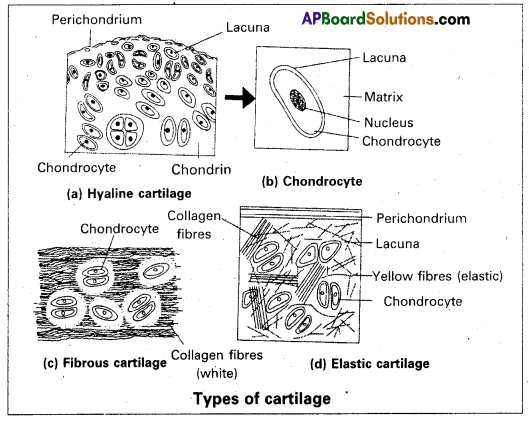
Describe the three types of cartilage.
Answer:
Cartilage is a solid, but semi-rigid (flexible) connective tissue. Cartilage is of three types, which differ form each other chiefly in the composition of the matrix. Cartilage is a solid, but semi-rigid (flexible) connective tissue. It resists compression. Matrix is firm but somewhat pliable. It has collagen fibres, elastic fibres (only in the elastic cartilage) and matrix-secreting cells called chondroblasts. These cells are enclosed in fluid-filled spaces called lacunae. Chondrocytes are the inactive cells of a cartilage. Cartilage is surrounded by a fibrous connective tissue sheath called perichondrium.
Cartilage is of three types, which differ from each other chiefly in the composition of the matrix.
1. Hyaline cartilage: It is bluish-white, translucent and glass-like cartilage. Matrix is homogeneous and shows delicate collagen fibres. It is the weakest and the most common type of all the cartilages. It is found in the walls of nose, larynx, trachea and bronchi.
2. Elastic cartilage: It is yellowish due to elastic fibres. Matrix has abundance of yellow elastic fibres in addition to collagen fibres. It provides strength and elasticity. Perichondrium is present. It is found in the pinnae of the external ears, Eustachian tubes and epiglottis.
3. Fibrous cartilage: Matrix has bundles of collagen fibres. The perichondrium is absent. It is the strongest of all types of cartilages. It occurs in the Intervertebral discs and pubic symphysis of the pelvis.
Question 14.
What are the modificätions observed in birds that help them in flight?
Answer:
So many modifications are observed in birds that help them in flight.
- Exo and endo skeletons and body structure features might have contributed for their successful arieal mode of life.
- The exoskeleton consists of epidermal feathers. Feathers are unique to birds. They are useful for flight, particularly the Quill feathers help in flight.
- Body is boat-shaped and streamlined.
- Forelimbs are modified into wings.
- Many bones are neumatic with extensions of air sacs.
- All modern flying birds are provided with powerful breast muscles (flight muscles) chiefly the pectoralis major and pectoralis minor.
- Lungs are associated with air sacs.
Question 15.
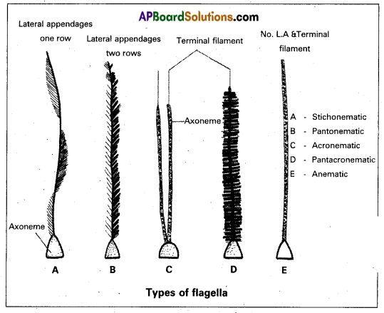
What are lateral appendages? Based on their presence and absence, write the various types of flagella giving at least one example for each type.
Answer:
Lateral appendages: Some flagella bear one or two or many rows of short, lateral hair-like fibrils called lateral appendages. They are of two types namely mastigonemes and ‘flimmers.
Types of Flagella: Based on the presence or absence and/or the number of rows of lateral appendages, five types of flagella are recognised.
- Stichonematic: This flagellum bears one row of lateral appendages on the axoneme. E.g. Euglena and Astasia.
- Pantonematic: This flagellum has two or more rows of lateral appendages on the axoneme. E.g. Percinema and Monas.
- Acronematic: This type of flagellum does not bear lateral appendages and the terminal part of the axoneme is naked without the outer sheath at its tip. E.g. : Chlamydomonas and Polytoma.
- Pantacronematic: This type of flagellum is provided with two or more rows of lateral appendages and the axoneme ends in a terminal naked filament. E.g. Urceolus.
- Anematic or simple: In this type of flagellum, lateral appendages and terminal filament are absent. Hence, it is , called anematic (a-no; nematic-threads). E.g. Chilomonas and Cyptomonas.
![]()
Question 16.
Distinguish between addiction and dependence.
Answer:
Addiction: It is a psychological attachment to certain effects such as euphoria. The most important thing one fails to realise it, the inherent addictive nature of tobacco, drugs and alcohol, with the repeated use of TDA, the tolerance level of the receptors present in our body increases. Consequently, the receptors respond only to higher doses leading to greater intake and addiction. However, it should be clearly borne in mind that use of TDA even once, can be a forerunner to addiction. Thus, the addictive potential of tobacco, drugs and alcohol pull the users into a vicious circle leading to their regular use (abuse) from which they may not be able to get out. In the absence of any guidance or counselling, people get addicted and become dependent on them.
Dependence: It is the tendency of the body of manifest a characteristic unpleasant condition (withdrawal syndrome). It the regular dose of drugs or alcohol is abruptly discontinued. The withdrawal syndrome is characterised by anxiety. Shakiness (tremors), nausea, and sweating which may be relieved when the regular use is resumed again. Dependence leads the patients to ignore all social norms.
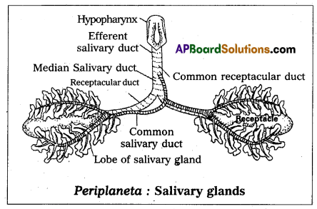
Question 17.
Draw a neat labelled diagram of the salivary apparatus of cockroach.
Answer:
Question 18.
Describe ‘Green House Effect’.
Answer:
The term Green House effect’ has been derived from a phenomenon that occurs in a greenhouse’. Greenhouse is a small
glass house and is used for growing plants, especially during winter. In a greenhouse, the glass panel allows the passage of light into it, but does not allow heat to escape (as it is reflected back). Therefore. the greenhouse warms up, very much like inside a car that has been parked in the sun for a few hours.
The greenhouse effect is a naturally occurring phenomenon that is responsible for heating of the Earth’s surface and atmosphere. It would be surprising to know that without greenhouse effect the average temperature of the Earth’s surface would have been a chilly – 18°C rather than the present average of 15 °C.
When sunlight reaches the outermost layer of the atmosphere, clouds and gases reflect about one-fourth of the incoming solar radiation and absorb some of it. Almost half of the incoming solar radiation falls on the Earth’s surface and heats it up. While a small proportion is reflected back.
Section – C
2 x 8 = 16
Note: Answer any two questions in 60 lines each:
Question 19.
Describe the life cycle of Plasomodium vivax in human with a neat labelled diagram.
Answer:
Life cycle of plasmodium in man (The human phase): In man, the plasmodium reproduces by asexual reproduction called schizogony. It occurs in liver cells (hepatocytes) as well as in RBC. In liver cells, it is called hepatic schizogony and in RBC it is called erythrocytic schizogony.
Hepatic Schizogony: This was discovered by Shott and Carnham. Whenever a mosquito infected by plasmodium bites a
man, nearly 2000 sporozoites are released into the blood of man through its saliva, within half an hour, they reach the hepatocytes where They undergo pre-erythrocytic and exo-erythrocytic cycles.
Pre-erythrocytic cycle: Whenever the sporozoites enter the liver cells they transform into trophozoites. They feed on the
contents of the hepatic cells, assume spherical shape and attain the maximum size, This stage is called schizont stage. It’s nucleus divides several times mitotically, followed by the cytoplasmic divisions resulting in approximately 12,000 daughter individuals called cryptozoites or the P’ generation merozoites. They enter the sinusoids of the liver by rupturing the cell membrane of the schizont and the liver cells. This entire process is completed approximately in 8 days. Now these first-generation merozoites have two options. i.e., they can enter either fresh liver cells and continue exo-erythrocytic cycle or they can enter RBC and continue erythrocytic cycle.
Exo-erythrocytiC cycle: If the cryptozoites enter the fresh liver cells, they undergo the changes similar to that of the pre-erythrocytic cycle and produce the second-generation merozoites called meta cryptozoites. These are of two types the smaller microme tacryptozoites and larger macro-metacry-protozites. This entire process is completed approximately in two days. The macro meta cryptozoites attack fresh liver cells and continue another exoerythrocytic cycle, whereas the micro meta cryptozoites always enter bloodstream and attack fresh RBC to continue erythrocytic cycle.
Prepatent period: The interval between the first entry of plasmodium into the blood in the form of sporozoites and the second entry of plasmodium into the blood in the form of cryptozoites is called prepatent period. It lasts approximately 8 days. During this period, the host does not show any clinical symptoms of the disease. It is only a means of multiplication.
Erythrocytic cycle: It was first described by Camilip Golgi. Hence it is also called golgi cycle. This cycle is initiated either by the cryptozoites of pre-erythrocytic cycle or the micro metocry ptozoites of exo-erythrocytic cycle in the fresh RBC, these stages assume spherical shape and transform into trophozoites. It develops a small vacuole which gradually enlarges in size pushing the cytoplasm and nucleus to the periphery. Now the plasmodium looks like a fisher ring. Hence this stage is called signet ring stage.
Soon it loses the vacuole, develops pseudopodia and becomes amoeboid stage with the help of pseudopodia. It actively feeds on the contents of the RBC and increases in size. As a result, the RBC grows almost double the size. This process is called hypertrophy. The malaria parasite digests the globin part of the ingested haemoglobin and converts the soluble haem into an insoluble crystalline haemozoin.
It is called the ‘malaria pigment which is called a disposable product. During this stage, small red-coloured dots appear in the cytoplasm of the RBC known as Schuffner’s dots. These are believed to be the antigens released by the parasite. Now the plasmodium loses the pseudopodia, further increases in size, occupies the entire RBC and becomes a schizont. It undergoes schizogony similar to that of the pre-erythrocytic cycle and produces 12 to 24 erythrocytic merozoites. They are arranged in the form of the petals of a rose in the RBC. Hence this stage is called the rosette stage. Finally the erythrocyte bursts and releases the merozoites along with haemozoin into the blood. This cycle is completed approximately in 48 hours.
Incubation period: The period between the entry of plasmodium into the blood in the form of sporozoite and the first
appearance of symptoms of malaria in man is called incubation period, it is approximately 10 to 14 days.
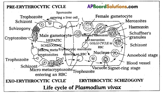
Formation of gametocytes: After repeated cycles of erythrocytic schizogony when the number of fresh RBC decreases, some merozoites enter the RBC and transform into gametocytes instead of continuing the erythrocytic cycle. This process generally takes place when the RBCs are present in spleen and bone narrow.
The gametocytes are of two types namely, smaller micro gametocytes or male gametocytes and larger macrogametocytes or female gametocytes. The gametocytes cannot undergo further development in man as the temperature and H of the blood of man are not suitable for further development. These gametocytes reach the blood circulation and wait to reach the next host. They degenerate and die if they are not transferred to mosquito within a week.
![]()
Question 20.
Describe the digestive system of cockroach with the help of a neat labelled diagram.
Answer:
The digestive system of cockroach consists of an alimentary canal and the associated glands. The preoral cavity surrounded by the mouth parts, is present in front of the mouth. The hypopharynx divides it into two chambers called cibagium (anterior) and salivarium (posterior).
Alimentary canal: The alimentary canal of cockroaches is a long tube and is coiled at some places. It extends between the mouth and the anus. It is divided into three regions namely foregut of stomodaeum, midgut or mesenteron and hindgut are internally lined by ectoderm. The mesenteron is lined by the endodermal cells.
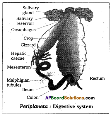
Foregut or stomodaeum: The foregut includes pharynx, oesophagus, crop and gizzard. It is internally lined by a chitinous cuticle. Mouth opens into the pharynx, which in turn leads into a narrow tubular oes oesophagus. The oesophagus opens behind into a thin walled distensible sac called crop. The crop serves as a reservoir for storing food. Its outer surface is covered by a network of tracheae. Behind the trop there is a thick-walled muscular proven- triculus or gizzard.
The chitinous inner living of the gizzard has six powerful teeth, which form an efficient grinding apparatus. Behind each’ tooth is a hairy pad, which bears backwardly directed bristles. Among these plates, food is thoroughly ground into fine particles. These food particles are filtered by the bristles. The gizzard thus acts both as a grinding mill and also as a sieve. There is a membranous projection ‘of the gizzard into the mesenteron in the form of a funnel called stomodeal valve. This valve prevents the entry (regurgitation) of food from the mesenteron back into the gizzard.
Midgut (mesenteron or ventriculus): The midgut is a short and narrow tube behind the gizzard. It is also called mesenteron or ventriculus. Between the ventriculus and the gizzard, arising from ventriculus there are six to eighth finger-like diverticular called hepatic caecae.
They are helpful in digestion and absorption of the digested food materials. Ventriculus is functionally divided into an anterior secretory part and a posterior absorptive part.
The secretory part of the ventriculus has many gland cells and it secretes several enzymes. The bolus’ of food in the mesenteron is enveloped by a chitinous and porous membrane called peritrophic membrane, which is secreted by the funnel-like stomodeal valve of the gizzard. Digested food is absorbed into the food through the peritrophic membrane in the posterior absorptive region of the ventriculus.
The peritrophic membrane protects the wall of the ventriculus from hard food particles in the food. A sphincture muscle controls the opening of the ventriculus into the hindgut. It prevents entry of undigested food from the hindgut into the midgut.
Hindgut or proctodaeum: The hindgut is a long coiled tube, consisting of three regions namely ileum, colon and rectum. It is interanlly lined by chitinous cuticle. The ileum that lies behind the mesenteron is a short tube. Six bundles of fine yellow, blind tubules called Malphigian tubules open into the ileum near the junction of mesenteron and ileum. Malphigian tubules are excretory in function. Ileum collects uric acid from the maiphigian tubules and undigested food from the mesenteron. Ileum opens behind into a long coiled tube called colon. Colon leads into a short and wide rectum which opens out through the anus. Rectum bears on its inner side six longitudinal chitinous folds called rectal papillae. They are concerned with the reabsorption of water from the undigested food.
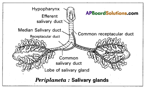
Digestive gland: The digestive glands associated with the alimentary canal of cockroach are salivary glands, hepatic caecae and glandular cells of ‘the mesenteron.
Salivary glands: There is a pair of salivary glands attached to the ventrolateral sides of the crop, one on each side. Each salivary gland has two lobes. Each lobe of salivary gland has many lobules called acini. Each acinus is a group of secretory cells called zymogen cells with a small ductule. The ductules of both the lobes of a salivary gland unite to form a common salivary duct on each side.
The two common salivary ducts are joined to form the median salivary duct. Between the two lobes of a salivary gland of each side is a sac called salivary receptacular duct or common reservoir duct. The midious salivary duct opens into the common receptacular duct. Later these two form an efferent salivary duct. The efferent salivary duct opens at the base of the hypopharynx. Acinar cells secrete saliva, which contains starch-digesting enzymes such as amylase.
Hepatic caecae: The hepatic caecae are also termed midguts caecae. They contain secretory and absorptive cells. Glandular cells of the mesenteron: The glandular cells of the mesenteron secrete enzymes such as maltase, invertase, proteases and lipase.
Question 21.
List out the major air pollutants and describe their effects on human beings.
Answer:
Air pollutants cause injury to all living organisms. They reduce growth and yield of crops. They are harmful to the respiratory system of humans and animals. Increase in the concentration of pollutants or duration of exposure increase the harmful effects on the organisms.
The major air pollutants:
1. Carbon monoxide (CO): It is produced mainly due to incomplete combustion of fossil fuels. Automobiles are a major cause of CO pollution in larger cities and towns. Automobile exhausts, fuels from factories, emissions from power plants, forest fires kind even burning of firewood contribute to CO pollution. Haemoglobin has greater affinity for CO and SO, CO competitively interferes with oxygen transport. CO symptoms such as headache and blurred vision at lower còncentrations. In higher concentrations, it leads to coma and death.
2. Carbon Dioxide (CO2): Carbon dioxide is the main pollutant that is leading to global warming. Plants utilize CO2 for photosynthesis and all living organisms emit carbon dioxide in the process of respiration. With rapid urbanization, automobiles, aeroplanes, power plants and other human activities that involve the burning of fossil fuels such as gasoline, carbon dioxide is turning out to be an important pollutant of concern.
3. Sulphur Dioxide (SO2): It is mainly produced by burning of fossil fuels. Melting of sulphur ores is another important source for SO2 pollution. Metal, smelting and other industrial processes also contribute to SO2 pollution. Sulphur dioxide and nitrogen oxides are the major causes of acid rains, which cause acidification of soils, lakes and streams and also accelerated corrosion of buildings and monuments. High concentrations of sulphur dioxide (SO2) can result in breathing problems in asthmatic children and adults. Other effects associated with long-term exposure to sulphur dioxide, include respiratory illness, alterations in the lungs defenses and aggravation of existing cardiovascular problems.
To control SO2 pollution, the emissions are filtered through scrubbers. Scrubbers are devices that are used to clean the impurities in exhaust gases. Gaseous pollutants such as SO2 are removed by scrubbers.
4. Nitrogen Oxides: Nitrogen oxides are considered to be major primary pollutants. The source is mainly automobile exhaust. The air polluted by nitrogen oxide is not only harmful to humans and animals but also dangerous for the life of plants. Nitrogen oxide pollution also results in acid rains and formation of photo-chemical smog. The effect of nitrogen oxides on plants include the occurrence of necrotic spots on the surface of leaves. Photosynthesis Is affected in crop plants and the yield is reduced. Nitrogen oxides combine with volatile organic compounds by the action of sunlight to form secondary pollutants called Peroxyacetyl nitrate (PAN) which are found especially in photochemical smog. They are powerful irritants to eyes and respiratory tract.
5. Particulate matter/Aerosols: Tiny particles of solid matter suspended in a gas or liquid constitute the particulate matter. Aerosols refer to particles and /or liquid droplets and the gas together (a system of colloidal particles dispersed in a gas) Combustion of ‘fossil fuels” (petrol, diesel etc) fly ash produced in thermal plants, forest fires, cement factories, asbestos mining and manufacturing units, spinning and ginning mills etc. are the main sources of particulate matter pollution. According to the Central Pollution Control Board (CPCB) particles of 2.5 micrometres or less in diameter are highly harmful to man and other air-breathing organisms.
![]()
An electrostatic precipitator is a widely used ‘filter’ for removing particulate matter from the exhaust of thermal power plants. It can remove 99% partiéulate matter. it has high-voltage electrodes which produce a ‘corona’ that releases electrons. These are collected by collecting plates which attract the charged particles. The air flowing between the plates is kept in low velocity so as to allow the dust particles to fall. Thus clean air is released into the atmosphere.