Varied difficulty levels in AP Inter 1st Year Zoology Model Papers and AP Inter 1st Year Zoology Question Paper June 2015 cater to students with diverse academic strengths and challenges.
AP Inter 1st Year Zoology Question Paper June 2015
Time : 3 Hours
Max. Marks: 60
General Instructions:
Note : Read the following instructions carefully
- Answer All questions of Section A. Answer ANY SIX questions in Section B and answer ANY TWO questions in Section C.
- In Section A questions from Sr. Nos. 1 to 10 are of “Very Short Answer Type”. Each question carries TWO marks. Every answer may be limited to 5 lines. Answer all these questions at one place in the same order.
- In Section ‘B’, questions from Sr. Nos. 11 to 18 are of “Short AnswerType”. Each question carries FOUR marks. Every answer may be limited to 20 lines.
- In Section ‘C’, questions from Sr. Nos. 19 to 21 are of “Long Answer Type”. Each question carries EIGHT marks. Every answer may be limited to 60 lines.
- Draw labelled diagrams wherever necessary in Sections ‘B’ and ‘C’.
Section – A (10 × 2 = 20)
Note : Answer all the questions in 5 lines each.
Question 1.
Differentiate between Protostomia and Deuterostomia.
Answer:
a) Protostomia: The eumetazoans in which blastopore develops into mouth are referred to as the protostomians. Eg : Annelida.
b) Deuterostomia : These are eucoelomates in which anus is formed from or near the blastopore. Eg : Echinodermata.
Question 2.
Distinguish between holocrine and apocrine gland.
Answer:
- Holocrine glands : In which the entire cell disintegrates to discharge the contents. Eg : sebaceous glands.
- Apocrine glands : In which the apical part of the cell is pinched off alpng with the secetory product. Eg : mammary gland.
Question 3.
What is haematocrit value ?
Answer:
The percentage of total volume occupied by RBCs is called haematocrit value.
![]()
Question 4.
What is cephalization ? How is it useful to its possessors ?
Answer:
Cephalization is concentration of nerve and sensory cells at the anterior end. As a result of cephalization, bilaterally symmetrical animals can sense the new environment into which they enter and respond more efficiently and quickly.
Question 5.
What is Aristotle’s Lantern ? Give one example of an animal possessing it.
Answer:
In the mouth of a sea-urchin a complex five jawed masticatory apparatus called Aristotle’s lantern is present.
Ex : Echinus sea urchin.
Question 6.
What are claspers ? Which group of fishes possesses them ?
Answer:
In the group chondrichthys males possess claspers on pelvic fins to facilitate internal fertilization Ex : shark.
Question 7.
Distinguish between proter and opisthe.
Answer:
During Transverse binary fission of paramoecium, two daughter paramoecia are formed. The upper or anterior one is proter which receive upper contractile vacuole cytopharynx and cytostome of its parent. The lower or posterior daughter is opisthe which receives the posterior contractile vacuole only.
Question 8.
Distinguish between synchronous and metachronous movements.
Answer:
- Synchronous movement : Cilia in a transverse row beat simultaneously in one direction. It is called synchronous movement.
- Metachronous movement : The sequential movement of cilia, in a longitudinal row, one after the other in one direction is called metachronous movement.
Question 9.
Define neoplasia. Give one example.
Answer:
Neoplasia: Some cause an abnormal growth of the host cells in a tissue to form new structures. This effect is called Neoplasia which leads to cancers. Eg: some viruses.
![]()
Question 10.
Define entropy.
Answer:
Energy lost or not available for work in a system is called ENTROPY.
Section – B (6 × 4 = 24)
Note : Answer any six questions in 20 lines each.
Question 11.
What is the evil quartet ?
Answer:
The following are the four major causes for accelerated rates of species extinction in the world. These causes are referred to as evil quartet.
a) Habitat loss and fragmentation : These are most important reasons for the loss of biodiversity.
b) Over exploitation : When need turns to greed, it leads to over exploitation.
c) Invasion of Alien species : When Alien species are introduced into a habitat, they turn invasive and establish themselves at the cost of indigenous species.
d) Co – extinctions : In an obligate association between a plant and an animal, if a plant becomes extinct, the animal also becomes extinct as seen in a parasitic and host association.
Question 12.
Describe the structure of a multipolar neuron.
Answer:
A neuron usually consists of a “cell body” with one to many dendrites and a single axon.
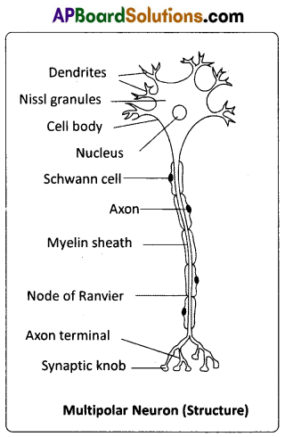
Cell body: It is also called perikaryon, cyton or soma. It contains abundant granular cytoplasm and a large spherical nucleus. The cytoplasm has Nissl bodies (they represent RER, the sites of protein synthesis), neurofibrils and lipofuscin granules (the products of cellular wear and tear, accumulating in lysosomes with age). A group of cell bodies in the central nervous system is called a ‘nucleus’, and in the peripheral nervous system, it is called a ‘ganglion’.
Dendrites :
Several short, branched processes which arise from the cyton are called dendrites. They also contain Nissl bodies and neurofibrils. They conduct nerve impulses towards the cell body (afferent processes).
Axon :
An axon is a single, long, cylindrical process that originates from a region of the cyton called axon hillock. Plasmalemma of an axon is called axolemma, and the cytoplasm is called axoplasm, which contains neurofibrils. However, Nissl bodies are absent. An axon may give rise to collateral branches. Distally it branches into many fine filaments called telodendria, (axon terminals), which end in bulb like structures called synaptic knobs or terminal boutons. Synaptic knobs possess ‘synaptic vesicles’ containing chemicals called neurotransmitters. Axon transmits nerve impulse away from the cyton (efferent process) to an interneuronal or neuromuscular junction called synapse.
Question 13.
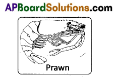
What are the chief characters of crustaceans ?
Answer:
- They are aquatic.
- Head and thorax fuse forming the cephalothorax (covered by chitinous carapace). In some the exoskeleton is hardened by calcium carbonate (crabs and lobsters).
- Cephalic region bears two pairs of antennae (antennules and antennae – unique feature), one pair of mandibles and two pairs of maxillae.
- Thoracic and abdominal appendages are ‘biramous’.
- Respiratory organs are gills (branchiae).
- Excretory organs are green glands or antennary glands.
- Sense organs include antennae compound eyes, statocysts, etc.
- Development is indirect and includes different larval forms. Examples : Palaemon (freshwater prawn), Cancer (crab), Balanus (rock barnacle), Sacculina (root-headed barnacle), Astacus (crayfish), Daphnia (waterflea).
![]()
Question 14.
What are the features peculiar to Ratitae birds ? Give two examples of Ratitae birds.
Answer:
Ratitae / Palaeognathae : They are modern flightless running birds. They are ‘discontinuous’ in their distribution like the lung fishes and marsupials. They are characterized by the presence of reduced wings, a raft like sternum without keel and males with penis. They do not possess syrinx, clavicles and usually preen gland.
E.g. Struthio camelus (African ostrich), Kiwi (National bird of New Zealand), Rhea (American ostrich), Dromaeus (Emu), Casuarius.
Question 15.
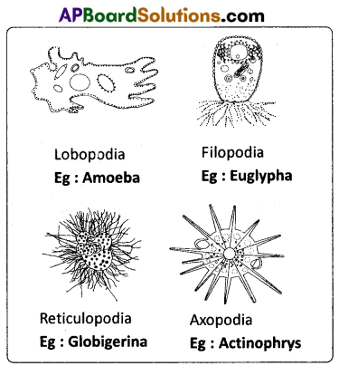
Give an account of pseudopodia.
Answer:
Pseudopodia : These are found in rhizopods. The pseudopodia are temporary extensions of cytoplasm that develop in the direction of the movement. These temporary structures are useful to move on the substratum as our legs do, hence the name ‘pseudopodia’. There are four kinds of pseudopodia namely lobopodia (blunt finger-like) as in Amoeba and Entamoeba, filopodia (fibre-like) as in Euglypha, reticulopodia (net-like) as in Elphidium and axopodia or heliopodia (sun ray-like) as in Actionophrys.
Question 16.
What is the need for parasites to develop special adaptations ? Mention sfome special adaptations developed by the parasites.
Answer:
Parasites have evolved special adaptations to meet the requirements and lead successful life in the hosts.
- In order to live in the host, some parasites have developed structures like hooks, suckers, rostellum, etc., for anchoring. E.g. Taenia solium.
- Some intestinal parasites have developed protective cuticle to withstand the action of the digestive enzymes of the host. E.g. Ascaris lumbricoides.
- Some intestinal parasites produce anti enzymes to neutralize the effect of host’s digestive enzymes, e.g. Taenia solium.
- Some parasites live as obligatory anaerobes as the availability of oxygen is very rare for them. e.g. Entamoeba histolytica, Taenia solium, etc.
- Some intestinal parasites live as facultative anaerobes i.e., if oxygen is not available, they live anaerobically and if oxygen is available, they respire aerobically, e.g. Ascaris lumbricoides.
- The morphological and anatomical features are greatly simplified while emphasizing their reproductive potential. For example, an Ascaris lays nearly two lakh eggs per day. In Taenia solium the body is divided into 700 to 900 proglottids of which each proglottid acts as a unit of reproductive system and releases approximately 35,000 eggs.
- The life cycles of endoparasites are more complex because of their extreme specialization. For example, life cycle of certain parasites like Fasciola hepatica (sheep liver fluke) is very complex involving many developmental stages and two intermediate hosts to increase the chances of reaching a new definitive host.
- Certain parasites like Entamoeba develop cysts to tide over the unfavourable conditions like desiccation while reaching the new host.
- Some parasites elude production of vaccines against them (smart*parasites!) as they keep changing their surface antigens form time to time, e.g. Plasmodium, HIV, etc.
Question 17.
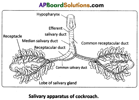
Draw a neat labelled diagram of the salivary apparatus of cockroach.
Answer:
![]()
Question 18.
What is summer stratification ? Explain.
Answer:
Summer stratification : During summer in temperate lakes, the density of the surface water decreases because of increase in its temperature (21 – 25°C). This ‘upper more warm layer’ of a lake is called epilimnion. Below the epilimnion there is a zone in which the temperature decreases at the rate of 1°C per meter in depth, and it is called thermocline or metalimnion. The bottom layer is the hypolimnion, where water is relatively cool, stagnant and with low oxygen content (due to absence of photosynthetic activity).
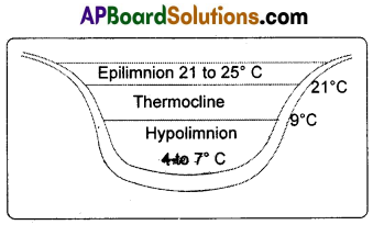
During autumn (also called Fall), the epiiimnion cools down, and the surface water becomes heavy when the temperatures 4°C, and sinks to the bottom of the lake. Overturns bring about ‘uniform temperature’ in lakes during that period. This circulation during the autumn is known as the fall or autumn overturn. The upper oxygen rich water reaches the hypoliminion and the nutrient rich bottom water comes to the surface. Thus there is uniform distribution of nutrients and oxygen in the lake.
Question 19.
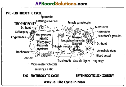
Describe the life cycle of Plasmodium vivax in man.
Answer:
Life cycle of Plasmodium in man (The human phase) : In man, the Plasmodium reproduces by asexual reproduction called schizogony. It occurs in liver cells (hepatocytes) as well as in RBC. In liver cells, it is called hepatic schizogony and in RBC it is called erythrocytic schizogony.
Hepatic schizogony : This was discovered by Shortt and Garnham. Whenever, a mosquito infected by Plasmodium bites a man, nearly 2000 sporozoites are released into the blood of man through its saliva. Within half an hour, they reach the hepatocytes where they undergo pre-erythrocytic and exo- erythrocytic cycles.
Pre-erythrocytic cycle : Whenever the sporozoites reach the liver cells, they transform into trophozoites. Theyfeed on the contents of the hepatic cells, assume spherical shape and attain the maximum size. This stage is called schizont stage. Its nucleus divides several times mitotically, followed by the cytoplasmic divisions resulting in approximately 12,000 daughter individuals called cryptozoites or the lstgeneration merozoites. They enter the sinusoids of the liver by rupturing the cell membrane of the schizont and the liver cells. This entire process is completed approximately in 8 days. Now these first generation merozoites have two options i.e., they can enter either fresh liver cells and continue exo-erythrocytic cycle or they can enter RBC and continue erythrocytic cycle.
Exo-erythrocytic cycle : If the cryptozoites enter the fresh liver cells, they undergo the changes similar to that of the pre-erythrocytic cycle and produce the second generation merozoites called metacryptozoites. These are of two types – the smaller micro-metacryptozoites and larger macro-metacryptozoites. This entire process is completed approximately in two days. The macro-metacryptozoites attack fresh liver cells and continue another exo – erythrocytic cycle, whereas the micro-metacryptozoites always enter blood stream and attack fresh RBC to continue erythrocytic cycle.
Prepatent period : The interval between ‘the first entry of Plasmodium into the blood in the form of sporozoites and the second entry of Plasmodium into the blood in the form of cryptozoites is called prepatent period. It lasts approximately 8 days. During this period, the host does not show any clinical symptoms of the disease. It is only a means of multiplication.
Erythrocytic cycle : It was first described by Camillo Golgi. Hence it is also called Golgi cycle. This cycle is initiated either by the cryptozoites of pre-erythrocytic cycle or the micro- metacryptozoites of exp – erythrocytic cycle. In the fresh RBC, these stages assume spherical shape and transform into trophozoites. It develops a small vacuole which gradually enlarges in size, pushing the cytoplasm and nucleus to the periphery. Now the Plasmodium looks like a finger ring. Hence this stage is called signet ring stage. Soon it loses the vacuole, develops pseudopodia and becomes amoeboid stage. With the help of pseudopodia, it actively feeds on the contents of the RBC and increases in size. As a result, the RBC grows almost double the size.
This process is called hypertrophy. The malaria parasite digests the globin part of the ingested haemoglobin and converts the soluble haem into an insoluble crystalline haemozoin. It is called the ‘malaria pigment’ which is a disposable product. During this stage, small red coloured dots appear in the cytoplasm of the RBC known as Schuffner s dots. These are believed to be the antigens released by the parasite.
Now the Plasmodium loses the pseudopodia, further increases in size, occupies the entire RBC and becomes a schizont. It undergoes schizogony similar to that of the pre-erythrocytic cycle and produces 12 to 24 erythrocytic merozoites. They are arranged in the form of the petals of a rose in the RBC. Hence, this stage is called the rosette stage. Finally the erythrocyte bursts and releases the merozoites along with haemozoin into the blood. This cycle is completed approximately in 48 hours.
Incubation Period : The period between ‘the entry of Plasmodium into the blood in the form of sporozoite and the first appearance of symptoms of malaria in man’ is called incubation period. It is approximately 10 to 14 days.
Formation of gametocytes : After repeated cycles of erythrocytic schizogony, when the number of fresh RBC decreases, some merozoites enter the RBC and transform into gametocytes instead of continuing the erythrocytic cycle. This process generally takes place when the RBCs are present in spleen and bone marrow.
The gametocytes are of two types namely, smaller microgametocytes or male gametocytes and larger macrogametocytes or female gametocytes. The gametocytes cannot undergo further development in man as the temperature and pH of the blood of man are not suitable for further development. These gametocytes reach the blood circulation and wait to reach the next host. They degenerate and die if they are not transferred to mosquito within a week.
![]()
Question 20.
Describe the respiratory system of cockroach with the help of neat and labelled diagrams.
Answer:
Respiratory System of Periplaneta : Due to the absence of respiratory pigment, the blood of cockroach is colourless and it cannot carry oxygen to different tissues. Therefore a tracheal system is developed to carry the air directly to the tissues. The respiratory system of cockroach consists of stigmata, tracheae and tracheoles.
Stigmata or spiracles : The tracheal system communicates with the exterior by ten pairs of openings called stigmata or spiracles. The first two pairs of spiracles are present in the thoracic segments, one pair in mesothorax and one pair in the
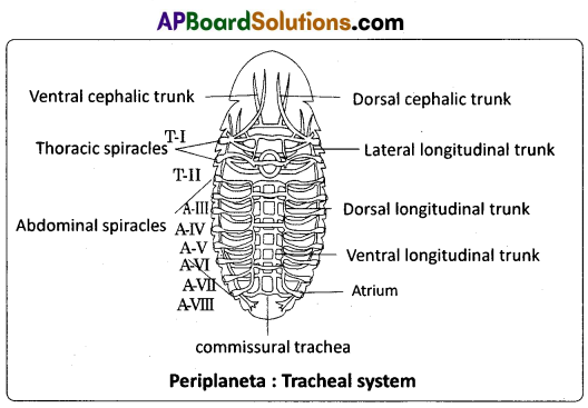
metathorax. The remaining eight pairs are present in the first eight abdominal segments. Spiracles are located in the pleura of their respective segments. The respiratory system in insects is classified on the basis of number and nature of spiracles. The spiracles of cockroach are polypneustic (as they are more than 3 pairs) and holopneustic (as all of them are functional). All spiracles are valvular and each of them is surrounded by a chitinous ring called peritreme. All spiracles bear small hair like structures called trichomes to filter the dust particles. Each spiracle opens into a small chamber called atrium.
Tracheae : From the atrium of each thoracic spiracle several horizontal tracheae run inside. They join with each other in the thorax to form many tracheal trunks like dorsal cephalic, ventral cephalic trunks and their branches. These branches enter all organs of the head. The thoracic region also contains lateral longitudinal trunks. The abdominal spiracles lead into atria.
From the atrium of each abdominal spiracle three tracheal tubes arise. All these tracheal tubes of one side open into three separate longitudinal tracheal trunks. They are lateral, dorsal and ventral longitudinal trunks. Lateral longitudinal trunks are the longest tracheal trunks. The three pairs of longitudinal tracheal trunks of both the sides are interconnected by many commissural tracheae. From all the tracheal trunks several branches are given out, which enter different organs. All tracheal branches entering into an organ end in a special cell called tracheole cell.
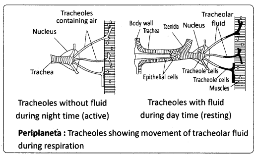
The wall of the tracheae is made of three layers. They are an outer basement membrane, a middle one cell thick epithelium and an inner layer of cuticle called intima. The intima is produced into spiral thickenings called taenidia. The taenidia keep the tracheae always open and prevent it from collapsing.
Tracheoles : The terminal cell of trachea is called tracheoblast or tracheole cell. It has several intracellular tubular extensions called tracheoles. Tracheoles are devoid of intima and taenidia. They are formed of a protein called trachein. Tracheolar fluid is present inside the tracheoles. The level of the tracheolar fluid varies with the metabolic activity of the insect. It is more when the insect is inactive and completely reabsorbed into the tissues, when the insect is more active. Tracheoles penetrate the cell and are intimately associated with mitochondria (to supply oxygen to them).
Mechanism of respriation : Respiration includes two events, viz., inspiration and expiration. The muscles helpful are dorsoventral muscles and ventral longitudinal muscles. Dorsoventral muscles are the principal muscles of respriation.
Inspiration : Taking in of air is inspiration. It is effected by the relaxation of the dorsoventral muscles and ventral longitudinal muscles. Due to the relaxation of the dorsoventral muscles, tergal plates are elevated and the volume of the body cavity increases. Due to the relaxation of the ventral longitudinal muscles, the telescoped segments come to the normal position. So the volume of the body cavity increases in the longitudinal axis. As air is drawn in due to the relaxation of the muscles, the process is a ‘passive’ process. During inspiration the thoracic spiracles are kept open and the abdominal spiracles are kept closed.
Expiration : Sending out air from the body is called expiration. On contraction the dorsoventral muscles depress the tergal plates. Body cavity decreases in size and pressure increases. Due to the contraction of the ventral longitudinal muscles, the segments are telescoped and the volume of the body cavity decreases in the longitudinal axis increasing the pressure further. As this process involves the contraction of muscles, expiration is described as active process. During expiration thoracic spiracles are closed and abdominal spiracles are kept open.
Exchange of gases : As air enters the tracheoles, oxygen from the air is taken into the cells and CO2 is released into haemolymph. The CO2 from the haemolymph mostly goes out through the inter- segmental membranes of the body wall. Cockroach and some other insects such as grasshoppers and beetles exhibit the phenomenon of discontinuous ventilation. In this mode of respiration continuous exchange of gases is interrupted by extended periods during which spiracles remain closed.
The expulsion of CO2 from the body occurs in bursts, when the spiracles are open. The exchange of gases depends on the metabolic rate and temperature. When air enters the tracheoles, oxygen diffuses faster into the tissues due to its high partial pressure. At the same time the carbon dioxide of tissues, instead of passing into the tracheal system, goes into the haemolymph. Carbon dioxide is carried more quickly into the haemolymph due to its greater solubility in it. This CO2 accumulates near the spiracles and diffuses into the artial chambers near the spiracles and goes out in bursts through the abdominal spiracles. Opening and closing of spiracles is influenced by CO tension in haemolymph and oxygen tension in the trachea.