Varied difficulty levels in AP Inter 1st Year Zoology Model Papers Set 1 cater to students with diverse academic strengths and challenges.
AP Inter 1st Year Zoology Model Paper Set 1 with Solutions
Time : 3 Hours
Max. Marks: 60
Instructions to Candidate:
- Answer all the questions of Section ‘A’. Answer any Six questions in Section ‘B and answer any two questions in Section ‘C’.
- In Section ‘A’, questions from Sr. Nos. 1 to 10 are of Very Short Answer Type. Each question carries two marks. Every answer may be limited to 5 lines. Answer all these questions at one place in the same order.
- In Section ‘B’, questions from Sr. Nos. 11 to 18 are of Short Answer Type. Each question carries four marks. Every answer may be limited to 20 lines.
- In Section ‘C’, questions from Sr.Nos. 19 to 21 are of Long Answer Type. Each question carries eight marks. Every
answer may be limited to 60 lines. - Draw labelled diagrams wherever necessary in Section ‘B’ and ‘C’.
Section – A
10 x 2 = 20
Question 1.
What is trinominal nomenclature? Give an example.
Answer:
The tririominal nomenclature is the extension of the binominal system of nomenclature. It is permits the designation of subspecies with a three worded name called ‘trinomen’. Ex: Homo Sapiens Sapiens, Corvus splendens splendens.
Question 2.
What is cephalization? How is it useful to its possessors?
Answer:
Cephalization: The concentration of nerve (Brain) and sensory cells at the anterior end of body is called as Cephalization. As a result of cephalization, these animals can sense the new environment and move efficient than the other animals in seaking food, locating mates and in avoiding or escaping from predators.
Question 3.
Distinguish between exocrine and endocrine glands with examples.
Answer:
Exocrine glands are provided with ducts. Secrete mucus, saliva, earwax, oil, milk, digestive enzymes and other cell products. Endocrine glands are ductless and their products are hormones which are not sent out via ducts but are carried to the target organs by blood. Ex: Pituitary gland.
![]()
Question 4.
Which is the strongest cartilage? In which regions of the human body do you find it?
Answer:
The fibrous cartilage is the strongest of all types of cartilage. It is occurs in the intervertebral discs and public Symphysis of the pelvis.
Question 5.
What is Aristotle’s Iantern? Give an example of an animal possessing it.
Answer:
In the mouth of sea Urchin a complex five-jawed masticatory apparatus called Aristotle’s Lantern. Ex: Echinus.
Question 6.
How do you justify the statement, heart in fish is a branchial heart?
Answer:
Heart of fishes is ‘two-chambered’ and is described as branchial heart as it supplies blood only to the gills.
Question 7.
Distinguish between lobopodium and filopodium. Give an example to each of them.
Answer:
Lobopodium: The blunt and finger-like tubular pseudopodia containing both ectoplasm and endoplasm is called lobopodium.
Ex: Amoeba proteus.
Filopodium: The slender filamentous pseudopodia with pointed tips, composed of only ectoplasm are called Filopodium. Ex: Euglypha.
Question 8.
Distinguish between proter and opisthe.
Answer:
During transverse binary fission in Paramecium, two daughter individuals are formed. The anterior one is called proter and the posterior is called opisthe.
Question 9.
In what way does tobacco affect respiration? Name the alkaloid found in tobacco.
Answer:
Tobacco increases the carbon monoxide (CO) level and reduces the oxygen level in the blood, the alkaloid found in the tobacco is “Nicotine”.
Question 10.
Define commensalism. Give an example.
Answer:
This is the interaction in which one species benefits and the other is neither harmed nor benefited. Ex: Barnacles growing on the back of a whale benefit while the whale derives no noticeable benefit.
![]()
Section – B
6 x 4 = 24
Question 11.
Write a short note on in-situ conservation.
Answer:
In-situ conservation (On-site conservation): In-situ conservation is the process of protecting an animal species in its natural habitat. The benefit is that it maintains recovering populations in the surroundings where they have developed their distinctive properties. Conservationists identified, certain regions by name ‘Biodiversity hot spots’ for maximum protection as they are characterized by very high levels of species richness and high degree of endemism. By definition ‘A biodiversity hot spot is a ‘Biogeographic Region’ with a significant reservoir of biodiversity that is under threat of extinction from humans. They are Earth’s biologically ‘richest’ and ‘most threatened’ terrestrial Ecoregions.
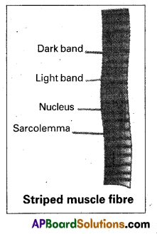
Question 12.
Describe the structure of a skeletal muscle.
Answer:
Skeletal (striped and voluntary) muscle: It is usually attached to skeletal structures by ‘tendons’. In a typical muscle such as the ‘biceps’ muscle/skeletal muscle fibre is surrounded by a thin connective tissue sheath, the endomysium. A bundle of muscle fibres is called a fascicle. It is surrounded by a connective tissue sheath called perimysium. A group of fascicles form a ‘muscle’ which is surrounded by an epimysium (outermost connective tissue sheath). These connective tissue layers may extend beyond the muscle to form a chord-like tendon or sheet-like aponeurosis.
A skeletal muscle fibre is a long, cylindrical and unbranched cell. It is a multinucleated cell with many oval nuclei characteristically in the “peripheral” cytoplasm (a syncytium formed by fusion of cells). Sarcoplasm has many myofibrils which show alternate dark and light bands. So it is called striped or striated muscle.
Question 13.
What are the chief characteristics of the crustaceans?
Answer:
- This includes prawns, crabs, lobsters, crayfishes etc.
- Mostly marine, a few are freshwater and some are adapted to terrestrial life.
- In most species, head and thorax fuse to form cephalon thorax.
- Cephalic appendages are five pairs – first antennae (antennules) second antennae, mandibles first maxillae and second maxillae.
- Thoracic and abdominal appendages are typically biramous.
- Respiration by gills.
- Excretory organs are green glands or antennal glands.
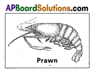
8. Sense organs include statocysts, compound eyes and antennae.
9. Gonopores are paired.
10. Development is direct or indirect involving several larval stages.
Basic larva is nauplius. Ex: Palaemon (Prawn); Cancer (Crab)
![]()
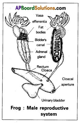
Question 14.
Describe the male reproductive system of frog.
Answer:
Male Reproductive System of Frog: The male reproductive system consists of a pair of yellowish and ovoid testes, which are attached to the kidneys and dorsal body wall by a double fold of peritoneum called mesorchium. Each testis is composed of innumerable seminiferous tubules which are connected to form 10 to 12 narrow tubules, the vasa efferentia.
They enter the kidneys and open into the Bidders canal which is connected to the ureter through transverse canals of the kidney. The urinogenital ducts of both the sides open into the cloaca.
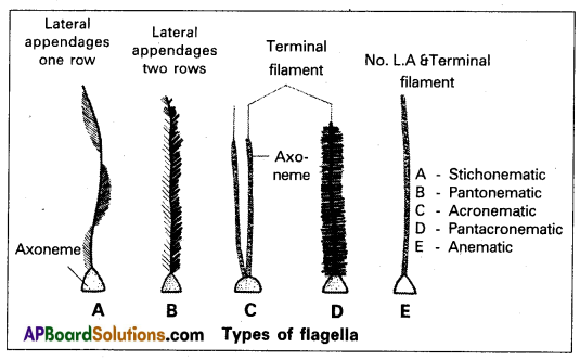
Question 15.
What are lateral appendages? Based on their presence and absence, write the various types of flagella giving at least one example for each type.
Answer:
Lateral appendages: Some flagella bear one or two or many rows of short, lateral hair-like fibrils called lateral appendages. They are of two types namely ‘mastigonemes and ‘flimmers’.
Types of Flagella: Based on the presence or absence and/or the number of rows of lateral appendages, five types of flagella are recognised.
- Stichonematic: This flagellum bears one row of lateral appendages on the axoneme. E.g; Euglena and Astasia.
- Pantonematic: This flagellum has two or more rows of lateral appendages on the axoneme. E.g. Peranema and Monas.
- Acronematic: This type of flagellum does not bear lateral appendages and the terminal part of the axoneme is naked without the outer sheath at its tip. E.g.: Chlamydomonas and Polytoma.
- Pantacronematic: This type of flagellum is provided with two or more rows of lateral appendages and the axoneme ends in a terminal naked filament. E.g. Urceolus.
- Anematic or simple: In this type of flagellum, lateral appendages and terminal filament are absent. Hence, it is called anematic (a-no;nematic-th reads). E.g. Chiomonas and Ciyptomonas.
Question 16.
Why is adolescence considered a vulnerable phase?
Answer:
Adolescence: It is the time period between the beginning of puberty and the beginning of adulthood. In other words, it is the bridge linking childhood and adulthood. The age between 12-18 years is considered adolescence period. It is both ‘a period and process during which a child becomes mature. It is accompanied by several biological and behavioural changes. Thus, adolescence is a very valuable phage of mental and psychological development of an individual.
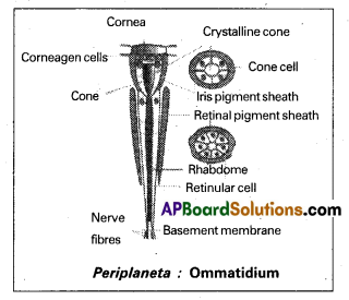
Question 17.
Draw a neat and labelled diagram of ommatidium.
Answer:
Question 18.
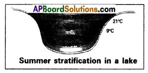
What is summer stratification? Explain.
Answer:
During summer in temperate lake, the density of the surface water decreases because of increase in its temperature (21-25°C). This ‘upper more warm layer’ of a lake is called epilimnion. Below the epilimnion, there is a zone in which the temperature decreases at the rate of 1°C per meter in depth and it is called thermocline or metalimnion. The bottom layer is the hypolimnion, where water is relatively cool, stagnant and with low oxygen content (due to absence of photosynthetic activity).
During autumn (also called fall). The epilimnion cools down and the surface water becomes heavy when the temperature is 4°C and sinks to the bottom of the lake overturns bring about ‘uniform temperature’ in lakes during that period. This circulation during the autumn. The upper oxygen-rich water reaches the hypolimnion and the nutrient-rich bottom water comes to the surface. Thus there is uniform distribution of nutrients and oxygen in the lake.
![]()
Section – C
2 x 8 = 16
Question 19.
Describe the structure and life cycle of Ascaris lumbricoides with the help of a neat and labelled diagram.
Answer:
Ascaris lumbricoids is commonly called the common roundworm. It lives in the small intestine of man, more frequently in children. It is cosmopolitan in distribution. Mode of infection is through contaminated food and water. Infective stage is the embryonated egg with the 2nd stage rhabditiform larva.
Structure: Sexes are separate and the sexual dimorphism is distinct. In both males and females. The body is elongated and cylindrical. Mouth is present at the extreme anterior end and is surrounded by three chitinous lips close to the mouth mid ventrally there is a small aperture called excretory pore.
Male: It has a curved posterior end which is considered the tail The posterior end possesses a cloacal aperture and a pair of equal
sized chitinous pineal spicules or pineal setae which serve to transfer the sperms during copulation.
Female: It has a straight posterior end the tail. The female genital pore or vulva is present mid-ventrally at about one-third the length from mouth. The anus is present a little in front of the tail end.
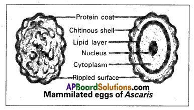
life history: Copulation takes place in the small intestine of man. After copulation, the female releases approximately two lakh eggs per day. Each egg is surrounded by a protein coat with rippled surface. Hence the eggs of Ascaris are described as mammillated eggs.
The protein coat is followed by a chitinous shell and a lipid layer internally. These eggs come out along with faecal matter. In the moist soil, development takes place inside the egg so that the 1-stage rhabditiform larva is produced. It undergoes the 1 moulting and becomes the 2-stage rhabditiform larva which is considered the stage infective to man. They reach the alimentary canal of man through contaminated food and water.
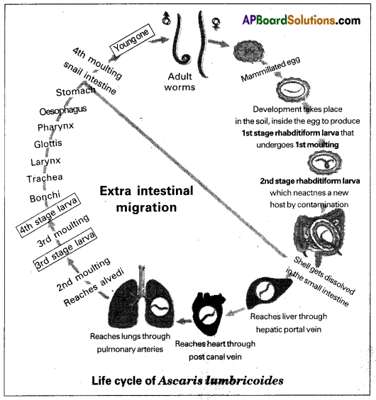
In the small intestine, the shell gets dissolved so that the 2-stage larva is released. Now it undergoes extra intestinal migration. First, it reaches the liver through the hepatic portal vein. From there it reaches the heart through the post caval vein, It goes to the lungs through the pulmonary arteries. In the alveoli of lungs, it undergoes the 2’ moulting to produce the 3-stage larva. It undergoes the 3 moulting so that the 4th stage larva is produced in the alveoli only.

It leaves the alveoli and reaches the small intestine again through bronchi, trachea, and larynx, glottis, pharynx, oesophagus and stomach. In the small intestine, It undergoes the 4th and final moulting to become a young one which attains sexual maturity within 8 to 10 weeks.
Question 20.
Describe the digestive system of cockroach with the help of a neat labelled diagram.
Answer:
The digestive system of cockroaches consists of an alimentary canal and the associated glands. The preoral cavity surrounded by the mouth parts, is present in front of the mouth. The hypopharynx divides it into two chambers called barium (anterior) and salivarium (posterior).
Alimentary canal: The alimentary canal of cockroach is a long tube and is coiled at some places. It extends between the mouth and the anus. It is divided into three regions namely foregut of stomodaeum, midgut or mesenteron and hindgut are internally lined by ectoderm. The mesenteron is lined by the endodermal cells.
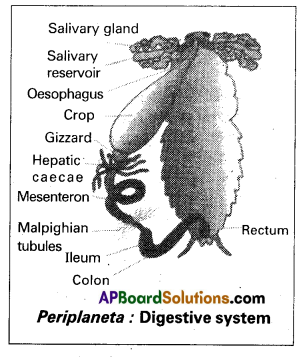
Forgut or stomodaeum. The foregut includes pharyngeal. Oeso phagus, crop and gizzard. it is internally lined by a chitinous cuticle. Mouth opens into the pharynx, which inturn leads into a narrow tubular oesophagus. The oesophagus opens behind into a thin walled distensible sac called crop. The crop serves as a reservoir for storing food. Its outer surface is covered by a network of tracheae. Behind the crop there is a thick-walled muscular proven- triculus or gizzard. The chitinous inner living of the gizzard has six powerful teeth, which form an efficient grinding apparatus. Behind each tooth is a hairy pad, which bears backwardly directed bristles. Among these plates, food is thoroughly ground into fine particles.
These food particles are filtered by the bristles. The gizzard thus acts both as a grinding mill and also as a sieve. There is a membranous projection of the gizzard into the mesenteron in the form of a funnel called stomodeal valve. This valve prevents the entry (regurgitation) of food from the mesenteron back into the gizzard. Midgut (mesenteron or ventriculus). The midgut is a short and narrow tube behind the gizzard. It is also called mesenteron or ventriculus. Between the ventriculus and the gizzard, arising from ventriculus there are six to eighth finger-like diverticula called hepatic cancer.
They are helpful in the digestion and absorption of the digested food materials. The ventriculus ¡s functionally divided into an anterior secretory part and a posterior absorptive part. The secretory part of the ventriculus has many gland cells and it secretes several enzymes. The ‘bolus of food in the mesenteron is enveloped by a chitinous and porous membrane called peritrophic membrane, which is secreted by the funnel-like stomodeal valve of the gizzard. Digested food is absorbed into the food through the peritrophic membrane in the posterior absorptive region of the ventriculus.
The peritrophic membrane protects the wall of the ventriculus from hard food particles in the good. The opening of the ventriculus into the hindgut is controlled by a sphincter muscle, It prevents entry of undigested food and uric from the hindgut into the midgut.
Hindgut or proctodaeum: The hindgut is a long coiled tube, consisting of three regions namely ileum, colon and rectum. It is interanlly lined by chitinous cuticle. The ileum that lies behind the mesenteron is a short tube. Six bundles of fine yellow, blind tubules called Malpighian tubules open into the ileum near the junction of mesenteron and ileum. Malpighian tubules are excretory in function.
Ileum collects uric acid from the malpighian tubules and undigested food from the mesenteron. The ileum opens behind into a long coiled tube called colon. Colon leads into a short and wide rectum which opens out through the anus. Rectum bears on its inner side six longitudinal chitinous folds called rectal papillae. They ae concerned with the reabsorption of water from the undigested food.
![]()
Digestive gland: The digestive glands associated with the alimentary, canal of cockroach are salivary glands, hepatic caecae and ‘ glandular cells of the mesenteron.

Salivary glands: There is a pair of salivary glands attached to the ventrolateral sides of the crop, one on each side. Each salivary gland has two lobes. Each lobe of salivary gland has many lobules called acini. Each acinus is a group of secretory cells called zymogen cells with a small ductule. The ductules of both the lobes of a salivary gland unite to form a common salivary duct on each side.
The two common salivary ducts are joined to form the median salivary duct. Between the two lobes of a salivary gland of each side is a sac called salivary receptacular duct or common reservoir duct. The midious salivary duct opens into the common receptacular duct. Later these two form an efferent salivary duct. The efferent salivary duct opens at the base of the hypopharynx. Acinar cells secrete saliva, which contains starch-digesting enzymes such as amylase.
Hepatic caecae : The hepatic caecae are also termed midguts caecae. They contain secretory and absorptive cells. Glandular cells of the mesenteron: The glandular cells of the mesenteron secrete enzymes such as maltase, invertase, proteases and liphase.
Question 21.
List the major ‘air pollutants and describe their effects on human beings.
Answer:
Air pollutants cause injury to all living organisms. They reduce growth and yield of crops. They are harmful to the respiratory system of humans and animals. Increase in the concentration of pollutants or duration of exposure increase the harmful effects on the organisms.
The major air pollutants:
1. Carbon monoxide (CO): It is produced mainly due to incomplete combustion of fossil fuels. Automobiles are a major cause of CO pollution in larger cities and towns. Automobile exhausts, phones from factories, emissions from power plants, forest fires and even burning of firewood contribute to CO pollution. Haemoglobin has greater affinity for CO and SO CO competitively interferes with oxygen transport. CO symptoms such as headache and blurred vision at lower concentrations. In higher concentrations, it leads to coma and death.
2. Carbon Dioxide (CO2): Carbon dioxide ¡s the main pollutant that is leading to global warming. Plants utilize CO2 for photosynthesis and all living organisms emit carbon dioxide in the process of respiration. With rapid urbanization, automobiles, aeroplanes, power plants and other human activities that involve the burning of fossil fuees such as
gasoline, carbon dioxide is turning out to be an important pollutant of concern.
3. Sulphur Dioxide (SO2): It is mainly produced by burning of fossil fuels. Melting of sulphur ores is another important source for SO2 pollution. Metal smelting and other industrial processes also contribute to SO2 pollution. Sulfur dioxide and nitrogen oxides are the major causes of acid rains, which cause acidification of soils, lakes and streams and also accelerated corrosion of buildings and monuments.
High concentration of sulphur dioxide (SO2) can result ‘in breathing problems in asthmatic children and adults. Other effects associated with long-term exposure to sulphur dioxide, include respiratory illness, ‘alterations in the lungs’ defenses and aggravation of existing cardiovascular problems.
To control SO2 pollution, the emissions are filtered through scrubbers. Scrubbers are devices that are used to clean the impurities in exhaust gases. Gases pollutants such as SO2 are removed by scrubbers.
4. Nitrogen Oxides: Nitrogen oxides are considered to be major primary pollutants. The source is mainly automobile exhaust. The air polluted by nitrogen oxide is not only harmful to humans and animals, but also dangerous for the life of plants. Nitrogen oxide pollution also results in acid rains and formation of photochemical smog. The effect of nitrogen oxides on plants, include the occurrence of necrotic spots on the surface of leaves.
Photosynthesis is affected in crop plants and the yield is reduced. Nitrogen oxides combine with volatile organic compounds by the action of sunlight to form secondary pollutants called Peroxyacetyl nitrate (PAN) which are found especially in photochemical smog. They are powerful irritants to eyes and respiratory tract.
5. Particulate matter/Aerosols: Tiny particles of solid matter suspended in a gas or liquid constitute the (particulate matter) (Aerosols) refer to particles and /or liquid droplets and the gas together (a system of colloidal particles dispersed in a gas) Combustion of “fossil fuels” (petrol, diesel etc) fly ash produced in thermal plants, forest fires, cement factories, asbestos mining and manufacturing units, spinning and ginning mills etc., are the main sources of particulate matter pollution.
According to the Central Pollution Central Board (CPCB) particles of 2.5 micrometres or less in diameter are highly harmful to man and other air-breathing organisms.
An electrostatic precipitator is a widely used ‘filter’ for removing particulate matter from the exhaust of thermal power plants. It can remove 99% particulate matter. It has high-voltage electrodes which produce a ‘corona’ that releases electrons. These are collected by collecting plates which attract the charged particles. The air flowing between the plates is kept in low velocity so as to allow the dust particles to fall. Thus clean air is released into the atmosphere.