Students get through AP Inter 1st Year Zoology Important Questions 2nd Lesson Structural Organisation in Animals which are most likely to be asked in the exam.
AP Inter 1st Year Zoology Important Questions 2nd Lesson Structural Organisation in Animals
Question 1.
The body of sponges does not possess tissue level of organization, though it is made up of thousands of cells. Comment on it.
Answer:
- The body of sponges is madeup of thousands of cells.
- But there is no co-ordination between cell layers because nerve and sensory cells are absent.
- Also they are arranged as loose cell aggregates and they donot form tissues.
- So, sponges belong to cellular level of organisation, but not tissue level.
Question 2.
What is ’tissue’ level of organisation among animals? Which metazoans do exhibit this organisation?
Answer:
- In animals, the cells which perform the same function are arranged into tissues.
- There is cellular co-ordination and it is due to the presence of nerve and sensory cells.
- The metazoans which exhibit tissue level of organisation is Cnidaria.
![]()
Question 3.
Animals exhibiting which level of organisation lead relatively more efficient way of life when compared to those of the other levels of organisation? Why?
Answer:
- Animals exhibiting ‘organ level of organisation’ lead relatively more efficient way of life.
- Reason: In this level, highly specialised sensory and nerve cells are present.
Question 4.
What is monaxial heteropolar symmetry? Name the group of animals in which it is the principal symmetry. [AP M-19]
Answer:
- The symmetry, by which any plane passing through the central axis of the body divides it into two identical parts, is called Monaxial heteropolar symmetry (or) radial symmetry.
- Ex: Diploblastic animals like Cnidaria.
Question 5.
Radial symmetry is an advantage to the sessile or slow moving organisms. Justify this statement. [AP M-19]
Answer:
- Animals having radial symmetry live in water and they receive stimuli from any direction.
- Accordingly, they can respond equally to any stimuli that come from all directions.
Question 6.
What is cephalization? How is it useful to its possessors? [TS M-15,20][AP May-17]
Answer:
- Cephalization is the formation of nerve and sensory cells at the anterior part of the body.
- Animals with cephalization can sense the new environment and move efficient than the other animals in seaking food, locating mates and in avoiding or escaping from predators.
Question 7.
Mention the animals that exhibited a ‘tube -within-a-tubef organisation for the first time? Name their body cavity. [AP M-15,18,20]
Answer:
- ‘Tube-within-a-tube’ organisation is first time formed in Nematoda.
- The body cavity in Nematodes is Pseudocoelom.
Question 8.
Why is the true coelom considered a secondary body cavity? [AP M-16] [TS M-15]
Answer:
- In eucoelomate animals, blastocoel is the primary cavity during development. [IPE-14]
- Later the blastocoel(primary body cavity) is replaced by true coelomfsecondary body cavity) derived from mesoderm.
- Hence the body cavity of eucoelomates is considered as the secondary body cavity.
![]()
Question 9.
What are retroperitoneal organs? [TS M-16,19] [AP 18]
Answer:
- The organs like kidneys in vertebrates are covered by the parietal peritoneum only on the ventral side.
- Such a peritoneum is called retroperitoneum and the organs lined by it are called retroperitoneal organs.
Question 10.
If the mesentoblast cell is removed in the early embryonic development of protostomes, what would be the fate of such animals?
Answer:
If Mesentoblast is removed then
- Mesoderm between ectoderm and endoderm does not form.
- Mesodermal organs donot develop.
- True body cavity does not appear.
- Hence, the animal cannot survive.
Question 11.
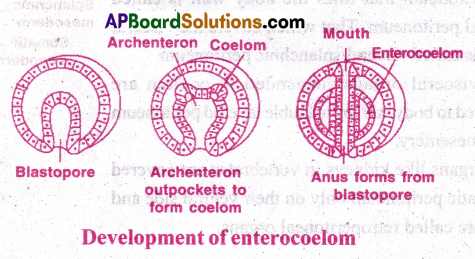
What is Enterocoelom? Name the enterocoelomate phyla in the animal kingdom? [TS M-15]
Answer:
- Enterocoelom is a type of true coelom that arises by mesodermal out pouching of the archenter on.
- Enterocoelomate phyla in kingdom Animalia are Echinodermates, Hemichordates, Chordates.
Question 12.
Stratified epithelial cells have limited role in secretion. Justify their role in our skin.
Answer:
- The main function of stratified epithelial cells is to provide protection against chemicals and mechanical stress.
- Thus, the stratified epithelial cells have limited role in secretion.
Question 13.
Distinguish between exocrine and endocrine glands with examples. [TS M-15]
Answer:
| Exocrine glands | Endocrine glands |
| 1) Exocrine glands are glands with ducts. 2) They secrete saliva, mucus, ear wax, oil, milk and digestive juices. 3) Ex: Salivary glands, Mucus glands |
1) Endocrine glands are ductless glands. 2) They secrete hormones which are directly released into blood. 3) Ex: Pituitary gland, Thyroid gland |
Question 14.
Distinguish between holocrine and apocrine glands.
Answer:
Holocrine
- Holocrine glands are exocrine glands.
- The entire gland cell disintegrates to discharge the contents.
- Ex: Sebaceous glands (oil glands of skin)
Apocrine
- Apocrine glands are exocrine glands.
- Only the terminal (apical) part of the cell is pinched off and released.
- Ex: Mammary glands.
Question 15.
Mention aný two substances secreted by mast cells and their functions. [AP, TS M-16]
Answer:
Mast cells secrete heparin, serotonin, histamine and bradykinin. [TS M-20]
- Heparin is an anticoagulant.
- Serotonln is vasoconstrictor
- Histamine and Bradykinin are vasodilators.
![]()
Question 16.
Distinguish between a tendon and a ligament. [TS M-22] [AP May-17] [AP M-15,17,19]
Answer:
- Tendons connect skeletal muscles to the bone. They contain ¿nly collagen fibres.
- Ligaments connect one bone with another bone. They contain collagen fibres along with few elastic fibres.
Question 17.
Distinguish between brown fat and white fat. [AP M-19]
Answer:
Brown Fat
- Brown fat is found in foetus and infants.
- Brown fat is metabolically active.
- Adipocyte has several lipid droplets.
White fat
- White fat is found in adults.
- White fat is metabolically inactive.
- Adipocyte has a single large lipid droplet.
Question 18.
What is the strongest cartilage? In which regions of the human body, do you find it? [AP M-16, 20] [TS M-15]
Answer:
- ‘Fibrous cartilage’ is the strongest cartilage.
- It is present in intervertebral discs and pubic symphysis of pelvis.
Question 19.
Distinguish between osteoblasts, and osteoclasts?
Answer:
Osteoblasts
- Osteoblasts are immature osteocytes.
- They produce collagen fibres and minerals for the bone.
Osteoclasts
- Osteoclasts are phagocytic cells of the bone.
- They remove minerals from the bone.
Question 20.
Define osteon. [TSM-20] [AP M-15]
Answer:
Osteon: In a dens& bone, a Haversian canal and the surrounding lamellae nd lacunae are collectively called Osteon or Haversian system. It works as transport system.
Question 21.
What are Volkmann’s canals? What is their role?
Answer:
- Volkmann’s canals are oblique or transverse canals which connect Haversian canals with marrow cavity.
- It helps in transport of materials.
Question 22.
What is a Sesamoid bone? Give an example. [TS M-22] [AP M-22]
Answer:
- Sesamoid bone is a tendon bone. It is formed by ossification in tendon.
- Ex: Patella (knee cap).
![]()
Question 23.
What is lymph? How does it differ from plasma?
Answer:
- Lymph is the interstitial colourless fluid which passes through the lymphatic vessels.
- Lymph differs from plasma because it lacks RBC, Platelets and large plasma proteins.
Question 24.
What is the haematocrit value? [AP M-19,22] [AP Mar, May-17]
Answer:
- The percentage of volume of RBC in total volume of blood is Haematocrit value.
- It is also called packed cell volume,
Question 25.
What are intercalated discs? What is their significance?
Answer:
- Intercalated discs: The dark lines across the cardiac muscle are called intercalated discs.
- Significance: They have gap junctions which help in quick transport of electrical impulses necessary for the heart beat.
Question 26.
“Cardiac muscle is highly resistant to fatigue”. Justify. [APM-20][TS May-17]
Answer:
Cardiac muscle is highly resistant to fatigue because it has numerous sarcosomes, many molecules of myoglobin and rich supply of blood for continuous aerobic respiration.
Question 27.
Distinguish between ’nucleus’ and ’ganglion’ with respect to the nervous system.
Answer:
- Nucleus: A group of cell bodies in the central nervous system is called nucleus.
- Ganglion: A group of cell bodies in the peripheral nervous system is called ganglion.
Question 28.
Distinguish between ’tracts’ and ’nerves’ with respect to the nervous system.
Answer:
- Tract: Bundles of axons in central nervous system is called tract.
- Nerve: Bundle of axons in peripheral nervous system is nerve.
Question 29.
Name the glial cells that form myelin sheath around the axons of central nervous system and peripheral nervous system respectively.
Answer:
- In Central Nervous system, Oligodendrocytes (glial cells) form the myelin cover around axons. A single oligodendrocyte can myelinate many axons of CNS.
- In Peripheral Nervous system, Satellite cells cover the cell bodies. Schwann cells form neurilemma around axon.
Question 30.
Distinguish between white matter and grey matter of’CNS’. Answer: In central nervous system the nerve matter appears in two colours.
White matter
- White colour is due to myelin.
- White matter consists of myelinated nerve fibres.
Grey matter
- Grey colour is due to nissl bodies.
- Grey matter consists of non-myelinated nerve fibres, dendrites and cytons.
![]()
Question 31.
What are microglia and what is their origin and add a note on their function.
Answer:
- Microglia are neuroglia cells. [AP, TSM-19]
- They are non conducting cells of nervous system.
- They are phagocytic and combat infection or injury of the nervous system.
- They are mesodermal in origin.
Question 32.
What are pseudounipolar neurons? Where do you find them?
Answer:
- Pseudo unipolar neurons are normal unipolar neurons.
- They are sensory neurons.
- The cell body (soma) is present in dorsal root ganglion of spinal nerve.
- A single process arises from body and divides into two. One is axon and the other is dendrite.
Short Answer Questions
Question 1.
Describe the four different levels of organization in metazoans?
Answer:
Metazoa are multicellular animals. They exhibit four levels of body organisation.
1) Cellular level of organisation:
- It is the lowest level of organisation.
- There is no co-ordination among cells.
- Nerve and sensory cells are absent.
- Cells look like ‘loose cell aggregates’.
- Outer layer is formed by pinacocytes and inner layer is formed by choanocytes. Ex: Sponges.
2) Tissue level of organisation:
- The body has two coordinating layers of cells (diploblastic).
- The outer layer is epidermis and inner layer is gastrodermis (tissues).
- In between the two layers there is jelly like mesoglea.
- Nerve cells and sensory cells are present. Ex: Cnidaria.
- These animals have radial symmetry.
3) Organ level of organisation:
- These are triploblastic animals having ectoderm, endoderm and mesoderm.
- Tissues having similar function aggregated cells to form organs.
Ex: Platyhelminthes (first time appeared)
4) Organ-system level of organisation:
- It is the highest level of organisation.
- The animals are triploblastic.
- Organ systems are formed for different functions.
- There is increasing complexity in the evolutionary development.
Ex: Flat worms, nematodes, annelids etc.,
Question 2.
In which group of bllaterians do you find solid bauplan? Why is it called so?
Answer:
- Solid bauplan is found in Platyhelminthes(flatworms).
- They are bilaterally symmetrical and acoelomate animals.
- Mesoderm is developed. It occupies the entire perivisceral space.
- There is no body cavity in the adult. So the animals are solid without any body cavity. Hence they are called acoelomate animals.
- The name solid bauplan is given due to the following reasons:
i) Their internal organs cannot move freely (Just like a solid structure).
ii) Diffusion of digested material is slow and less efficient.
![]()
Question 3.
Mention the advantages of coelom over pseudocoelom.
Answer:
Advantages of coelom over pseudocoelom:
- Coelom is perivisceral cavity developed from mesoderm.
- Pseudocoel is extension of blastocoel.
- Visceral organs of eucoelomates are muscular and move independently. Ex: Peristalsis.
- Regional specialization of alimentary canal in stomach, gizzard, oesophagus took place in eucoelomates due to mesodermal contact with endoderm. It is called primary induction.
- Regional specialization is not possible because endoderm doesnot come into contact with mesoderm in pseudocoelomates.
- Gametes and excretory products are released into coelom in some coelomates.
Question 4.
Describe the formation of schizocoelom and enterocoelom.
Answer:
Coelom formation differs in different groups of animals.
Two important types are schizocoelom and enterocoelom.
1) Schizocoe formation:
- Mesoderm is formed from 4d blastomere or mesentoblast.
- Mesentoblast divides in to blocks between ectoderm and endoderm.
- A split appears in each block and one block fuses with ectoderm and the other with endoderm.
- The blocks near the ectoderm form the somatic peritoneum. The blocks near the endoderm form visceral peritoneum.
- The space between somatic and visceral peritoneum is the coelom. As it is formed by splitting up of mesoderm it is called schizocoel. Ex: Annelida, Arthropoda & Mollusca .
- In Arthropoda and mollusca the true coelom is reduced and fused with blastocoel. It gets filled with blood and called haemocoel.
2) Enterocoel formation:
- Enterocoel is formed in Echinodermata, hemichordate and chordate.
- In these animals the mesoderm is located in archenteron wall as two patches.
- The mesoderm of archenteron out pouches to form coelom and occupies the entire perivisceral space.
- As the coelom is formed from archenteron it is called enterocoel.
Question 5.
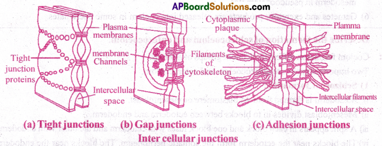
Describe briefly about the three types of intercellular junctions of epithelial tissues.
Answer:
Epithelium forms outer covering and inner lining of various organs and skin.
The epithelial cells are held together by three types junctions. They are tight junctions, desmosomes and gap junctions.
1) Tight Junction:
- The plasma membranes of adjacent cells are tightly pressed and bound by specific proteins.
- They prevent leakage of water into the surrounding cells and makes the skin water resistant.
Ex: In sweat gland the water passes only to outside but not into the adjacent cells.
2) Desmosomes:
- Epithelial and muscle cells have desmosomes.
- Desmosomes act as rivets binding the cells to form strong sheets.
- There are intermediate filaments made of keratin to support desmosomes in the cytoplasm.
3) Gap Junctions:
- They are present in cardiac tissue and also in some other tissues.
- They provide continuous cytoplasmic channels for transport of materials from one cell to another.
- In cardiac muscle, they allow rapid conduction of electrical impulses.
Question 6.
Give an account of glandular epithelium.
Answer:
- Glandular Epithelium (G.E) is a tissue responsible for formation of glands.
- Location: G.E forms the covering of all major glands.
- Based on the combination of cells G.E are two types (i) Unicellular (ii) Multicellular
- Unicellular glands are isolated glandular cells. Ex: Goblet cells of gut.
- Multicellular glands consist of clusters of cells. Ex: Salivary glands.
- Function: Secretion is the main function of G.E.
- Based on the secretions G.E are two types (i) Exocrine glands (ii) Endocrine glands.
- Exocrine glands: They are glands with ducts. Ex: Salivary glands, Mammary glands.
- Endocrine glands: They are ductless glands. Ex: Pituitary, Thyroid
![]()
Question 7.
Give a brief account of the cells of areolar tissue.
Answer:
Areolar tissue:
- Areolar tissue is a loose connective tissue.
- It is present in many parts of the body as packing tissue.
- It has fibres, cells and empty spaces called ‘areolae’.
- The cells of this tissue are fibroblasts, mast cells, macrophages, adipocytes and plasma cells.
Fibroblasts: They produce fibres. The inactive cells are called fibrocytes.
i) Fibres of Areolar tissue: These are 3 types.
- Collagen fibres: These are white fibres made of protein collagen.
- Reticular fibres: These are thin, branched network fibres made of protein collagen.
- Elastin fibres: These are yellow, branched fibres made of protein elastin.
ii) Mast cells: Secrete Heparin (anti coagulant) histamine, bradykinin (vasodilators) serotonin
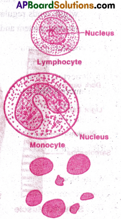
iii) Macrophages1 Phagocytic amoeboid cells which act as scavengers. The macrophage connected to the tissue are called histiocytes and others are wandering macrophages. Macrophages are formed from monocytes.
iv) Plasma cells are formed from B-lymphocytes. They produce antibodies.
v) Adipocytes are fat storing cells.
Question 8.
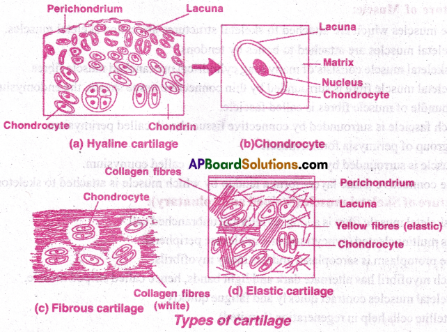
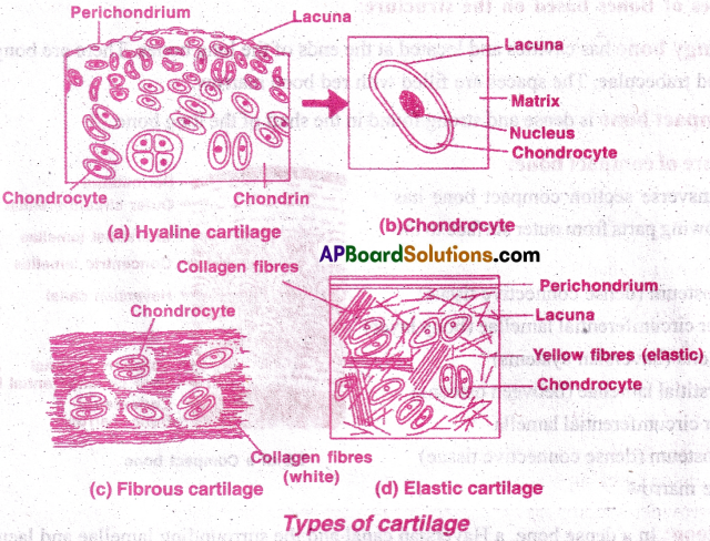
Describe the three types of cartilage. [ AP M-20] [TS M- 18,20]
Answer:
Cartilage is solid flexible connective tissue.
- It has collagen fibres, elastic fibres, chondroblasts enclosed in lacunae and surrounded by perichondrium.
- Cartilage has no blood supply.
- Growth and regeneration of cartilage takes place by the activity of perichondrial cells.
- Perichondrium has blood capillaries.
Types of Cartilage: There are three types of cartilage.
1) Hyaline cartilage:
- Bluish white, translucent cartilage.
- It has delicate collagen fibres.
- It is the weakest and most common cartilage.
Ex: Walls of nose, costal cartilage, trachea, bronchus and larynx. - It forms the embryosac endoskeleton of the vertebrates, endoskeleton of cyclostomes and cartilaginous fishes.
2) Elastic cartilage:
- It is yellow due to elastic, fibres in addition to collagen fibres.
- It provides strength and elasticity. Ex: Pinna, Eustachian tube and epiglottis.
3) Fibrous cartilage:
- Matrix has bundles of collagen, if It is strongest cartilage.
- Perichondrium is absent. Ex: Intervertebral discs and pubic symphysis.
![]()
Question 9.
Explain Haversian system. [AP, TS May-17] [APM-16] [IPE-14]
Answer:
Haversian system (Osteon) is a unit of compact bone. It consists of the following.
- Haversian canal: This canal runs parallel to marrow cavity of bone. It contains an artery, a vein and a lymph vessel.
- Concentric Lamellae: There are concentric rings of bone lamellae around the haversian canal.
- Lacunae: Small fluid filled spaces called lacunae are present in between lamellae. These spaces enclose osteocytes (inactive)
- Canaliculi: They are minute canals connecting various lacunae and haversian canal. The protoplasmic processes of osteocytes extend through canaliculi.
- Volkman’s canals: They connect haversian canal to marrow cavity.
Question 10.
Write short notes on lymph. [TS M-22]
Answer:
- Lymph is a colourless fluid.
- The interstitial fluid that passes through lymphatic system is called lymph.
- Lymph contains plasma and lymphocytes.
- It contains less amount of nutrients and oxygen but has more C02 and other metabolites.
- Lymph is formed in interstitial space. When blood passes through capillaries then nutrients, oxygen and other materials diffuse into interstitial space, due to hydrostatic pressure:
- Most of the fluid in interstitial space enters blood. But a small portion reach the lymph vessels and becomes lymph.
- Finally, lymph reaches the blood through subclavian veins, near heart.
- Lymphatic system provides an ‘accessory route’ by which interstitial fluid flows back to blood.
Question 11.
Describe the structure of a Skeletal Muscle. [APM-19] [TS M-15]
Answer:
Structure of Muscle:
- The muscles which are attached to skeletal structures are called skeletal muscles.
- Skeletal muscles are attached to bones by tendons.
- A skeletal muscle consists of many long cylindrical unbranched muscle fibres.
- Skeletal muscle fibre is surrounded by thin connective tissue sheath, the endomysium.
- A bundle of muscle fibres is called fascicle.
- Each fascicle is surrounded by connective tissue sheath called perimysium.
- A group of perimysia form a muscle.
- Muscle is surrounded by connective tissue sheath called epimysium.
- The connective tissue layers form a tendon by which muscle is attached to skeleton.
Structure of Skeletal muscle (Striped and voluntary):
- A skeletal muscle fibre is a long, cylindrical unbranched cell.
- It is multinucleated (syncytium) and nuclei are peripheral.
- The protoplasm is sarcoplasm and has many myofibrils.
- Each myofibril has alternate dark and light bands, hence called striped muscle.
- Skeletal muscles contract quickly and fatigue quickly.
- Satellite cells help in regeneration (Limited).
Question 12.
Describe the structure of a cardiac muscle.
Answer:
Structural of cardiac muscle (Striped and involuntary):
- Cardiac muscle is the heart muscle(myocardium)
- The myocardial cells are short, cylindrical, mononucleate and striated.
- The muscle fibres are branched.
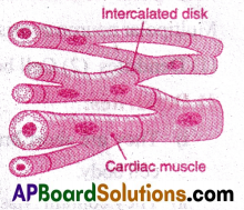
- The muscle cells are connected by gap junctions for quick conduction of electrical impulses.
- There are dark lines called intercalated discs which are characteristic of heart muscle.
- These discs help in rapid conduction of electrical impulses, resulting in heart beat.
- Heart beat (contraction of muscle) is involuntary and carried on by pace maker.
- The rate of heart beat is under the control of autonomous nervous system and hormones like epinephrine and adrenaline.
- The cardiac muscle is highly resistant to fatigue.
- The cardiac muscle is a functional syncytium.
![]()
Question 13.
Given an account of the supporting cells of nervous tissue.
Answer:
- Supporting cells of nerve tissue are called neuroglial cells.
- They are non-conducting cells.
- They undergo division through out life.
- Neuroglial cells of CNS are as follows:
i) Oligo dendrites form myelin sheath.
ii) Astrocytes form network that bind neurons and capillaries, thereby providing blood brain barrier.
iii) Ciliated ependymal cells line the cavities (ventricles) of brain and help the movement of cerebro spinal fluid.
iv) Microglia cells are phagocytic and mesodermal in origin (attend to infectious and injury). - Neuroglia cells of peripheral nervous system are as follows:
i) Satellite cells surrounding the cell bodies in ganglia.
ii) Schwann cells forming neurilemma around axons.
Question 14.
Describe the structure of a multipolar neuron.
Answer:
Multipolar neurons are the common neurons of the body. Multipolar neuron consists of (1) Dendrites (2) Cell body (3) Axon.
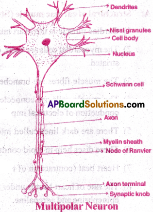
1) Dendrites:
- They are short branched processes which arise from cell body.
- They contain Nissl granules and neurofibrils.
- They conduct nerve impulse towards the cell body (af¬ferent)
2) Cell body:
- It is also called cyton or soma or perikaryon.
- ii) It includes nissl granules (RER) for protein synthesis.
- iii) It contains larger spherical nucleus, Neurofibrils & Lipofuscin granules.
3) Axon:
- It is a long cylindrical thread like structure which arises from cell body.
- The junction between cell body and axon is called Axon hillock
- The plasma lemma is called axolemma.
- The cytoplasm is called axoplasm.
- Microfibrils are present. Nissl bodies are absent.
- The terminal part of axon is branched into small filaments called telodendria.
- Telodendria have knob like endings called synaptic knobs or terminal boutons.
- Synoptic knobs contain neuro transmitters (acetyl choline).
- Axon carries nerve impulse from the cell body to the next neuron (efferent).
Question 15.
Write short notes on (a) Platelets and (b) Synapse.
Answer:
(a) Platelets:
- Platelets are also called thrombocytes.
- They are a kind of formed elements.
- Blood platelets are non nucleated, round or oval biconvex discs.
- Their number range from 2,50,000 to 5,00,000 per cubic mm.
- Platelets are fragments of mega karyocytes.
- Their average life span is 5 to 9 days.
- They secrete thromboplastin which is important for blood clotting.
- They clump at the damaged part of the blood vessel and form a plug preventing blood loss.
(b) Synapse:
- Synapse is a junction between two adjacent neurons. It plays an important role in nerve transmission.
- It consists of synaptic knob of preceding neuron, a gap or functional bridge and dendrites of succeeding neuron.
- During transmission of nerve impulse the neurotransmitters of synaptic knob is released into gap. It functions as temporary bridge for transmission.
- After transmission the neuro transmitter breaks and recombines in synaptic knob.
![]()
Very Short Answer Questions
Question 1.
What is coelom? Explain the different types of coelom with suitable examples and neat labelled diagrams?
Answer:
Coelom is the fluid filled space between visceral organs and body wall and lined by mesoderm. Coelom is found in triploblastic animals except flat worms (platy helminthes).
I) Types of Coelom based on arrangement of mesoderm:
- Acoelom
- Pseudocoelom
- Eucoelom
1) Acoelom:
- The condition of the body in which the body cavity is absent is called Acoelom. The animals in which
the body cavity is absent are called acoelomates.
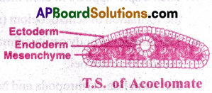
- In these animals, the entire blastocoel is present between the ectoderm and endoderm. It is occupied by mesodermal originated tissue called mesenchyme. Hence they exhibit solid body plane. Ex: Flatworms
2) Pseudocoelom:
- The body cavity which is not lined by mesodermal epithelia is called pseudocoelom. The animals with pseudocoelom are called pseudocoelomates.
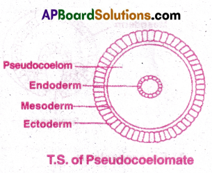
- During their embryonic development, mesoderm occupies only a part of blastocoel adjoining the ectoderm. The unoccupied portion of blastocoel persists as pseudocoelom, which is filled with pseudocoelomic fluid.
- These are the first animals to exhibit a ‘tube within-a- tube’ organisation. Ex: Aschelminthes.
3) Eucoelom:
- The body cavity which is lined by mesodermal peritonea is called eucoelom. The animlas lined by mesodermal epithelia are called Eucoelomates.
- The peritoneum that lines the body wall is called pariental peritoneum. That which covers the visceral organs is called visceral/splanchnic peritoneum.
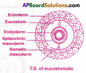
- Some visceral organs suspended in coelom are connected to body wall by a double layered peritoneum called mesentery.
- Some organs like kidneys in vertebrates are covered by somatic peritoneum only on their ventral side and hence are called retroperitoneal organs.
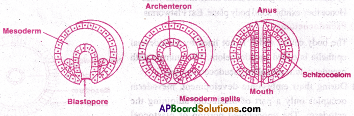
II) Types of Coelom based on formation of Coelom:
- Schizocoelom
- Enterocoelom
1) Schizocoelom:
- The body cavity which is formed by ‘splitting of mesoderm’ is called schizocoelom.
The animals possessing it are called schizocoelomates. - All schizocoelomates are protostomes and they show holoblastic, spiral and determinate cleavage.
- The 4d blastomere or mesentoblast cell of early embryo divides into mesodermal blocks between the ectoderm and endoderm.
- Then a split appears in each mesodermal block and enlarges. All these blocks fuse leading to the formation of schizocoelom (split coelom) replacing the blastocoel.
- The functional body cavity of molluscs and arthropods is filled with blood (haemolymph) and is. called haemocoel.
Ex: Annelida, Arthropoda and Mollusca
![]()
2) Enterocoelom:
- The body cavity which is formed from the evaginated mesodermal pouches of archenteron are enterocoelom and the animal possessing it are called enterocoelomates.
- All enterocoelomates are deuterostomes and they show radial and indeterminate cleavage.
- In these animals, mesodermal pouches that evaginate from the wall of archenteron into the blastocoel are fused with one another to form the enterocoelom.
Ex: Echinoderms, Hemichordates and chordates.
Question 2.
What is symmetry? Describe the different types of symmetry in the animal kingdom with suitable examples.
Answer:
The regular arrangement of body parts in a geometrical design relative to the axis of the body is called symmetry.
Paired body parts with symmetry are arranged equidistant on either side of the principal axis.
Types of Symmetry:
- Radial symmetry
- Bilateral symmetry
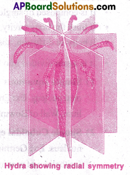
1) Radial Symmetry:
- It is also called monaxial heteropolar symmetry.
- The shape of the body is cylindrical or umbrella or star shaped.
- The body can be cut into two equal parts through any plane that passes through oro-aboral axis.
- Such animals can receive stimuli from all directions.
Ex: Cnidaria, ctenophores (Biradial) - These animal are either sedentary, sluggish or planktonic. Very few exhibit swimming.
- Symmetry in Echinodermata is penta radial.
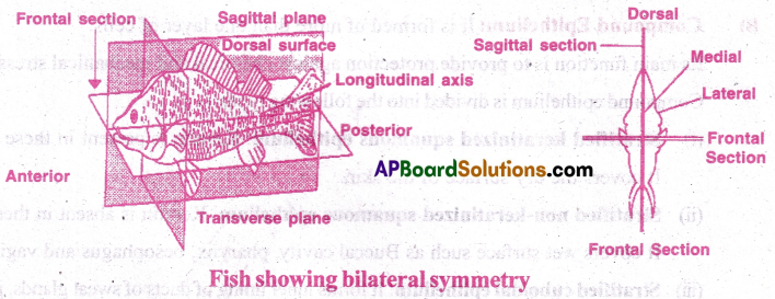
2) Bilateral Symmetry:
- Highly advanced and active animals have bilateral symmetry.
- It is the principal symmetry of triploblastic animals.
- The body can be cut into two identical parts (antimeres) in only one plane (median sagittal plane) that passes through the central axis (anterior – posterior axis).
- Bilaterally symmetrical animals are more active because of cephalization.
- Symmetry in gastropods: The larva of gastropoda is bilaterally symmetrical.
- Due to torsion in metamorphosis the symmetry is lost and adult becomes asymmetric.
Asymmetry: Primitive animals do not have definite shape. They cannot be cut into two equal halves. Ex: Most of the sponges.
![]()
Question 3.
Classify and describe the epithelial tissues on the basis of structural modification of cells with examples.
Answer:
Types of Epithelia: |Based on number of layers and shapes of cells
A) Simple epithelium
B) Compound epithelium
C) Glandular epithelium (Based on secretion of cells)
A) Simple Epithelium:
- It is composed of single layer of cells.
- It lines the body cavity, ducts and vessels.
- It helps in diffusion, absorption filtration and secretion of substances.
- It is again divided into three types based on the shape of the cells.
1) Simple squamous epithelium: It has single layer of flat tile like cells with a central oval nucleus. Ex: Endothelium of blood vessels, Peritoneum, pleura, pericardium- Mesothe- lium, Wall of Bowman’s capsule, lining of alveoli of lungs.
2) Simple Cuboidal epithelium: It is formed of single layer of cuboid cells with a central nucleus. Ex: Germinal epithelium, epithelium of proximal and distal convoluted tubules, Epithelium of proximal convoluted tubule has microvilli.
3) Simple Columnar epithelium: It is composed of single layer of tall cells with nucleus at the base. Mucus secreting goblets cells may be present. It is of two types.
i) Ciliated Columnar epithelium:
Columnar cells have cilia at their free surface.
Ex: Fallopian tubules, ventricles of brain, central canal of spinal cord, bronchioles etc.
ii) Non-ciliated columnar epithelium: Columnar cells do not bear cilia.
Ex: Lining of stomach and intestine
B) Compound Epithelium: It is formed of more than one layer of cells.
Its main function is to provide protection against chemical and mechanical stress.
Compound epithelium is divided into the following types:
- Stratified keratinized squamous epithelium: Keratin is present in these cells.
It covers the dry surface of the skin. - Stratified non-keratinized squamous epithelium: Keratin is absent in these cells. It covers wet surface such as Buccal cavity, pharynx, oesophagus and vagina.
- Stratified cuboidal epithelium: It forms inner lining of ducts of sweat glands, pancreas.
- Transitional Epithelium: It forms the wall of the urinary bladder.
C) Glandular epithelium:
- Glandular Epithelium (G.E) is a tissue responsible for formation of glands.
- Location: G.E forms the covering of all major glands.
- Based on the combination of cells G.E are two types (i) Unicellular (ii) Multicellular
- Unicellular glands are isolated glandular cells. Ex: Goblet cells of gut.
- Multicellular glands consist of clusters of cells. Ex: Salivary glands.
- Function: Secretion is the main function of G.E.
- Based on the secretions G.E are two types (i) Exocrine glands (ii) Endocrine glands.
- Exocrine glands: They are glands with ducts. Ex: Salivary glands, Mammary glands.
- Endocrine glands: They are ductless glands. Ex: Pituitary, Thyroid
Question 4.
Describe the various types of connective tissue proper with suitable examples.
Answer:
- Connective tissue binds and supports various tissues and organs inside the body.
- It is made up of cells, different kinds of fibres, water, polysaccharides and proteins.
Types of Connective tissue proper; A) Loose connective tissue B) Dense connective tissue.
A) Loose connective tissue: The cells and fibres are loosely arranged. The ground substance
is semi fluid. It is again divided into 3 types.
- Areolar
- Adipose
- Reticular tissue.
1) Areolar tissue: It is the binding or packing tissue of the body. It is widely distributed inside the body. It forms the subcutaneous tissue. It has five different types of cells.
- Fibroblasts: These cells produce collagen and elastin fibres. When they become inactive they are called fibrocytes.
- Mastcells: These are Secretary cells which produce heparin (anticoagulant),Histamine, Bradykinin (Vasodilators) and Serotonin (Vasconstrictor).
- Macrophages: These are phagocytes and are internal scavengers.They are formed from monocytes. When they are fixed to tissues they are called histiocytes and other free cells are called ‘wandering cells’.
- Plasma cells: These ceils produce antibodies which are formed from B-lymphocytes.
- Adipocytes: These are fat storing cells.
Fibres of Areolar tissue: These are 3 types.
- Collagen fibres: These are white fibres made of protein collagen.
- Reticular fibres: These are thin, branched network fibres made of protein collagen.
- Elastin fibres: These are yellow, branched fibres made of protein elastin.
2) Adipose tissue:The cells are adipocytes. It is fat storing tissue. In aquatic animals it forms blubber and provides thermal insulation (whales, sea cows).
In camels, it is present in hump. In palms and soles, it acts as shock absorber.
- White Adipose tissue is adult tissue. It is inactive. The adipocyte has a single large fat droplet (monolocular).
- Brown adipose tissue is found in embryos and infants. It is active and produce heat necessary for the survival of just born baby. The adipocytes has many droplets (multilocular). This tissue has large number of capillaries and hence brown in colour.
3) Reticular tissue:The cells are reticular cells (fibro blasts). They form network and support lymphoid organs (bone marrow, spleen and lymph nodes) and basement membrane.
![]()
B) Dense connective tissue: It has few cells and more fibres. Ground substance is very little. Based on arrangement of fibres it is divided into two types.
i) Dense regular connective tissue: The collagen fibres are parallel and in bundles.
Ex: Tendons connect muscle to bone. Ligaments connect bones to other bones.
ii) Dense Irregular connective tissue: The collagen fibres are irregularly arranged. Ex: Periosteum, endosteum, pericardium, heart valves, joint capsules and deeper dermis.
- Elastic connective tissue: It is made up of yellow elastic fibres. The tissue can recoil to the original shape. Ex: Walls of arteries, vocal cords, trachea, bronchi & ligaments of vertebrae.
- Also, there is mucous connective tissue in embryos. It is present in umbilical cord as Wharton’s jelly.
Question 5.
What is a skeletal tissue? Describe the various types of skeletal tissue.
Answer:
Skeletal tissue:
- Skeleton gives support and protection to the body.
- It provides attachment surface for the muscle so that movement is possible.
Types of Skeletal tissue: I) Cartilage II) Bone.
I) Cartilage:
- It is solid flexible connective tissue.
- It has collagen fibres, elastin fibres, chondroblasts.
- The chondroblasts are present inside the fluid filled spaces called lacunae.
- The chondroblasts later become inactive chondrocytes.
- The cartilage has no blood vessels.
- The cartilage is surrounded by a fibrous connective tissue sheath called perichondrium.
- Blood capillaries are present in perichondrium.
- Growth, repair and regeneration of cartilage takes place by perichondrial cells.
Types of cartilage based on the matrix:
i) Hyaline cartilage:
- It is bluish white, translucent cartilage.
- Matrix has delicate collagen fibres.
- It forms the embryonic skeleton of bony vertebrates.
- It forms the adult endoskeleton of cyclostomes and elasmobranchs (sharks, rays & skates)
- It is present at the tips (place of joint) of long bones and called articular cartilage.
- Sternal parts of ribs are cartilaginous.
- It is also present in nasal septum, tracheal rings, larynx and bronchi.
- It is the most common and weakest cartilage.
ii) Elastic cartilage:
- It is yellow due to yellow elastin fibres.
- Collagen fibres are also present. Ex: Pinnae, Eustachian tubes and epiglottis
iii) Fibrous cartilage:
- There are more bundles of collagen fibres.
- Perichondrium is absent. Ex: Intervertebral discs and pubic symphysis.
II) Bone:
- Bone is highly calcified, solid, hard and rigid connective tissue.
- It forms the major part of vertebrate endoskeleton.
- It provides support and shape to the body.
- Bone is the homeostatic reservoir of calcium, magnesium and phosphorus.
- Bone is highly vascular.
- The outer fibrous connective tissue of a bone is periosteum.
- The inner connective tissue around marrow cavity is endosteum.
- Between periosteum and endosteum there is hard matrix and living cells.
- The bone cells are osteoblasts, osteocytes and osteoclasts.
- Osteoblasts secrete collagen fibres. They mineralize the bone.
- Osteocytes are in active mature bone cells.
- Osteoblasts become osteocytes.
- Osteoclasts demineralize the bone.
![]()
A) Types of bones based on the method of formation:
- Cartilage bones (replacing or endochondral) are formed by ossification of cartilage .
Ex: Limb bones, vertebrae and girdles. - Investing bones (membrane or dermal bones) are formed by ossification of mesenchyme. Ex: Bones of cranium.
- Sesamoid bones are formed by ossification of tendon.
Ex: Patella (knee cap) - Visceral bones are formed by ossification of soft tissues.
Ex: Os cordis (in heart of ruminants) Ospenis (In glans penis of rodents, bats and carnivores)
B) Types of Bones based on the structure:
- Spongy bone has cavities and located at the ends of the long bones. There are bony ridges called trabeculae. The spaces are filled with red bone marrow.
- Compact bone is dense and strong found in the shaft of the long bone.
Structure of compact bone:
In a transverse section compact bone has the following parts lfom outer surface to the centre.
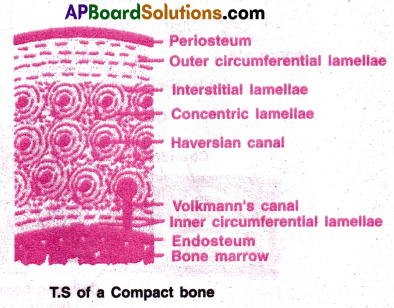
- Periosteum (dense connective tissue)
- Outer circumferential lamellae (bony layer)
- Osteons (haversian systems)
- Interstitial lamellae (between osteons)
- Inner circumferential lamella
- Endosteum (dense connective tissue)
- Bone marrow.
iii) Osteon: In a dense bone, a Haversian canal and the surrounding lamellae and lacunae are collectively called Osteon or Haversian system. It works as transport system.
Question 6.
Give an account of the ‘formed elements’ of Blood. [TS M-22]
Answer:
Formed elements of blood:
- Blood is composed of plasma and formed elements.
- Blood cells are formed from yolksac mesoderm in the embryos.
- Other haemopoietic organs are liver and spleen.
- In adults red bone marrow produces blood cells.
- The formed elements are (I) Red blood cells (II) White blood cells (III) Platelets )
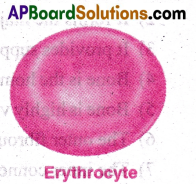
Red blood corpuscles (Erythrocytes):
- Erythrocytes of mammals are circular, enucleate, biconcave discs (elliptical in camels) Biconcave shape increases the surface area for exchange of gases.
- Erythrocyte is 7.8 mm in diameter.
- The number ofRBC per cubic millimeter ofblood is about 5 million in man and 4.5 million in a woman.
- The reduction in number of RBC, leads to anaemia.
- Vitamin B12 and folic acid are necessary for maturation of RBC.
- Life span of human RBC is 120 days.
- RBC are destroyed in spleen and liver.
![]()
White blood corpuscles (Leucocytes):
- White blood cells are either round or irregular (amoeboid) in shape.
- They can move through wall ofblood vessels.
- Their number is 6000 to 10000 per cubic mm of blood.
Formation
Formation of WBC is Leucopoiesis.
- Increase in number of WBC is leucocytosis. It takes place during infections and allergy.
- Abnormal increase of WBC causes Leukemia (blood cancer)
- Fall in count of WBC is leucocytopenia.
- WBC are of two types based on cytoplasmic granules i. Granulocytes ii. Agranulocytes.
i) Granulocytes: Cytoplasm of WBC has granules.
- Nucleus is lobed and hence called polymorph – nuclear leucocytes.
- These are three types based on staining properties.
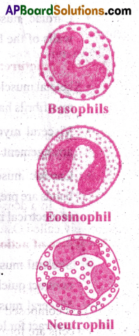
a) Basophils: They take up basic stains
- Their Nucleus is irregularly lobed.
- Their number is only 0.4% of leucocytes.
- They produce heparin and histamine.
- They take up the function of mast cells when necessary.
b) Eosinophils: They are also called acidophils.
- They take up acid stains.They remove antigen- antibody complexes.
- Their number is 2.3% of leucocytes.
- Their Nucleus is bilobed.
c) Neutrophils: They are most common WBC.
- Their Nucleus is many lobed.
- They are microscopic policemen because of active phagocytosis.
- Their number is 62% leucocytes.
ii) Agranulocytes: Cytoplasmic granules are absent.
- Their Nucleus is not lobed.
- There are 2 types of agranulocytes. (a) Lymphocytes (b) Monocytes.
a) Lymphocytes: Small spherical cell with large round nucleus and small amount of cytoplasm.
- B-lymphocytes produce antibodies.
- T lymphocytes play a role in immunological reactions.
- Their number is 30% of leucocytes.
b) Monocytes: The nucleus is kidney shaped.
- They are large and motile.
- They engulf bacteria and cell wastes.
- They become macrophages in connective tissue.
- Their number is 5.3% of total leucocytes.
![]()
III) Blood platelets:
- They are colourless, enucleated, round or oval discs.
- Their count is 2,50,000 to 4,50,000 per cubic mm.
- They are formed from megakaryocytes by fragmentation.
- Their life span is 5-9 days.
- They secrete thromboplastin which play a role in blood clotting. Blood platelets
Question 7.
Compare and contrast the three types of muscular tissues. [AP M-22]
Answer:
Comparison of three types of muscles skeletal, visceral and cardiac muscles, is done under the following headings:
1) Attachment:
- Skeletal muscles are attached to skeleton by tendons.
- Visceral muscles are present in walls of the visceral organs. They are arranged in sheets.
- Cardiac muscles are present in myocardium of the heart attached to septa and lateral walls of the heart.
2) Cell structure:
- Skeletal muscle cell is long, cylindrical and unbranched. It is multinucleate (syncytium). The myofibrils have alternate dark and light bands.
- Visceral myocyte is spindle shaped. It is uninucleate. The myofibrils have irregular arrangement of actin and myosin so that there are no dark and light bands.
- Cardiac muscle is cylindrical, uni or binucleate and branched. Alternate dark and light bands are present. Intercalated discs are characteristic of cardiac muscle for quick transport of electrical impulse.
3) Nature of action and control:
- Skeletal muscles are voluntary and imder the control of somatic nervous system. They contract quickly and fatigue quickly.
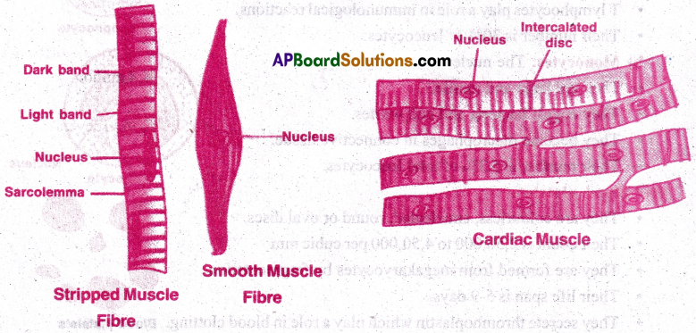
- Visceral muscles are involuntary. Their contractions are slow and prolonged. They can contract for long periods without fatigue. They are under the control of Autonomic nervous – system.
- Cardiac muscles are involuntary. Their contractions are under the control of pace maker which sends regular electric impulses. The rate of heart beat is changed by autonomic nervous system and hormones. The muscle is highly resistant to fatigue.
Multiple Choice Questions
Question 1.
Areolar tissue connects
1. Muscle with bone
2. Skin with muscle
3. Bone with bone
4. Cartilage with bone
Answer:
2. Skin with muscle
Question 2.
Bones are mainly made up of
1. calcium and phosphorus
2. calcium and sulphur
3. calcium and magnesium
4. calcium and iron
Answer:
1. calcium and phosphorus
![]()
Question 3.
Cell division does not occur in
1. Neuroglia cells
2. Neurons
3. Epithelial cells
4. Chondroblasts
Answer:
2. Neurons
Question 4.
Difference between blood and lymph is
1. blood has RBC and WBC while lymph has no cells
2. blood has RBC and WBC while lymph has only WBC
3. blood has WBC while lymph has RBC
4. blood has dissolved salt while lymph as no cells
Answer:
2. blood has RBC and WBC while lymph has only WBC
Question 5.
Elastic cartilage is found in
1. Epiphyses
2. Epistemum
3. Epiglottis
4. Epidermis
Answer:
3. Epiglottis
Question 6.
Fall in WBC count is called
1. Leukemia
2. Leucocytosis
3. Leucopoiesis
4. Leucocytopenia
Answer:
4. Leucocytopenia
Question 7.
Glial cells that help in providing blood brain barrier are
1. Astrocytes
2. Ependymal cells
3. Microglia
4. Oligodendrocytes
Answer:
1. Astrocytes
Question 8.
Heart valves and joint capsules are made up of
1. Dense regular connective tissue
2. Elastic connective tissue
3. Dense irregular connective tissue
4. Adipose tissue
Answer:
3. Dense irregular connective tissue
Question 9.
Iris muscles are
1. Ectodermal striated muscles
2. Mesodermal striated muscles
3. Ectodermal smooth muscles
4. Mesodermal smooth muscles
Answer:
3. Ectodermal smooth muscles
![]()
Question 10.
Largest smooth muscle is present in
1. leg
2. thigh
3. uterus of pregnant woman
4. urethra
Answer:
3. uterus of pregnant woman
Question 11.
Ligaments connect
1. Bones with muscles
2. Mesodermal striated muscles
3. Muscles with muscles
4. Mesodermal smooth muscles
Answer:
2. Mesodermal striated muscles
Question 12.
The number of RBCs per cubic millimeter of blood in a healthy woman is about
1. 4.5 million
2. 5 million
3. 5.5 million
4. 4 million
Answer:
1. 4.5 million
Question 13.
Cells that line cavities of brain and spinal cord are
1. Oligodendrocytes
2. Microglia cells
3. Ependymal cells
4. Astrocytes
Answer:
3. Ependymal cells
Question 14.
Tissue specialised for fat-storage is
1. Areolar tissue
2. Adipose tissue
3. Reticular tissue
4. Elastic connective tissue
Answer:
2. Adipose tissue
Question 15.
Acid-base buffers in blood plasma are
1. Nucleoproteins
2. Plasma proteins
3. Fatty acids
4. Chromo proteins
Answer:
2. Plasma proteins
Question 16.
Embryonic skeleton of bony vertebrates is made up of
1. Elastic cartilage
2. Fibrous cartilage
3. Calcified cartilage
4. Hyaline cartilage
Answer:
4. Hyaline cartilage
Question 17.
Intercalated discs occur in
1. Smooth muscle
2. Skeletal muscle
3. Cardiac muscle
4. Voluntary muscle
Answer:
3. Cardiac muscle
![]()
Question 18.
pH of blood plasma is
1. 7.4
2. 7.0
3. 8.4
4. 6.0
Answer:
1. 7.4
Question 19.
Strongest cartilage is
1. Hyaline cartilage
2. Costal cartilage
3. Elastic cartilage
4. Fibrous cartilage
Answer:
4. Fibrous cartilage
Question 20.
Blubber is present in
1. Whales and sea cows
2. Camels and llamas
3. Whales and cows
4. Goats and sheep
Answer:
1. Whales and sea cows
Question 21.
Antibodies are produced by
1. Histiocytes
2. Mast cells
3. Plasma cells
4. Fibroblasts
Answer:
3. Plasma cells
Question 22.
Large amount of extracellular matrix is present in
1. Epithelial tissue
2. Connective tissue
3. Muscular tissue
4. Nervous tissue
Answer:
2. Connective tissue
Question 23.
Thin connective tissue sheath that covers a nerve fibre is called
1. Endomysium
2. Endoneurium
3. Perineurium
4. Epineurium
Answer:
2. Endoneurium
Question 24.
In central nervous system of vertebrates, myelinated nerve fibres occur in
1. Grey matter
2. White matter
3. Ganglia
4. Cranial and spinal nerves
Answer:
2. White matter
![]()
Question 25.
Stratified epithelia are mainly helpful for
1. Secretion
2. Absorption
3. Filtration
4. Protection
Answer:
4. Protection
Question 26.
A complete gut formed for the first time in
1. Nematodes
2. Annelids
3. Tapeworms
4. Chordates
Answer:
1. Nematodes
Question 27.
Open type of circulatory system is present in the members of
1. Cephalochordata
2. Urochordata
3. Cephalopoda
4. Vertebrata
Answer:
2. Urochordata
Question 28.
A skeletal muscle fibre is
1. Long, fusiform and unbranched
2. Long, cylindrical and unbranched
3. Long, cylindrical and branched
4. Short, cylindrical and unbranched
Answer:
2. Long, cylindrical and unbranched
Question 29.
The weakest cartilage is
1. Hyaline cartilage
2. Elastic cartilage
3. Fibrous cartilage
4. Calcified cartilage
Answer:
1. Hyaline cartilage
Question 30.
Cardiac muscle cells are
1. Short, cylindrical and mononucleate or binucleate
2. Short, cylindrical and multi nucleate
3. Long, cylindrical and mononucleate or binucleate
4. Short, fusiform and mononucleate
Answer:
1. Short, cylindrical and mononucleate or binucleate
Question 31.
Blood platelets are
1. Enucleated complete cells
2. Enucleated cell-fragments
3. Nucleated complete cells
4. Nucleated cell-fragments
Answer:
2. Enucleated cell-fragments
![]()
Question 32.
Neurons are derived from –
1. Ectoderm
2. Endoderm
3. Mesoderm
4. Ecto-mesoderm
Answer:
1. Ectoderm
Question 33.
Nucleus of monocytes is
1. 2 to5 lobed
2. Kidney-shaped
3. Bilobed
4. Spherical
Answer:
2. Kidney-shaped
Question 34.
The nature of cardiac muscle is
1. Unstriated and involuntary
2. Striated and voluntary
3. Unstriated and voluntary
4. Striated and involuntary
Answer:
4. Striated and involuntary
Question 35.
Blood plasma is
1. Slightly acidic
2. Strongly acidic
3. Slightly alkaline
4. Strongly alkaline
Answer:
3. Slightly alkaline
Question 36.
Tendons connect
1. Cartilages with cartilages
2. Cartilages with bones
3. Bones with other bones
4. Skeletal muscles with bones
Answer:
4. Skeletal muscles with bones
Question 37.
Marrow cavity occurs in the
1. Diaphy sis of long bone
2. Diaphysis of short bone
3. Epiphyses of long bone
4. Epiphyses of short bone
Answer:
1. Diaphy sis of long bone
Question 38.
The lowest level of organisation in metazoans is
1. Protoplasmic level
2. Organ level
3. Tissue level
4. Cellular level
Answer:
4. Cellular level
Question 39.
Tissue level of organisation is exhibited by
1. Sponges
2. Coelenterates
3. Flatworms
4. Annelids
Answer:
2. Coelenterates
Question 40.
Incomplete gut is present in
1. Nematodes
2. Annelids
3. Flatworms
4. Molluscans
Answer:
3. Flatworms
Question 41.
The stopping of bleeding is called
1. Homeostasis
2. Haemostasis
3. Haemopoiesis
4. Haemolysis
Answer:
2. Haemostasis
![]()
Question 42.
Blood platelets are formed from
1. Proerythroblasts
2. Reticulocytes
3. Histiocytes
4. Megakaryocytes
Answer:
4. Megakaryocytes
Question 43.
A bundle of muscle fibres is known as
1. Fascicle
2. Funiculus
3. Myofibril
4. Fascia
Answer:
1. Fascicle
Question 44.
Plasma cells are the descendants of
1. Monocytes
2. T-lymphocytes
3. Basophils
4. B-lymphocytes
Answer:
4. B-lymphocytes
Question 45.
Patella or knee cap is made up of
1. Investing bone
2. Sesamoid bone
3. Dermal bone
4. Replacing bone
Answer:
2. Sesamoid bone
Question 46.
In a bone, osteons are interconnected by
1. Haversian canals
2. Volkmann’s canals
3. Bidder’s canals
4. Inguinal canals
Answer:
2. Volkmann’s canals
Question 47.
Epithelium of urinary bladder is
1. Pseudostratified epithelium
2. Transitional epithelium
3. Ciliated epithelium
4. Columnar epithelium
Answer:
2. Transitional epithelium
Question 48.
One of the most widely distributed connective tissue in the body is
1. Areolar tissue
2. Adipose tissue
3. Reticular tissue
4. Neural tissue
Answer:
1. Areolar tissue
Question 49.
Skeletal muscles are
1. Unstriped and involuntary
2. Striped and involuntary
3. Unstriped and voluntary
4. Striped and voluntary
Answer:
4. Striped and voluntary
Question 50.
An abnormal rise in the RBC count is termed
1. Erythrocytopenia
2. Leucocytosis
3. Polycythemia
4. Leucocytopenia
Answer:
3. Polycythemia
![]()
Question 51.
The smallest and the most abundant serum protein is
1. Albumin
2. Fibrinogen
3. Globulin
4. Prothrombin
Answer:
1. Albumin
Question 52.
Plasma proteins are produced in the
1. Brain
2. Kidney
3. Heart
4. Liver
Answer:
4. Liver
Question 53.
The average life span of blood platelets is about
1. 120 days
2. 1 to 2 days
3. Many years
4. 5 to 9 days
Answer:
4. 5 to 9 days
Question 54.
The process of formation of WBC is called
1. Erythropoiesis
2. Leucocytosis
3. Leucopoiesis
4. Leucocytopenia
Answer:
3. Leucopoiesis
Question 55.
Arthritis occurs due to the degeneration of
1. Articular cartilages
2. Laryngeal cartilages
3. Costal cartilages
4. Nasal septal cartilage
Answer:
1. Articular cartilages
Question 56.
The major organic substance in the bone matrix is
1. Calcium carbonate
2. Elastin
3. Calcium phosphate
4. Collagen
Answer:
4. Collagen
Question 57.
The bones formed by the ossification of soft tissue are called
1. Replacing bones
2. Visceral bones
3. Sesamoid bones
4. Dermal bones
Answer:
2. Visceral bones
Question 58.
Cephalization is associated with
1. Radial symmetry
2. Bilateral symmetry
3. Asymmetry
4. Biradial symmetry
Answer:
2. Bilateral symmetry
![]()
Question 59.
An axon differs from a dentdrite by the –
1. absence of neurofibrils
2. presence of Nissl bodies
3. presence of neurofibrils
4. absence of Nissl bodies
Answer:
4. absence of Nissl bodies
Question 60.
Most of the neurons in our body are
1. Unipolar
2. Multipolar
3. Bipolar
4. Pseudounipolar
Answer:
2. Multipolar
Question 61.
Star-shaped glial cells are
1. Astrocytes
2. Ependymal cells
3. Microglia
4. Oligodendrocytes
Answer:
1. Astrocytes
Question 62.
Number of planes of symmetry in a star fish is
1. One
2. Many
3. Two
4. Five
Answer:
4. Five
Question 63.
Unicellular organisms exhibit ………..organisation
1. Cellular level
2. Protoplasmic level
3. Tissue level
4. Organ system level
Answer:
2. Protoplasmic level
Question 64.
The multicellular animals having ………. type of body organisation can lead more efficient way of life
1. A cellular level
2. Cellular level
3. Tissue level
4. Organ system level
Answer:
4. Organ system level
Question 65.
Multicellular animals do not exhibit
1. Biradial symmetry
2. Spherical symmetry
3. Radial symmetry
4. Bilateral symmetry
Answer:
2. Spherical symmetry
Question 66.
Tube with in a tube type of organisation is seen in
1. Cnidarians
2. Flat worms
3. Nematodes
4. Sponges
Answer:
3. Nematodes
![]()
Question 67.
Body cavity is formed by replacing blastocoel in
1. Cnidaria
2. Platyhelminthes
3. Nematoda
4. Annelida
Answer:
4. Annelida
Question 68.
First key transition in the evolution of animal body plan is
1. Formation of tissue
2. Formation of coelome
3. Formation of symsmetry
4. Formation of organs
Answer:
1. Formation of tissue
Question 69.
The cell junctions called tight, adhering and gap junctions are found in
1. connective tissue
2. epithelial tissue
3. neural tissue
4. muscular tissue
Answer:
2. epithelial tissue
Question 70.
The epithelial tissue present on the inner surface of bronchioles and Fallopian tubes is
1. glandular
2. ciliated
3. squamous
4. cuboidal
Answer:
2. ciliated
Question 71.
The kind of epithelium which forms the inner walls of blood vessels is
1. cuboidal epithelium
2. columnar epithelium
3. ciliated columnar epithelium
4. squamous epithelium
Answer:
4. squamous epithelium
Question 72.
Mucous connective tissue is present in
1. Umbilical cord
2. Vocal cords
3. Mucous membrane
4. none of these
Answer:
1. Umbilical cord
Question 73.
The mast cells are found in
1. adipose tissue
2. yellow elastic tissue
3. white fibrous tissue
4. areolar tissue
Answer:
4. areolar tissue
![]()
Question 74.
Haversian canal is a
1. canal for passing water
2. canal for passing blood
3. characteristic of mammalian bone
4. characteristic of the canal
Answer:
3. characteristic of mammalian bone
Question 75.
Red muscle fibres are rich in
1. myoglobin and cytochrome
2. lactic acid and acetic acid
3. haemoglobin and glucose
4. only myosin
Answer:
1. myoglobin and cytochrome
Question 76.
Cardiac muscles are different from that of skeletal muscles as they
1. are striated
2. lactic acid and acetic acid
3. have intercalated disc
4. only myosin
Answer:
3. have intercalated disc
Question 77.
The longest cell in the human may be
1. nerve cell
2. cells of leg muscles
3. bone cell
4. cells of alimentary canal
Answer:
1. nerve cell
Question 78.
Axons serve to
1. bring impulse to cytons
2. take away impulse from cytons
3. bring blood to heart
4. take blood to brain
Answer:
2. take away impulse from cytons
Question 79.
In which of the following tissue, Schwann’s cells are found
1. Muscle tissue
2. Connective tissue
3. Nervous tissue
4. Epithelial tissue
Answer:
3. Nervous tissue
![]()
Question 80.
Collagen is
1. fibrous protein
2. globular protein
3. lipid
4. carbohydrate
Answer:
1. fibrous protein