Thoroughly analyzing TS Inter 2nd Year Zoology Model Papers and TS Inter 2nd Year Zoology Question Paper May 2018 helps students identify their strengths and weaknesses.
TS Inter 2nd Year Zoology Question Paper May 2018
Time: 3 Hours
Maximum Marks: 60
Instructions:
Note : Read the following instructions carefully.
- Answer all the questions of Section ‘A’ Answer any six questions in Section ‘B’ and answer any two questions in Section ‘C’.
- In Section ‘A’, questions from Sr. Nos. 1 to 10 are of Very Short Answer Type. Each question carries two marks. Every answer may be limited to 5 lines. Answer all these questions at one place in the same order.
- In Section ‘B’, questions from Sr. Nos. 11 to 18 are of Short Answer Type. Each question carries four marks. Every answer may be limited to 20 lines.
- In Section ‘C’, questions from Sr. Nos. 19 to 21 are of Long Answer Type. Each question carries eight marks. Every answer may be limited to 60 lines.
- Draw labelled diagrams wherever necessary in Sections B’ and ‘C’.
Section – A (10 × 2 = 20)
Note : Answer all the questions in 5 lines each:
Question 1.
What are renal pyramids and renal papillae ?
Answer:
The conical shaped medullary regions of kidney are the renal pyramids. Tips of the renal pyramids which open into pelvis are renal papillae.
Question 2.
Write the difference between actin and myosin.
Answer:
| Actin | Myosin. |
| 1. Actin is a thin contractile protein. | 1. Myosin is a thick contra-ctile protein. |
| 2. It is present in light bands and is called an isotropic band. | 2. It is present in dark bands and is called an anisotropic band. |
| 3. Each actin filament is made of two ‘F’ actin molecules helically wound around each other, tropomyosin and a complex protein called troponin. | 3. Each myosin is made up of monomeric protein called meromyosins. Each mer-omyosin has two parts namely head, and arm (or) neck. |
Question 3.
Name the cranial sutures and their locations.
Answer:
- Coronal suture – between the frontal and parietal bones.
- Lambdoid suture – between the parietal and occipital bones.
Question 4.
What is erythropoietin ? What is its function ?
Answer:
Erythropoietin is a hormone secreted the juxtaglomerular cells of the kidney. It plays an important role in the erythropoiesis i.e., in formation of RBC. Erythropoietin controls the formation of RBC by regulating the differentiation and proliferation of erythroid progenitor cells in the bone marrow.
![]()
Question 5.
Name the structure of gut which is vestigial in human beings, but well developed in herbivores. Mention the type of tissue with which it is mostly formed.
Answer:
Appendix is vestigial part in human beings. It is a narrow finger like tubular projection, arises from the caecum. In herbivores it is a functional part and useful in the digestion of cellulose materials. The appendix contain a high concentration of lymphoid tissues. These are highly specialized structures which are a part of the immune system.
Question 6.
What are Islets of Langerhans ?
Answer:
The endocrine region of pancreas is called Islets of langerhans where it contain 1 to 2 millions Islets of langerhans. There are two main types of cells α – cells and β – cells, α – cells produce the hormone glucagon, whereas β – cells produce insulin.
Question 7.
Mention the names of any four connecting links that you have studied.
Answer:
Connecting links clearly explain the path of evolution.
- Peripatas between ahnelida and arthropoda.
- Prototherians between reptilia and mamnalia.
Question 8.
Define atavism with an example.
Answer:
Sudden appearance of some vestigial organs in a better developed condition is called atavism. Eg : Tailed human baby.
Question 9.
Distinguish between a drone and worker in a honey bee colony.
| Drones | worker bees |
| 1) These are fertile males. | 1) These are sterile female. |
| 2) These are developed from unfertilized ova by male parthenogenesis. | 2) These are developed from fertilized eggs. |
| 3) These are short lived. | 3) These live for two and three months. |
Question 10.
What does ADA stand for ? Deficiency of ADA causes which disease ?
Answer:
ADA stands for adenosine deaminase. Deficiency of adenosine deaminase (ADA) causes severe combined immuno deficiency (SCID).
![]()
Section – B (6 × 4 = 24)
Note : Answer any six questions in 20 lines each:
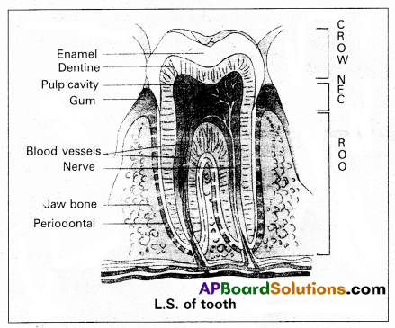
Question 11.
Draw a-neat labelled diagram of L.S. of a tooth.
Answer:
Question 12.
How is respiratory movements regulated in man ?
Answer:
In human beings the respiratoiy movements are regulated by neural system.
1. A special centre present in the medulla region of brain, called ‘respiratory rhythm centre’ is primarily responsible for this regulation.
2. Another centre present in the pons of the brain stem called ‘pneumotaxic centre’ can moderate the functions of the respiratory rhythm centre. Neural signal from and this centre can reduce the duration of inspiration and thereby alter the respiration rate.
3. A chemo-sensitive area is situated adjacent to the respiratory rhythm centre which is highly sensitive to CO2 and H+. Increase in these substances can activate this centre, which intum can send signals to the respiratory rhythm centre to make necessary adjustments in the respiratory process by which these substances can be eliminated.
4. Receptors associated with aortic arch and carotid artery also recognize changes in CO2 and H+ concentration and send necessary signals to the respiratory rhythm centre for necessary actions.
The role of oxygen in the regulation of the respiratory rhythm is quite insignificant.
Question 13.
Give an account of the retina of the human eye.
Answer:
Retina is the inner coat of the eye. It consist of a pigmented epithelium and a neural portion. The pigmented epithelium is a sheet of melanin containing epithelial cells. The neural portion has three layers namely photoreceptor layer, bipolar cell layer and ganglion cell layer.
Photoreceptor layer consist of rods and cones. Rods contain a protein called rhodopsin. Rods are concerned with dim light. Cones contain a visual pigment called iodopsin and they are important in daylight vision and colour vision. There are three types of cones and are response to red, green and blue colours.
The centre of the posterior portion of the retina is called yellow spot. A depression present in the yellow spot is called ‘Forea’ contractile and it contains only cones. Forea is responsible for sharp vision. The region of retina which is devoid of rods and cones is known as blind spot (or) optic disc, which form the optic nerve called 2nd cranial nerve.
Question 14.
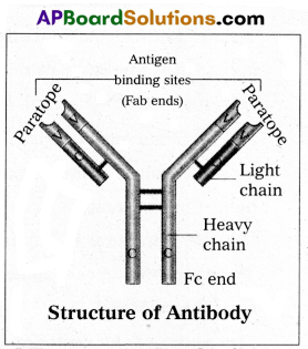
Write a short note on immunoglobulins.
Answer:
Whenever pathogen enters our body, the B-lymphocytes produce an army of proteins called antibodies to fight with them. They are highly specialised for binding with specific antigens. The part of an antibody that recognises an antigen is called the paratope antigen binding site.
Based on their mobility, antibodies are of two types.
1. Circulating or free antibodies : These are present in the body fluids like serum, lymph etc.
2. Membrane bound antibodies : These are present on the surface of the mature B-cells as well as the memory cells.
Structure : Immunoglobulin is a ‘Y’ shaped molecule with four polypeptide chains of which two are long identical heavy chains (H) and two are small, identical light chains (L). These two chains are linked by disulfide bonds. One end of the antibody molecule is called Fab end (Fragment-antigen binding) and the other end is called Fc end (Fragment-Crystaline). Based on the structure, the antibodies are of five types namely Ig G, Ig A, Ig M, Ig D and Ig E.
Question 15.
What is the necessity of sex education in schools ?
Answer:
Governmental and non-governmental agencies have taken various steps to educate people on reproduction-related issues using audio-visual and print media. Introduction of sex education in schools will provide right information about the reproductive organs, adolescence and related changes, safe and hygienic sexual practices, sexually transmitted diseases such as HIV etc, would help people, especially those in adolescent age group lead a reproductively healthy life.
Question 16.
Write the salient features of HGP (Human Genome Proj ect).
Answer:
Salient features of HGP :
- The human genome comprised of 3164.7 million nucleotide bases.
- Human genome contains 30,000 genes.
- Each gene consist of an average 3000 bases.
- Functions of 50% of genes discovered are unknown.
- All proteins are coded by less than 2% of the genome.
- Majority of the genome consisted by repeated sequences.
- Chromosome one has highest number of genes i.’e., 2,968 genes and Y chromosome has the fewest genes i.e., 231 genes.
- It is also identified that 1.4 millions locations, where Single base DNA difference (SNPs) occurs in humans. This information promises to revolutionise the process of finding chromosomal locations for disease associated sequences and tracing human history.
![]()
Question 17.
Explain Darwin’s theory of natural selection with industrial melanism as an experimental proof.
Answer:
Darwin’s theory of natural selection does not explain what exactly evolution is, but explains how evolution might have occurred in nature. A classical example for natural selection is industrial melanism, exhibited by peppered moth-Biston betularia. These moths were available in two colours grey and black. Grey moths were abundant before industrial revolution in all over England. The reason for the existence of large number of grey moths during that period was camouflage on the trunks of trees. But after the establishment of industries in England, black coloured moths were more and grey forms were less. This is due to pollution from industries in the form of soot turned barks of trees into black.
So grey moths were easily identified and were more predated by birds. Thus grey moths decreased in number, black moths increased in the population.
Thus natural selection favoured the melanic moths (black) to reproduce more successfully. Natural selection of darker forms in response to industrial pollution is known as industrial melanism.
Question 18.
List the various steps involved in MOET.
Answer:
The following are the steps involved in Multiple Ovulation and Embryo Transfer (MOET):
- A cow is administrated hormones, with FSH like activity.
- This induces follicular maturation and super ovulation.
- In Super ovulation instead of one egg, which they produce per cycle, they produce 6 – 8 eggs.
- The cow is either mated with elite bull or artificially inseminated.
- The embryos are at 8 – 32 called stages are recovered non-surgically and transferred to surrogate mother, when the embryo develops into complete animal.
Now the genetic mother is ready for another round of super ovulation. This technology is in use for cattle, sheep, rabbits, buffaloes etc. to produce high yielding ones.
Section – C (2 × 8 = 16)
Note : Answer any two questions in 60 lines each:
Question 19.
What are multiple alleles ? Describe multiple alleles with the help of ABO blood groups in man.
Answer:
Generally a gene has two ‘alternative forms called allele. Sometimes a gene may have more than two alleles. These are referred to as multiple alleles. When more than two alleles exist in a population of a specific organism, the phenomenon is called multiple allelism. Multiple alleles cannot be observed in the genotype of a diploid individual, but can be observed in a population.
The number of genotypes that can occur for multiple alleles is given by the expression where ‘n’ = number of alleles.
ABO blood groups are the best example for multiple allelism in human beings.
The ABO blood group system was proposed by Karl Landsteiner. The blood groups A, B, AB and O types are characterised by the presence or absence of antigens on the surface of RBC. Blood type ‘A’ person have antigen A on their RBCs and anti-B antibodies in the plasma. Blood type ‘B’ person have antigen B. On their RBCs and anti-A antibodies in the plasma. Blood type ‘AB’ person have antigens A and B on the RBCs and no antibodies in the plasma. Blood type ‘O’ person have no antigens on their RBCs and both anti-A, and anti-B antibodies are present in the plasma.
| Blood group | Antigen on RBC | Antibodies in Plasma |
| A | A | b |
| B | B | a |
| AB | AB | — |
| O | — | a, b |
Bernstein discovered that these phenotypes were inherited by the interaction of three ‘autosomal alleles’ of the gene named T, located on chromosome 9. IA, IB and IO are the three alleles of the gene I. The alleles IA and IB are responsible for the production of the respective antigens A and B. The allele IO does not produce any antigen. The alleles IA and IB are dominant to the allele IO but co-dominant to each other (IA = IB > IO).
A child receives one of the three alleles from each parent, giving rise to six possible genotypes and four possible blood types. The genotypes are IA IA, IA IO, IB IB, IB IO, IA IB, IO IO. The phenotypic expressions of IA IA and IA IO, are A-type blood, the phenotypic expression of IB IB and IB IO are B-type blood, and that of IA IB, is AB blood type. The phenotype IO IO is ‘O-type’ blood.
Question 20.
Describe the structure of the heart of a human with the help of a neat labelled diagram.
Answer:
Human heart is a hallow muscular, cone shaped, and pulsating organ situated between lungs. It is about the size of a closed fist.
The heart is covered by double walled pericardium, which consists of outer fibrous pericardium and inner serous pericardium. The serous pericardium is double layered, outer parietal layer and inner visceral layer. These two layers are separated by pericardial space, which is filled with pericardial fluid. This fluid reduces friction between the two membranes and allow free movement of the heart.
Human heart has four chambers with two smaller upper chambers called atria and two larger lower chambers called ventricles. Atria and ventricles are separated by a deep transverse groove called coronary sulcus.
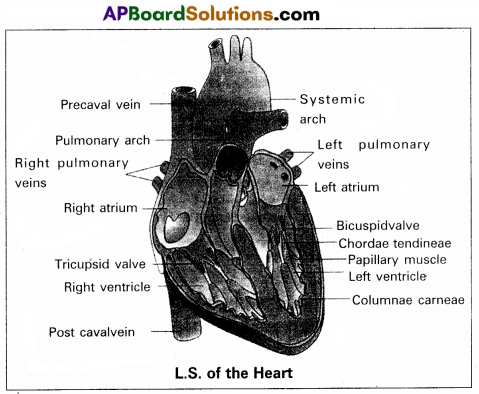
i) Atria :
Atria are thin walled receiving chambers. The right one is larger than the left.
The two atria are separated by thin inter-atrial septum. It has a small pore known as Foramen Ovale’ in fetal stage. Later it is closed and appears as depression (oval patch) known as ‘Fossa ovale’. If, the foramen ovale does not close properly it is called a patent foramen ovale.
The right atrium receives deoxygenated blood from different parts of the body, through three caval veins like two precaval veins and one post caval vein.
The right atrium also receives blood from wall of the heart through coronary sinus, whose opening into the right atrium is guarded by the valve of Thebesius.
Opening of the post caval vein is guarded by the Eustachian valve. It is functional in fetal stage and directs the blood from post caval vein into left atrium through foramen ovale. But it is non-functional in adult.
The openings of the precaval veins into the right atrium have no valves.
Left atrium receives oxygenated blood from lungs’ through a pair of pulmonary veins, which open into the left atrium through a common pore. Atrio-ventricular septum separates atria and ventricles. It has right and left atrio-venticular apertures.
Tricuspid valve guards the right atrio-ventricular aperture. Bicuspid valve guards the left atrio-ventricular aperture.
ii) Ventricles :
These are the thick walled blood pumping chambers, separated by an interventricular septum. The wall of the left ventricle is thicker than that of the right ventricle as the left ventricle must force the blood to all the parts of the body.
The inner surface of the ventricles is raised into muscular ridges called columnae cameae. Some of them are large ‘ and conical and known as papillary muscles. Colla-genous cords are known as chordae tendinae are present between atrio-ventricular valves and papillary muscles. They prevent the cusps of valves from bulging too far into atria during ventricular systole.
Nodal tissue : A specialized cardiac musculature called the nodal tissue is also distributed in the heart.
1) Sino-artrial node (SAN) – Present in the right upper comer of right atrium.
2) Atrio-ventricular node (AVN) – Present in the lower left comer of right atrium.
iii) Aortic arches : Human heart has two aortic arches.
1) Pulmonary arch: Arises from the left anterior angle of the right ventricle. It carries deoxygenated blood to lungs. It’s opening from right ventricle is guarded by pulmonary valve made with 3 semiluminar valves.
2) Left systemic arch : Arises from the left ventricle to ’ distribute oxygenated blood to various parts in the body. Its opening is also guarded by aortic valve made with a set of 3 semilunar valves.
A fibrous strand, known as ligamentum arteriosm is present at the point of contact of the systemic and pulmonary arches. It is the remnant of the ductus arteriosus, which connects the systemic and pulmonary arches in the embryonic stage.
![]()
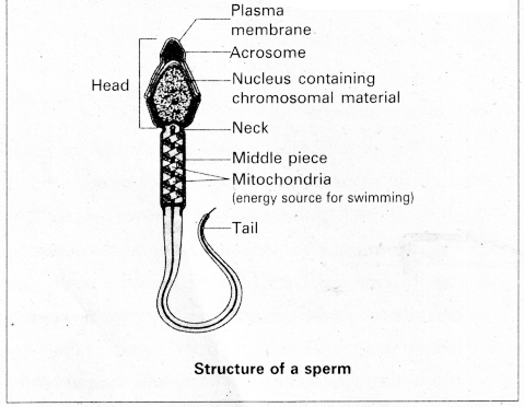
Question 21.
Describe male reproductive system of a man. Draw a labelled diagram of it.
Answer:
The male reproductive system or male genital system consists of a number of sex organs that are a part of the human reproductive process. The sex organs which are located in the pelvic region include a pair of testes, accessory ducts, glands and external genitalia.
1) Testes : The testes are a pair of oval pinkish male sex organs suspended in abdominal cavity within a pouch called scrotum. The scrotum helps in maintaining the low temperature of the testes (2 – 2.5°C) necessary for spermatogenesis. The cavity of scrotal sac is connected to the abdominal cavity through the ‘inguinal canal’. Testes is held in position in the scrotum of the ‘guber- naculum’, a fibrous cord that connects the testis with the bottom of scrotum and a ’spermatic cord’, formed by the vas deferens, nerves, blood vessels and other tissues that run from abdomen down to each testicle, through inguinal canal. Each testis is enclosed in a fibrous envelope, ‘tunica albuginea’, which extends inwards into testis and divide it into lobules. Each lobule contains 1 to 3 highly coiled seminiferous tubules. A pouch of serous membrane ’tunica vaginalis’ covers the testis.
Miniferous tubules : Each seminiferous tubule is lined by ‘germinal epithelium’ which consists of undifferentiated male gum cells called ‘spermatogonial mother cells’ and it also bears ‘nourishing cells’ called ‘sertoli cells’.
- Spermatogonial cells (or) primary spermatocytes undergo meiotic division, producing spermatozoa or sperms by a process spermatogenesis.
- Sertoli cells provide nutrition to spermatozoa and produce a .hormone ‘inhibin’, which inhibits secretion of FSH.
The region outside the tubules, contain interstitial cells of ‘Leydig cells’. They produce androgens, the most important in testosterone. It controls the development of secondary sexual characters and spermatogenesis. The seminiferous , tubules open into vasa efferntia through the rete testis. Rete testis is a network of tubules is of the testis carrying spermatozoa from the seminiferous tubules to the vasa efferentia.
2) Epididymis : The’ vasa efferntia leave the testis and open into a narrow, tightly coiled tube called ‘epididymis’ located along the posterior surface of each testis. The epididymis provides a storage space for sperms and gives them time to nature.
It is differentiated into three regions.
a) Caput epididymis.
b) Corpus epididymis.
c) Cauda epididymis.
The caput epididymis receives spermatozoa via the vasa efferntia of the mediastinum testis. It is mass of a connective tissue at the back of the testis that encloses the rete testis.
3) Vasa deferentia: The vas deferens or ductus deferens is a long, narrow mascular tube. The mucosa of the ductus deferens consists of a pseudo stratified columnar epithelium and lamina propia. It starts from the tail of epididymis, passes through the inguinal canal into the abdomen and loops over the urinary bladder. It receives a duct from seminal vesicle.
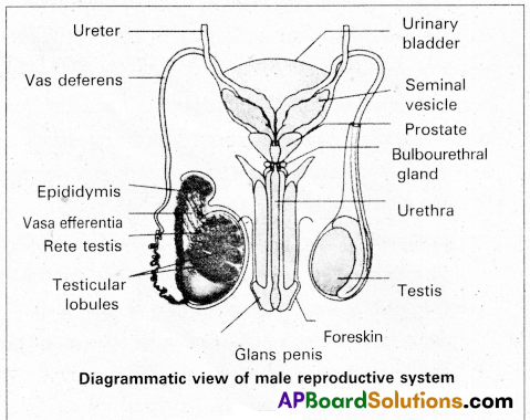
The vas deferens and the duct of the seminal vesicle units to form a ‘short ejaculatory duct1 or ‘ductus ejaculatorius’.
The two ducts, carrying spermatozoa and the fluid secreted by the seminal vesicles, converge in the centre of prostate and open into urethra, which transports the sperms to outside.
4) Urethra : In male, Urethra is the shared terminal duct of the reproductive and urinary systems. The urethra originates from urinary bladder and extends through the penis to its external opening called ‘urethral meatus’. The urethra provides an exit for urine as well as semen during ejaculation.
5) Penis : Urethra opens into the major copulatory organ of male, the ‘penis’. The penis and scrotum constitute the male external genitalia. The penis serves as a urinal duct and intromittent organ the transfers spermatozoa to the vagina of a female.
The penis is made up of three columns of tissue : two upper Corpora cavernosa on the dorsal aspect and one Corpus spongiosum on the ventral side. Skin and a subcutaneous layer encloses all three columns, which consists of special tissue that helps in erection of penis. The enlarged and bulbous end of penis is called glans penis’, which is covered by a loose fold of skin (foreskin) called prepuce.
Male accessory glands : Male accessory glands are :
(a) Seminal vesicles
(b) Prostate glands
(c) Bulbourethral glands.
a) Seminal vesicles : These are a pair of simple tubuiar glands present postero-inferior to the urinary bladder in the pelvis. Each seminal vesicle enters prostate gland through vas deferens. The vesicles produce seminal fluid rich is fructose, proteins, citric acid, in organic phosphorus, potassium and prostaglandins. All these serve sperm cells.
b) Prostate gland : It is located directly beneath the urinary bladder. The gland surrounds the ‘Prostatic urethra’, and sends its secretions through prostatic ducts. The prostatic secretion activates spermatozoa and provides nutrition. In man, the prostate contributes 15 – 30% of the semen.
c) Bulbourethral glands : These are also called cowper’s glands located beneath the prostate gland at the beginning of the internal portion of the penis. They add an alkaline fluid to semen and the fluid secreted by them lubricates urethra. It acts as flushing agent washing out the acidic urinary residues that remain in the urethra, before the semen is ejaculated.