Thoroughly analyzing AP Inter 2nd Year Zoology Model Papers and AP Inter 2nd Year Zoology Question Paper March 2015 helps students identify their strengths and weaknesses.
AP Inter 2nd Year Zoology Question Paper March 2015
Time: 3 Hours
Maximum Marks: 60
Instructions:
Note : Read the following instructions carefully.
- Answer all questions of Section – A. Answer ANY SIX questions in Section – B and answer ANY TWO questions in Section – C.
- In Section – A questions from SI. Nos. 1 to 10 are of Very Short Answer Type. Each question carries TWO marks. Every answer may be limited to 5 lines. Answer all these questions at one place in the same order.
- In Section – B, questions from SI. Nos. 11 to 18 are of Short Answer Type. Each question carries FOUR marks. Every answer may be limited to 20 lines.
- In Section – C, questions from SI. Nos. 19 to 21 are of Long Answer Type. Each question carries EIGHT marks. Every answer may be limited to 60 lines.
- Draw labelled diagrams wherever necessary in Sections – B and C.
Section – A
Note : Answer ALL the questions. (10 × 2 = 20)
Question 1.
What is Chyme ?
Answer:
The food is mixed thoroughly with the acidic gastric juice of the stomach by the churning movements of its muscular wall and the product is called Chyme. (The semi digested paste like acidic food of stomach is called chyme).
Question 2.
What are the columns of ‘Bertin’ ?
Answer:
The renai pyramids are separated by the projections of the cortex called Columns of Bertin (renal column).
Question 3.
Write the difference between Actin and Myosin.
Answer:
a) The light band in a myofibril contains actin and two regulatory proteins called troponin and tropomyosin. Actin filaments are thinner compared to myosin filaments.
b) The dark band in a myofibril contains (A band) myosin. Myosin filaments are thick and non-contractile.
![]()
Question 4.
What is “Insulin Shock” ?
Answer:
Hyper secretion of insulin leads to decreased level of glucose in the blood (hypoglycemia) resulting in insulin shock.
Question 5.
What is acromegaly ? Name the hormone responsible for this disorder.
Answer:
Hyper secretion of growth stimulating harmone (somatotropin) in adults results in an abnormality called acromegaly. It is characterized by enlargement of bones of Jaw, hand “and feet, thickened nose, lips and eye lids and gorilla like appearance of the person affected.
Question 6.
What is organ of corti ?
Answer:
The internal ear consists of 3 parts cochlea, vestibule and semicircular canals. The cochlear epithelium forms a sensory ridge called organ of corti on basilar membrane. The organ of corti contains hair cells that act as “auditory receptors”.
Question 7.
What are the functions of sertoli cells of the seminiferous tubules and the Leydig cells in man ?
Answer:
a) Sertoli cells: Nourishes the growing sperms and also produce a harmone called inhibin.
b) Leidig cells : Present in seminiferous tubules produce androgens, the most important of which is testosterone.
Question 8.
What is compaction in the human development ?
Answer:
In the morula due to unequal cleavage smaller and larger blastomeres are formed. The morula passes through a process called compaction. Now the embryo has a superficial flat cell layer and inner cell mass. Inner cell mass gives rise to the embryo proper. This is the first sign of cell differentiation in the human embryo.
Question 9.
What is electrocardiography and what are the normal components of ECG ?
Answer:
Electro Cardiography (ECG) is a commonly used, non invasive procedure for recording electrical changes in the heart. A normal ECG consists
- waves
- Intervals
- Segments and
- Complexes.
Question 10.
Define the terms layer and broiler.
Answer:
a) The birds which are raised exclusively for the production of eggs are called Layers.
b) The birds which are raised only for their meat are called Broilers.
![]()
Section – B (6 × 4 = 24)
Note : Answer ANY SIX questions.
Question 11.
Describe the process of digestion of proteins in the stomach.
Answer:
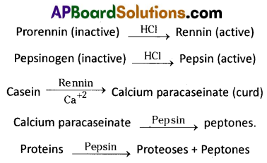
The gastric glands of stomach secrete acidic gastric juice. Gastric juice contains HCl, prorenin, pepsinogen and bicarbonates.
Proenzymes pepsinogen and prorennin, on exposure to HCl are converted into the active enzymes, pepsin and rennin respectively.
Pepsin converts proteins into proteoses and peptones. Rennin is found in gastric juice of infants. It acts on milk protein casein in the presence of calcium ions and converts it into calcium paracaseinate (curd) and proteoses. Pepsin again acts on calcium para- caseinate and converts it into peptones. Certain other cells in the wall of stomach produce bicarbonate, a base to buffer the acidic contents of the stomach. Bicarbonates and mucus produced by stomach wall forms a physical barrier to prevent HCI from damaging the wall of stomach.
Question 12.
Describe disorders of respiratory system.
Answer:
Disorders of the Respiratory System :
i) Asthma is a difficulty in breathing caused due to inflammation of bronchi and bronchioles. It is characterized by the spasm- of smooth muscles present in the walls of the bronchi and bronchioles. Symptoms include coughing, difficulty in breathing and wheezing. In the case of asthma, the allergen causes release of histamine and other inflammatory substances which cause constriction of the bronchi.
ii) Emphysema is a chronic disorder in which alveolar walls are damaged and their walls coalesce due to which respiratory surface area of exchange of gases is decreased. The lung shows larger but fewer alveoli and more fibrous and less elastic. One of the major causes of this is ‘smoking’ of tobacco.
iii) Bronchitis is the inflammation of the bronchi, resulting in the swelling of mucous lining of bronchi, increased mucus production and decrease in the diameter of bronchi. Symptoms include chronic cough with thick mucus/ sputum (phlegm).
iv) Pneumonia is infection of lungs caused by bacteria such as Streptococcus pneumoniae and also by certain viruses, fungi, protozoans and mycoplasmas. Symptoms include inflammation of lungs, accumulation of mucus in alveoli, and impaired exchange of gases, leading to death if untreated.
v) Emphysema, chronic bronchitis and asthma come under Chronic Obstructive Pulmonary Diseases (COPDs).
Occupational Respiratory disorders : These are caused by
exposure of the body to the harmful substances from certain industries, especially those involving grinding or stone breaking. Long term, exposure of the body to such substances can give rise to inflammation of respiratory passage and lungs leading to several disorders.
- Asbestosis : It occurs due to chronic exposure to asbestos dust in the people working in asbestos industry.
- Silicosis : It occurs because of long term exposure to ‘silica dust’ in the people working in mining industries, quarries, etc.
- Siderosis : It occurs due to deposition of iron particles in tissues. It can cause different types of siderosis such as pneumoconiosis due to inhalation of iron particles, hyperferremia and hemosiderosis (which causes recurrent alveolar hemorrhage).
- Black – lung disease : It is a lung disease that develops from inhalation of coal dust. It is common in long time coal mine workers.
Question 13.
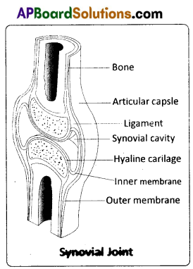
Draw a labelled diagram of T.S. of the spinal cord of man.
Answer:
Synovial joint is covered by a double layered synovial capsule. The outer layer consists of dense fibrous irregular connective tissue with more collagen fibres. This layer is continuous with the periosteum and resists stretching and prevents the dislocation of joints. Some fibres of these membranes are arranged in bundles called ligaments.
The inner layer of synovial capsule is formed of areolar tissue and elastic fibers. It secretes a viscous synovial fluid which contains hyaluronic acid, phagocytes, etc. and acts as a lubricant’ for the free movement of the joints. Synovial joints indude Bad and Socket joint, Hinge joint, Pivot joint, Gliding joint, Condyloid joint, Saddle joint.
![]()
Question 14.
Explain the mechanism by which HIV multiplies and leads to AIDS.
Answer:
After getting into the body of a person, the HIV enters the TH cells, macrophages or dendritic cells. In these cells the ssRNA of HIV synthesizes a DNA strand ‘complementary’ to the viral RNA, using the enzyme reverse transcriptase. The reverse transcriptase also catalyses the formation of the second DNA strand ‘complementary’ to the first strand forming the double stranded viral DNA. This viral DNA gets incorporated into the DNA of the host cells DNA by a viral enzyme (integrase) and it is in the form of a ‘provirus’. Transcription of DNA results in the production of RNA, which can act as the ‘genome’ for the new viruses or it can
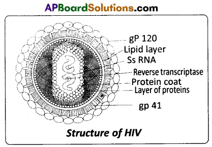
be translated into viral proteins. The various components of the viral particles are ‘assembled’ and the HIV are produced. The infected human cells continue to produce virus particles and in this way they act like HIV generating factories. New viruses ‘bud off from the host cell. This leads to a progressive decrease in the number of TH cells in the body of an infected person leading to the immuno-deficiency in him. Even though HIV attacks cells with CD4 marker, for reasons not known, only TH cells are destroyed and not the ‘macrophages’. The gp 120 molecules on the surface of HIV attach to CD4 receptors of human cells, mostly the TH cells (gp120 fits the CD4 marker). Attack on certain types of cells/ tissues only by viruses such as HIV is refered to as ’tissue tropism’.
Question 15.
Describe the Genic Balance Theory of sex determination.
Answer:
Genic balance theory of sex determination was proposed by Calvin Bridges with reference to sex determination in Drosophila. He proposed that both the X – chromosomes and autosomes together play a role in sex determination in Drosophila, where as Y – chromosome has no role. This theory explains that jengsJor maleness are located on autosome and for femaleness on X – chromosome in Drosophila. Y – chromosome in lacks male – determining factor but contains gametic information essential to male fertility . Sex in Drosophila is determined by ratio of X – chromosome to the number of haploid sets of autosomes.
Sex ratio = \(\frac{\text { No. of X- chromosomes }}{\text { No. of sets of autosomes }}\)
The chromosomal complement, X/A ratio and sex of Drosophila are tabulated below.
| Chromosomal complement | X/A ratio | Sex |
| AAX | 0.5 | Male |
| AAXX | 1.0 | Female |
| AA XXX | 1.5 | Metafemale |
| AAA XX | 0.67 | Intersex |
| AAA X | 0.33 | Metamale |
![]()
Question 16.
Write a short note on the theory of mutations.
Answer:
Mutation theory : It was proposed by Hugo de Vries, a Dutch botanist who coined the term ‘mutation’. Mutations are sudden, random inheritable changes that occur in organisms. He found four different forms in Oenothera lamarckiana (commonly called ‘evening primrose’) such as O. brevistylis- small style, O. levifolia- smooth leaves. O. gigas- the giant form, O. nanella- the dwarf form (mutant varieties). T.H. Morgan studied the inheritance pattern of mutations in Drosophila melanogaster. Darwin called mutations (large variations) sports of nature or saltations, whereas Bateson called them discontinuous variations.
Salient Features of Mutation theory :
- Mutations occur from time to time in naturally breeding populations.
- They are discontinuous and are not accumulated over generations.
- They are full-fledged, and so there are no’intermediate forms’.
- They are subjected to Natural Selection.
Question 17.
Distinguish between homologous and analogous organs.
Answer:
Homologous organs : The organs which have similar structure and origin but not necessarily the same function are called homologous organs. The evolutionary pattern that describes the occurrence of similarity in origin and internal structure is called homology. Such organs show adaptive radiation, hence ‘divergent evolution’, e.g. the appendages of vertebrates such as the flippers of whale, wings of bat, forelimbs of horse, paw of cat and hand of man, have a common pattern in arrangement of bones eventhough their external form and function may vary to suit their mode of life. It explains that all vertebrates might have had a common ancestor.
Analogous organs : The organs which have dissimilar structure and origin but perform the same function are called analogous organs. Analogous organs suggest ‘convergent evolution’, e.g. wings of a butterfly and wings of a bird.
Question 18.
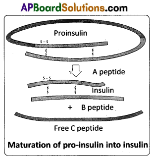
Explain in brief structure of Insulin.
Answer:
Structure of insulin: Human insulin is made up of 51 amino acids arranged in two polypeptide chains – chain A (21 amino acids) and chain B (30 amino acids), which are linked together by disulphide linkages. In mammals, including humans, insulin is synthesised as a pro- hormone (like a pro-enzyme, which needs to be processed before it becomes fully mature and functional hormone) which contains an extra stretch called the c peptide. This c peptide is not present in the mature insulin and is removed during maturation into insulin.
The main challenge for the production of insulin in the laboratory using rDNA technique was getting insulin assembled into its mature form.
Section – C (2 × 8 = 16)
Note : Answer ANY TWO of the following questions.
Question 19.
Write a note on the working of the heart of a man.
Answer:
The cardiac events that occur from the beginning of one heart beat to the beginning of the next constitute a cardiac cycle. This cardiac cycle consists of three phases, namely atrial systole, ventricular systole and cardiac diastole.
To begin with, all the four chambers of the heart are in a relaxed state / joint diastole stage. Blood from the pulmonary veins and venae cavae flows into the respective atria. As the A – V valves are in open condition, blood flows into the left and right ventricles, through the left and right atrioventricular apertures. The semilunar valves of the pulmonary and aortic arches are closed at this stage.
Atrial systole : The SAN now generates an action potential which stimulates both the atria to contract simultaneously causing the ‘atrial systole’. It lasts about 0.1 sec. This increases the flow of blood into the ventricles by about 30%. It means atrial systole accounts for about 30% of the filling of the ventricles, the remaining blood flows into the ventricles before the atrial systole.
Ventricular systole: The action potentials from the SAN reach the AVN from where they are conducted through the bundle of His, its branches and the Purkinje fibres to the entire ventricular musculature. This causes the simultaneous ventricular systole. It lasts for about 0.3 sec. The atria undergo relaxation coinciding with the ventricular systole. Ventricular systole increases the pressure causing the closure of the AV valves preventing the ‘backflow’ of blood. It results in the production of the first heart sound known as ‘Lub’ . As the ventricular pressure increases further, the semilunar valves guarding the pulmonary artery and the aorta are forced open. This allows the blood in the ventricles to flow into the aortic arches and enter the circulatory pathway.
Cardiac diastole : The ventricles now relax and the ventricular pressure falls causing the closure of the semilunar valves which prevents the back flow of blood. This result in the production of the second heart sound known as ‘Dup’. As the ventricular pressure declines further, the AV valves are pushed open by the pressure in the atria exerted by the blood, which flowed into them through the larger veins. The blood now once again flows freely into the ventricles. All the heart chambers are now again in a relaxed state (joint diastolic phase). Soon, another cardiac cycle sets in.
Cardiac output: The volume of blood pumped out by each ventricle, for each heart beat, is known as the stroke volume. The volume of blood pumped out by the heart from each ventricle per minute is termed cardiac output.
Cardiac output = Stroke volume × No. of beats per minute = 70 ml / beat × 72 beats / minute = 5040 ml/ mtn. or approximately 5 litres.
![]()
Question 20.
Describe female reproductive system of a woman with the help of a labelled diagram.
Answer:
The female reproductive system consists of a pair of ovaries along with a pair of oviducts, uterus, vagina and the external genitalia located in the pelvic region. These parts of the system along with a pair of the mammary glands are integrated structurally and functionally to support the processes of ovulation, fertilization, pregnancy, birth and child care.
Ovaries : Ovaries are the primary female sex organs that produce the female gametes (ova) and several steroid hormones (ovarian hormones). A pair of ovaries is located one on each side of the lower abdomen. The double layered fold of peritoneum connecting the ovary with the wall of the abdominal cavity is known as the mesovarium.
The ovaries are covered on the outside by a layer of simple cuboidal epithelium called germinal (ovarian) epithelium. This is actually the visceral peritoneum that envelops the ovaries. Underneath this layer there is a dense connective tissue capsule, the tunica albuginea. The ovarian stroma is distinctly divided into an outer cortex and an inner medulla. The cortex appears more dense and granular due to the presence of numerous ovarian follicle in various stages of development. The medulla is a loose connective tissue with abundant blood vessels, lymphatic vessels, and nerve fibers.
Fallopian tubes (Oviducts): Each fallopian tube extends from the periphery of each ovary to the uterus, and it bears a funnel shaped infundibulum. The edges of the infundibulum possess finger like projections called fimbriae, which help in collection of the ovum after ‘ovulation’. The infundibulum leads to a wider part of the oviduct called ampulla. The last part of the oviduct, isthmus has a narrow lumen and it joins the uterus. Fallopian tube is the site of fertilization. It conducts the ovum or zygote towards the uterus by peristalsis. The fallopian tube is attached to the abdominal wall by a peritoneal fold called mesosalpinx.
Uterus : The uterus is single and it is also called womb. It is a large, muscular, highly vascular and inverted pear shaped structure present in the pelvis between the bladder and the rectum. The uterus is connected to the abdominal wall by the peritoneal fold called mesometrium. The lower, narrow part through which the uterus opens into the vagina is called the cervix. The cavity of the cervix is called cervical canal which along with vagina forms the birth canal.
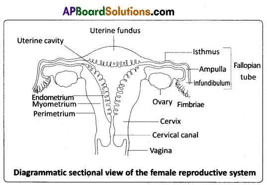
The wall of the uterus has three layers of tissue. The external thin membranous perimetrium, the middle thick layer of smooth muscle called myometrium and inner glandular lining layer called endometrium. The endometrium undergoes cyclic changes during menstrual cycle while the myometrium exhibits strong contractions during parturition.
Vagina : The vagina is a large, median, fibro – muscular tube that extends from the cervix to the vestibule (the space between the labia minora). It is lined by non – keratinised stratified squamous epithelium. It is highly vascular, and opens into the vestibule by the vaginal orifice.
Vulva : The term vulva (vulva = to wrap around) or pudendum refers to the external genitals of the female. The vestibule has two apertures – the upper external urethral orifice of the urethra and the lower vaginal orifice of vagina. Vaginal orifice is often covered partially by a membrane called hymen which is a mucous membrane. Vestibule is bound by two pairs of fleshy folds of tissue called labia minora (inner) and larger pair called labia majora (outer). Clitoris is a sensitive, erectile structure, which lies at the upper junction of the two labia minora above the urethral opening. The clitoris is homologous to the penis of a male as both are supported by corpora cavernosa internally. There is a cushion of fatty tissue covered by skin and public hair present above the labia majora. It is known as mons pubis.
Accessory reproductive glands of female : These glands include Bartholin’s glands, Skene’s glands and Mammary glands.
Bartholin’s glands : The Bartholin’s glands (Greater vestibular glands) are two glands located slightly posterior and to the left and right of the opening of the vagina. They secrete mucus to lubricate .the vagina and are homologous to the bulbourethral glands of the male reproductive system.
Skene’s glands : The Skene’s glands (Lesser vestibular glands) are located on the anterior wall of the vagina, around the lower end of the urethra. They secrete a lubricating fluid when stimulated. The Skene’s glands are homologous to the prostate glands, of the male reproductive system.
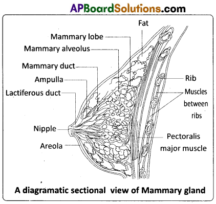
Mammary glands : A functio-nal mammary gland is characteristic of all female mammals. The mammary glands are paired structures ( breasts) that contain glandular tissue and variable amount of fat. The glandular tissue of each breast is divided into 15 – 20 mammary lobes containing clusters of cells called alveoli. The cells of the alveoli secrete milk, which is stored in the cavities (lumens) of the alveoli. The alveoli open into mammary tubules. The tubules of each lobe join to form a mammary duct. Several mammary ducts join to form a wider mammary ampulla which is connected to lactiferous duct through which milk is sucked out by the baby.
![]()
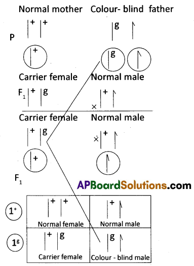
Question 21.
What is crisscross inheritance ? Explain the inheritance of one sex linked recessive character in human.
Answer:
The crisscross pattern of inheritance (skip generation inheritance) is one in which a gene responsible for the sex linked recessive character is transmitted from a male parent.to a male grand child through a carrier female of the first generation. Colour blindness is the best example for criss – cross inheritance in human being.
Colour blindness : It is a sex-linked recessive disorder. Retina of the eye in man contains the cells sensitive to red and green colours. This phenotypic trait is genetically controlled. Its alleles are located on the X – chromosome. When a woman with normal vision (homozygous) marries a colour – blind man, all the sons and daughters are normal, but daughters are carriers (heterozygous). If a carrierjwoman marries a man with normal vision, all the daughters and half of the sons have normal vision and another half of sons are colour – blind. Colour – blind trait is inherited from a male parent to his grand sons through carrier daughter, which is an example of crisscross pattern of inheritance.
Haemophilia : Haemophilia A is recessive X – linked genetic disorder involving lack of the functional clotting Factor – VIII and represents 80% of haemophilia cases. Haemophilia B is also a recessive X – linked genetic disorder involving lack of the functional clotting Factor IX. When a person with hemophilia is injured, bleeding is prolonged because a firm clot is slow to form. Haemophilia follows the characteristic crisscross pattern of inheritance like that of colour – blindness.
Duchenne Muscular dystrophy : Duchenne muscular dystrophy (DMD) is a recessive X – linked form of muscular dystrophy, affecting around 1 in 3,600 boys. The disease is characterized by a progressive weakening of the muscles and loss of coordination. Affected individuals rarely live past their early 20s. The disorder is caused by a mutation in the dystrophin gene (the largest known gene in humans) located on the X – chromosome, which codes for the protein dystrophin, an important structural component within muscle tissue (connects sarcoiemma and the outer most
layer of muscle filaments and supports muscle fiber strength). If the mother is known to be a carrier of this gene, about half of her male children are expected to be affected. All female children born to a carrier mother are expected to be normal, since the possibility of their being homozygous for this sex – linked recessive gene is virtually non – existent.