Thoroughly analyzing AP Inter 2nd Year Zoology Model Papers Set 6 helps students identify their strengths and weaknesses.
AP Inter 2nd Year Zoology Model Paper Set 6 with Solutions
Time: 3 Hours
Maximum Marks: 60
General Instructions:
Note : Read the following instructions carefully.
- Answer all questions of Section – A. Answer ANY SIX questions in Section – B and answer ANY TWO questions in Section – C.
- In Section – A questions from SI. Nos. 1 to 10 are of Very Short Answer Type. Each question carries TWO marks. Every answer may be limited to 5 lines. Answer all these questions at one place in the same order.
- In Section – B, questions from SI. Nos. 11 to 18 are of Short Answer Type. Each question carries FOUR marks. Every answer may be limited to 20 lines.
- In Section – C, questions from SI. Nos. 19 to 21 are of Long Answer Type. Each question carries EIGHT marks. Every answer may be limited to 60 lines.
- Draw labelled diagrams wherever necessary in Sections – B and C.
Section – A (10 × 2 = 20)
Note : Answer ALL the questions.
Question 1.
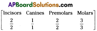
Give the dental formula of adult human beings.
Answer:
Dental formula of adult human being is = \(\frac{2123}{2123}\)
Question 2.
Name two hormones secreted by duodenal mucosa.
Answer:
a) Enterocrinin
b) Secretin
c) Cholecystokinin.
Question 3.
Define cardiac cycle and cardiac output.
Answer:
a) The cardiac events that occur from the beginning of one heart beat to the beginning of the next constitute a cardiac cycle.
b) The volume of blood pumped out by the heart from each ventricle per minute is termed as cardiac output.
![]()
Question 4.
What is triad system ?
Answer:
T – tubule and the two terminal cisternae at its sides form the triad system in a skeletal muscle fibre.
Question 5.
Human skull is described as dicondylic skull. Give the reason.
Answer:
In human skull two occipital condyles are present one on each side of the foramen magnum hence called dicondylic skull.
Question 6.
Which hormone is called anti diuretic hormone ? Write the name of the gland that secretes it.
Answer:
Vasopressin is called antidiuretic hormone (ADH). It is secreted by posterior lobe of pituitary gland.
Question 7.
Write the names of any four mononuclear phagocytes.
Answer:
Mono nuclear phagocytes are
- Histiocytes
- Kupffer cells
- microglia
- osteoclasts
- Synovial cells (any four).
Question 8.
Where are the testis located in Man ? Name the protective covering of each testis.
Answer:
Testis are located outside the abdomen with in a pouch called scrotum. Each testes is enclosed in a fibrous envelope, the tunica albuginea.
Question 9.
Mention any two advantages of in breeding.
Answer:
a) In breeding increases homozygosity.
b) It helps in the accumulation of superior genes and elimination of less defective genes.
Question 10.
List but any four features of cancer cells.
Answer:
a) These are characterised by indefinite growth,
b) Cells are without contact inhibition,
c) Cancer cells are characteristed by evading apoptosis,
d) These cells a typical of parent autonomous and aggressive.
Section – B (6 × 4 = 24)
Note : Answer ANY SIX questions.
Question 11.
What are the major transport mechanisms for CO2 ? Explain.
Answer:
Transport of Carbon Dioxide : CO2 is transported in three ways.
i) in dissolved state : 7 per cent of CO2 is carried in a dissolved state (physical solution) through plasma.
CO2 + H2O → H2CO3
ii) As carbamino compounds : About 20 – 25 per cent of CO2 combines directly with free amino group of the haemoglobin and forms carbamino-haemoglobin in a reversible manner.
Hb – NH2 + CO2 → Hb – NHCOO– + H+
This binding of CO2 is related to the partial pressure of CO2. pO2 is a major factor which could affect this binding. When pCO2 is high and pO2 is low as in the tissues, binding of more carbon dioxide occurs. When pCO2 is low and pO2 is high as in the alveoli, dissociation of CO2 from carbamino – haemoglobin takes place, i.e., CO2 which is bound to haemoglobin from the tissues is delivered at the alveoli. Carbamino compounds are also formed by the union of CO2 with plasma proteins.
iii) As Bicarbonates: About 70 per cent of CO2 is transported as bicarbonate. RBCs contain a very high concentration of the enzyme, carbonic anhydrase and a minute quantity of the same is present in the plasma too. This enzyme facilitates the following reaction in both the directions.
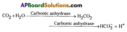
At the tissues where partial pressure of CO2 is high due to catabolism, CO2 diffuses into the blood (RBC and Plasma) and forms carbonic acid which dissociates into HCO3– and H+. At the alveolar site where pCO2 is low, the reaction proceeds in the opposite direction leading to the formation of CO2 and water. Thus CO2 is mostly trapped as bicarbonate at the tissues and transported to the alveoli where it is released out as CO2.
![]()
Question 12.
Give an account of the retina of the human eye.
Answer:
Retina (Nervous tunic): This is the third and inner coat of the eye. It consists of a pigmented epithelium (non – visual portion) and a neural portion (visual portion). The pigmented epithelium is a sheet of melanin – containing epithelial cells that lie between the choroid and the neural portion of the retina. The neural portion of the retina has three layers of retinal neurons namely: photoreceptor layer (the layer closest to the choroid coat), bipolar cell layer and ganglion ceil layer.
Photoreceptor layer consists of two types of photoreceptor cells called rods and cones. Rods contain a purplish – red protein called the rhodopsin or visual purple, which contains a derivative of vitamin – A and they are important in twilight (scotopic vision – the vision of the eye under low light conditions).
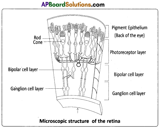
Cones contain a visual pigment (iodopsin, made of a protein called photopsin) and they are important in daylight (photopic) vision and colour vision. There are three types of cones, each having different sensitivity and they provide ‘optimal response’ to red, green and blue colours.
The centre of the posterior portion of the retina is called the macula lutea or yellow spot. A small depression present in the centre of the yellow spot is called fovea centralis, and it contains only cones. Fobea is responsible for sharp, central vision, which is useful while walking, reading, driving etc. The axons of the ganglion cells extend posteriorly and exit the eye ball as the optic nerve. The site of the retina where the optic nerve exits the eye ball is called optic disc or blind spot which is devoid of photoreceptor cells (no image is formed at that spot).
Question 13.
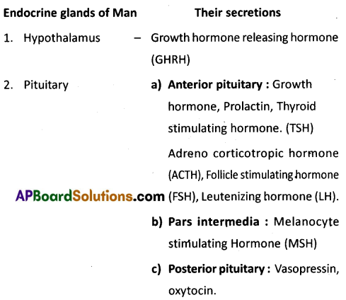
List out the names of endocrine glands present in human beings and mention the hormones they secrete.
Answer:
Question 14.
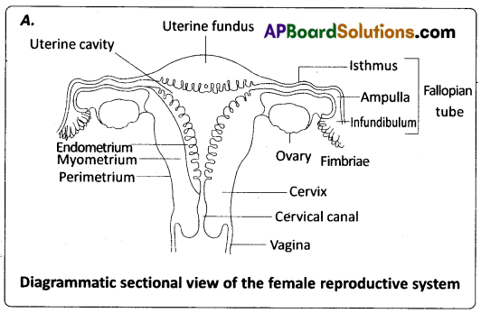
Draw a labelled diagram of the female reproductive system.
Answer:
Question 15.
Describe erythro blastosis foetalis.
Answer:
Destruction of RBC of Rh positive foetus by anti Rh antibodies produced by Rh negative mother due to immunological incompatibility is called Erythroblastosis foetalis or Haemolytic disorder of newborn (HDNB). This is due to genetically incompatible marriage involving Rh positive father and Rh negative mother. At the time of birth the Rh positive foetal blood mixes with the Rh negative blood of mother, through the ruptured placenta. The Rh antigens sensitize the mother to produce anti Rh antibodies (IgG antibodies) and memory cells. This first Rh positive by is unaffected because it is delivered by the time mother is sensitized. During the next pregnancy bearing Rh positive foetus these antibodies increase in concentration due to memory cells and cross the placenta, enter the blood of baby and destroy the RBC. Haemolytic anaemia is the symptom in this disorder.
Question 16.
Write a short note on the theory of mutations.
Answer:
Mutation theory : It was proposed by Hugo de Vries, a Dutch botanist who coined the term ‘mutation’. Mutations are sudden, random inheritable changes that occur in organisms. He found four different forms in Oenothera lamarckiana (commonly called ‘evening primrose’) such as 0. brevistylis – small style, 0. levifolia – smooth leaves. 0. gigas- the giant form, O. nanella- the dwarf form (mutant varieties). T.H. Morgan studied the inheritance pattern of mutations in Drosophila melanogaster. Darwin called mutations (large variations) sports of nature or saltations, whereas Bateson called them discontinuous variations.
Salient Features of Mutation theory:
- Mutations occur from time to time in naturally breeding populations.
- They are discontinuous and are not accumulated over generations.
- They are full-fledged, and so there are no ‘intermediate forms’.
- They are subjected to Natural Selection.
Question 17.
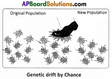
What is meant by genetic drift ? Explain genetic drift citing the example of founder effect.
Answer:
Genetic Drift: The change in the frequency of a gene that occurs merely by chance and not by selection, in small populations, is called genetic drift or Sewail Wright effect. Suppose, for a gene with two alleles, the frequency of a particular allele is 1% (q = 0.01), the probability of losing that allele by chance from the small population is more. The end result is either Fixation (p or q = 1) or Loss (p or q = 0) of that allele. The probability of reaching the end point depends on the size of the population. Genetic drift tends to reduce the amount of genetic variation within the population mainly by removing the alleles with low frequencies, it can be exemplified by the Founder Effect and Bottleneck Effect.
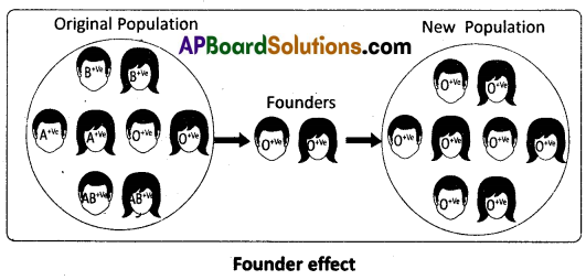
Founder effect: Ifa small group of individuals from a population start a new colony in an isolated region, those individuals are called the founders of the new population. The allelic frequencies of their descendants are similar to those of the founders rather than to their ancestral parent population, e.g. presence of O+ve blood group in nearly 100% of the Red-lndians. It means the forefathers of the Red Indian tribe were predominantly O+ve ve and they isolated themselves reproductively from other populations.
![]()
Question 18.
Honey bees are economically important. Justify.
Answer:
Economic importance of Honey bees : The bee products like Honey, wax, propolis and bee venom are used in various ways.
- Honey is a rich source of fructose, water, glucose, minerals and vitamins.
- Bee’s wax is used in the preparation of cosmetics, polishes of various kinds and candles.
- Propolis isused in the treatment of inflammation and superficial burns.
- Bee’s venom, which is extracted from the sting of worker bees, is used in the treatment of rheumatoid arthritis.
- Pollination : Bees are the pollinators of our crop plants such as sunflower, brassica, apple and pear.
Section – C (2 × 8 = 16)
Note : Answer ANY TWO of the following questions.
Question 19.
Describe the Excretory system of man, giving the structure of a nephron.
Answer:
Human Excretory System : In humans, the excretory system consists of a pair of kidneys, a pair of ureters, a urinary bladder and urethra.
Kidneys : Kidneys are reddish brown, bean shaped structures, situated on either side of the vertebral column between the levels of the last thoracic and third lumbar vertebrae, in a ‘retroperitoneal position’. The right kidney is slightly lower than the left one due to the presence of liver.
The outer surface of the kidney is convex and the inner surface has a deep notch called hilum, the point at which the renal artery and nerves enter and the renal vein and ureter leave. Each kidney is surrounded by a tough, fibrous capsule that protects its delicate inner surface.
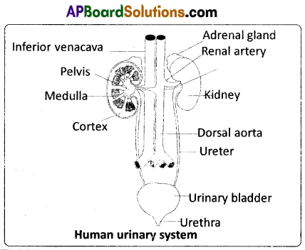
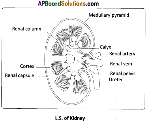
Internal structure : A longitudinal section of the human kidney shows two distinct regions, the outer cortex and the inner medulla. The medulla is divided into multiple cone shaped masses of tissue called renal pyramids. The renal pyramids are separated by the projections of the cortex called columns of Bertin (renal column). The base of each pyramid originates at the border between the cortex and the medulla and terminates in the renal papilla. Renal papillae project into cup like calyces, formed by the funnel shaped pelvis, which continues out as the ureter.
Ureters : These are slender whitish tubes which emerge from the pelvis of the kidneys. Their walls are lined by ‘transitional epithelium’. The ureters run downwards and open into the urinary bladder.
Urinary bladder: It is a median storage sac, situated in the lower abdominal cavity. It has thick, muscular, distensible wall lined by ‘transitional epithelium’. The neck of the bladder leads into the urethra, which has an internal urethral sphincter (made of smooth muscles) and external urethral sphincter (made of striped muscles). Urethra opens near the vaginal orifice in the female and through penis in the males.
Structure of a Nephron : Each kidney has nearly one million nephrons which are the ‘structural’ and ‘functional’ units. Each nephron has two parts – the ‘Bowman’s capsule’ and the renal tubule. The Bowmans’ capsule encloses a tuft of capillaries called glomerulus, formed by the afferent renal arteriole – a fine branch of the renal artery. Blood from the glomerulus is carried away by an efferent renal arteriole of a lesser diameter. The blind end of the tubule forms a double walled cup called Bowman’s capsule, which surrounds the glomerulus.
The inner wall of the Bowman’s capsule has certain unique cells called podocytes which wrap around each capillary. The podocytes are arranged in an intricate manner so as to leave some minute spaces called ‘filtration slits’ or ‘slit pores’. The endothelial cells of the capillaries have numerous pores or ‘fenestrations’. The glomerulus along with the Bowman’s capsule constitutes the Malphigian body or renal corpuscle.
The tubule continues further and forms a highly coiled proximal convoluted tubule (PCT). A hairpin shaped Henle’s loop, which has descending and ascending limbs, is the next part of the tubule. The proximal part of the ascending limb is thin and the distal part is thick. The thick ascending limb continues into the distal convoluted tubule (DCT). The DCT continues as the ‘initial collecting duct’ in the cortex. Some initial collecting ducts unite to form a straight collecting duct, which passes through the medullary pyramid. In the medulla, the tubes of each pyramid join and form the duct of Bellini, which finally opens on the tip of the renal papilla. The contents of the duct of Bellini are discharged into the renal pelvis through the renal calyx.
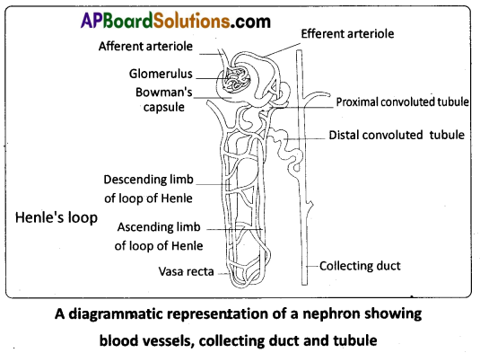
The Malpighian corpuscle, PCT and DCT of a nephron are situated in the cortical region of the kidney, whereas the loop of Henle is in the medulla. In a majority of nephrons, the loop of Henle is too short and extends only very little into the medulla. Such nephrons are called cortical nephrons. In some of the nephrons, the loops of Henle are very long and run deep into the medulla. These nephrons are called juxtamedullary nephrons.
The efferent arteriole emerging from the glomerulus forms a fine capillary net work called the peritubular capillaries, around the renal tubule. The portion of the peritubular capillaries that surrounds the loop of Henle is called the vasa recta. The vasa recta is absent or highly reduced in the cortical nephrdns. The juxtamedullary nephrons possess well developed vasa recta.
![]()
Question 20.
Write an essay on different events that occur during development of a human.
Answer:
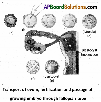
Cleavage : The first phase of embryonic development, the cleavage is holoblastic (because of microlecithal condition of egg), radial, indeterminate and unequal. The mitotic division (cleavage) starts as the zygote moves through the isthmus of the oviduct towards the uterus. The daughter cells are called blastomeres.
Morula : The embryo with 8 – 16 blastomeres looks like a ’mulberry’ and is called the morula. Morula is a solid mass of cells. The morula develops in the fallopian tube and reaches the uterus for further development. It is still surrounded by the zona pellucida. Due to unequal cleavage smaller and larger blastomeres are formed. The morula passes through a process called compaction. Now the embryo has a superficial flat cell layer and inner cell mass. The outer/supeficial layer becomes. the trophoblast or trophectoderm. The trophoblast serves to attach the embryo to the uterine wall by forming trophoblastic villi. The inner cell mass constitutes formative cells. The inner cell mass gives rise to the embryo proper and is, therefore, also called the embryoblast. This is the first sign of cell differentiation (cells becoming different) in the human embryo.
Blastocyst : Some fluid now passes into the morula from the uterine cavity, and partially separates the cells of the inner cell mass from those of the trophoblast. As the quantity of the fluid increases, the morula acquires the shape of a cyst. The cells of the trophoblast become flattened, and the inner cell mass comes to be attached to the inner side of the trophoblast on one side only. The morqja now becomes a blastocyst.
The cavity of the blastocyst is the blastocoel or segmentation cavity or primary body cavity. The side of the blastocyst to which the inner cell mass is attached is called the embryonic or animal pole, while the opposite side is the bembryonic pole. The cells of the trophoblast above the region of inner cell mass are called cells of Rauber.
Implantation: The zona pellucida present around the blastocyst gradually disappears and the cells of the trophoblast stick to the uterine endometrium. The trophoblast invades the endometrium of the uterus. The blastocyst gets implanted into the uterine mucosa till the whole of it comes to lie within the thickness of the endometrium. This is called interstitial implantation. In humans, implantation begins on the 6th day after fertilisation. The process of implantation is aided by proteolitic enzymes produced by the trophoblast. The uterine mucosa also aids the process.
Afterthe implantation of the embryo,the uterine endometrium is differentiated into what is called decidua. The portion of the decidua where the placenta is to be formed (i.e., deep to the developing blastocyst) is called the decidua basalis. The part of the decidua that separates the embryo from the uterine lumen is called the decidua capsularis. The part lining the rest of the uterine cavity is called the decidua parietalis. At the end of pregnancy the decidua is shed off, along with the placenta and membranes.
Formation of Bilaminar Embryonic Disc : Implantation of the blastocyst is completed by the end of the second week. The inner cell mass forms into a disc called embryonic disc. Soon, the ‘cells of Rauber’ disappear exposing the embryonic disc. From the lower part of inner embryonic disc, some cells get separated by delamination and eventually form a layer of cells on the inner surface of the embryonic disc i.e., on the surface facing the cavity. This cell layer develops into the hypoblast, which is the future extra embryonic endoderm. The remaining part of the embryonic disc is called epiblast. Now the embryonic disc is called bilaminar embryonic disc.
The cells of the hypoblast increase in number and spread along the inner surface of the trophoblast. This hypoblast layer below the trophoblast finally encloses a cavity called yolk sac or umbilical vesicle. Meanwhile, the thickness of the embryonic disc increases towards the caudal end. Gradually the embryonic disc becomes oval.
Gastrulation : Gastrulation involves proliferation, differentiation and movement of cells within the embryo. Along the longitudinal axis of the embryonic disc, a primitive streak is formed. A longitudinal furrow known as primitive groove forms along the middle of the primitive streak. On either side of it are the primitive folds. Anteriorly primitive streak has a shallow primitive pit. The anterior end of the primitive streak becomes thickened. This thickened part of the streak is called the primitive knot or primitive node or Hensen’s node.
Trilaminar Embryo – Formation of Primary Germ Layers : ingression, the future endodermal cells from the epiblast, replaces the hypoblast and forms the endoderm of the embryo. The future mesodermal cells converge towards the primitive folds, involute through the primitive groove and reach between epiblast and endoderm. The remaining epiblast is now known as the ectoderm. This invasion of the epiblast cells into the space between the epiblast and hypoblast is called gastrulation. Thus, the ectoderm, mesoderm and endoderm are all derived from the epiblast. The process of gastrulation converts the bilaminar embryonic disc into a trilaminar embryonic disc.
Extraembryonic Membranes : During the development of the human embryo, as in ail other amniotes four extraembryonic or foetal membranes are formed. They are chorion, amnion, allantois, and yolk sac. From the blastodisc, amniotic folds called head fold, tail fold and lateral fold are developed as somatopleure. As the folds fuse they are differentiated into outer chorion and inner amnion. Between the amnion and the embryo, there is an amniotic cavity filled with amniotic fluid. Amnion protects the
embryo as the amniotic fluid acts as a shock absorber and also prevents the embryo form desiccation. The chorion develops a rich supply of blood vessels and forms an intimate association with the endometrium of the uterus.
Allantois and yolk sac are derived from the splanchnopleure. Allantois is formed from the hind gut as an evagination. It stores the waste materials. Allantois and chorion are fused to form chorio allantoic membrane which constitutes the placenta. Yolk sac encloses a fluid filled cavity. It is connected to the mid gut. It has no nutritive role.
After gastrulation is complete and any extra embryonic membranes are formed, the next stage of embryonic development begins with organ formation.
Organogenesis : During organogenesis, regions of the three embryonic germ layers develop into the rudiments of organs. Whereas gastrulation involves mass movements of cells, organogenesis involves more localized changes.
Formation of the Notochord and Neural Tube : The chorda mesodermal cells converge and involute through the Hensen’s node and extend forwards as notochordal process. This is later transformed into a solid rod – the notochord, the embryonic axial skeleton which is replaced by the vertebral column. The notochordal mesoderm induces the overlying ectodermal cells to form the neural plate. This is a good example of induction. At first the neural plate is oval but later elongates oves the underlying notochord along the longitudinal axis of the embryo. The plate invaginates towards the notochord to form a neural groove, which deepens progressively to form a tube by fusion of the lateral neural folds. The process of formation of neural tube is referred to as neurulation.
Differentiation of Mesoderm and Formation of Coelom : The intra embryonic mesoderm spreads in all directions between the outer ectoderm and inner endoderm. The longitudinal column of mesoderm adjacent to the notochord and neural tube on either side is called epimere. The mesoderm around the gut is the hypomere. The mesoderm in between these two is the mesomere. The epimere becomes segmented into cubical blocks called somites or metameres. Each somite differentiates into myotome, sclerciome and dermatome. The sclerotome forms the vertebral column The dermatome forms the dermis of the skin and other connective tissues. The myotome forms the voluntary muscles of the body. The mesomere forms the urinogenital organs and their. The hypomere splits into outer somatic and inner splanchnic mesodermal layers. Intra embryonic coelom is formed between these two layers. It gives rise to pericardial, pleural, peritoneal cavities etc.
![]()
Question 21.
Describe chromosomal theory of Sex determination.
Answer:
Sex Determination: The mechanism of sex determination has always been a puzzle to geneticists. In fact, the cytological observations made in a number of insects led to the development of the concept of genetic / chromosomal basis of sex determination.
Sex Chromosomes : In most of the animals a pair of chromosomes is responsible for the determination of sex. These two chromosomes are called sex chromosomes or allosomes. The chromosomes other than the sex chromosomes are called autosomes. The first indication that sex chromosomes were distinct from the Other chromosomes came from the experiments conducted by Henking. He could trace a specific nuclear structure all through spermatogenesis in wasps, and it was also observed by him that 50 percent of the spermatozoa received this structure after spermatogenesis, whereas the other 50 percent did not receive it. Henking gave the name X – body to this structure, but he could not explain its significance. Further investigations by other scientists led to the conclusion that the X – body of Henking was in fact a chromosome and that is why it was given the name X – chromosome. Stevens and Wilson first identified Y – chromosome as a sex determining chromosome in the mealworm, Tenebrio molitor. The revealed that the chromosomal basis of sex depended 6n the presence or absence of the Y – chromosome.
Heterogametic Sex Determination: Heterogametic sex refers to the sex of a species in which the sex chromosomes are not similar. The process of sex determination by allosomes is called genetic or chromosomal sex determination. In the heterogametic sex determination, one of the sexes produces ‘similar’ gametes and the other sex (heterogametic sex) produces ‘dissimilar / unlike gametes. The sex of the young one is determined at the time of syngamy (fertilization). It depends on which gamete of the two dissimilar gametes unites with the other gamete produced by the ‘homogametic parent’.
Male Heterogamety : In this method of sex determination, the males (heterogametic) produce dissimilar gametes while females (homogametic) produce similar gametes. Male heterogamety is of two kinds, XX – XO type and XX – XY type.
XX – XO type : In some insects such as bugs, grasshoppers and cockroaches, females are with two X – chromosomes and males are with one X – chromosome in each somatic cell. Me Clung discovered this type in grasshoppers. The unpaired X – chromosome determines the male sex. The karyotype of the female (homogametic) is AAXX and that of the male (hetero¬gametic) is AAXO.
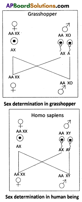
All the ova contain ‘AX’ complement of chromosomes and the sperms are of two types. One half of the sperms have ‘AX’ complement and the other half have’A’ complement of chromosomes. The sex of the offspring depends on the types of sperm that fertilizes the ovum.
XX – XY type: In human beings and some insects such as Drosophila, both females and males have the same number of chromosomes. The karyotype of the female is AAXX and that of the male is AAXY. Females are ‘homogametic’ with ‘XX’ chromosomes.
They produce similar ova having one X – chromosome each. Males are ‘heterogametic’ with X and Y – chromosomes. They produce two kinds of sperms; one half of them with X – chromosome and the other half with Y – chromosome. The sex of the offspring depends on the fertilizing sperm. The XX – XY type is also found in most other mammals.
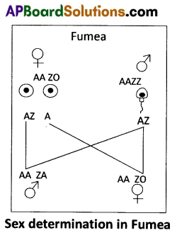
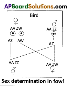
Female Heterogamety: In this method of sex determination, the males produce ‘similar gametes’ while females produce ‘dissimilar gametes’. Female heterogamety is of two kinds, ZO – ZZ type and ZW – ZZ type.
ZO – ZZ type: In mothsand some butterflies, female is heterogametic with one Z-chromosome (ZO) and male is homogametic with two Z – chromosomes (ZZ). The karyotype of female is AAZO and male is AAZZ. Females produce two kinds of ova, half of them with a Z – chromosome and the other half with no sex.
chromosome. Males produce similar type of sperms. The sex of he off spring depends on the type of ovum that is fertilized.
ZW – ZZ type : In birds, reptiles, some fishes, etc., the females are heterogametic with ZW – allosomes and males are homogametic with ZZ – allosomes. The karyotype of the female is AAZW and that of the male is AAZZ. All sperms are similar with the allosome -Z. Ova are of two different kinds; one half of the ova are with the allosome – Z and the other half with the allosome – W. The sex of the offspring depends on the type of ovum that is fertilized.
Sex Determination in Humans : It has already been mentioned that the sex determining mechanism in case of human is XX – XY type. Out of 23 pairs of chromosomes present, 22 pairs are exactly same in both males and females; these are the autosomes. A pair of X- chromosome is present in the female, whereas the presence of an X and Y – chromosome are determinant of the male characteristic. During spermatogenesis among males, two types of gametes are produced. 50 percent of the total sperm produced carry the X – chromosome and the rest 50 percent has Y – chromosome besides the autosomes.
Females, however, produce produce only one type of ovum with an X – chromosome. There is an equal probability of fertilisation of the ovum by the sperm carrying either X or Y-chromosome. In case the ovum is fertilised by a sperm carrying X – chromosome, the zygote develops into a female and the fertilisation of ovum with Y – chromosome carrying sperm results into a male offspring. Thus, it is evident that it is the genetic makeup of the sperm that determines the sex of the child. It is also evident that in each pregnancy there is always 50 percent probability of either a male or a female child.