
AP State Syllabus AP Board 9th Class Biology Solutions Chapter 3 Animal Tissues Textbook Questions and Answers.
AP State Syllabus 9th Class Biology Solutions 3rd Lesson Animal Tissues
9th Class Biology 3rd Lesson Animal Tissues Textbook Questions and Answers
Improve Your Learning
Question 1.
What do you understand by the term tissue? (AS 1)
Answer:
Tissue is a group of cells similar in structure and function.
Eg : Nerve tissue, epithelial tissue, muscle tissue etc.
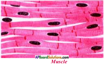
Question 2.
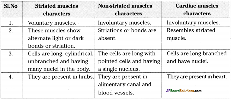
Show the difference between the three types of muscle fibres with diagrams. (AS 1)
Answer:
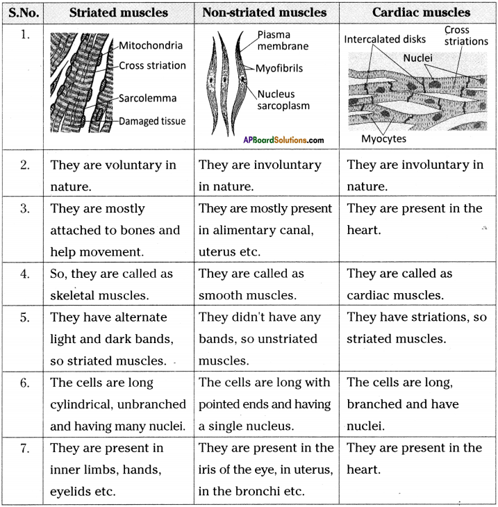
Question 3.
What is the specific function of the cardiac muscle? (AS 1)
Answer:
Specific function of the cardiac muscle :
- Cardiac muscle present in the heart.
- It is responsible for pumping of blood.
![]()
Question 4.
Differentiate between striated and unstriated muscles on the basis of their shape and site/location in the body. (AS 1)
Answer:
| Striated muscle | Unstriated muscle |
| Shape : Cells in striated muscle are long cylindrical and unbranched. |
Cells in unstriated muscle are long with pointed ends. |
| Site / Location : These are located in limbs and attached to skeleton. |
These are located in Alimentary canal, blood vessels, Iris of the eye, in uterus and in the bronchi of lungs. |
Question 5.
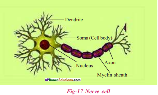
Draw a neatly labelled diagram of a neuron. (AS 3)
Answer:
Question 6.
Name the following. (AS 1)
a) Tissue that forms the inner lining of our mouth.
b) Tissue that connects muscle to bone in humans.
c) Tissue that transports food in animals.
d) Tissue that stores fat in our body.
e) Connective tissue present in the brain.
Answer:
a) Epithelial cells
b) Tendon
c) Connective tissue/blood
d) Adipose tissue
e) Areolar tissue
![]()
Question 7.
Identify the types of tissue in the following : Linings of the organs, skin, bone, internal lining of kidney tubule. (AS 1)
Answer:
| Linings of the organs | Epithelial tissue |
| Skin | Stratified squamous epithelium(epithelial tissue) |
| Bone | Connective tissue |
| Internal lining of Kidney tubule | Cuboidal epithelial tissue. |
Question 8.
If the platelets are not present in the blood, what happens? (AS 2)
Answer:
- If the platelets are not present in the blood, blood loss may be more from the injury.
- Whenever a blood vessel is injured, at the site of injury formation of a blood clot will not takes place.
- The wound will not be sealed by the clot.
Question 9.
If you touch at elbow, you get a shock like feeling. Why? (AS 7)
Answer:
- In human beings ulnar nerve runs from the shoulder to the hand.
- The ulnar nerve comes close to the surface near the elbow.
- Due to the superficial location it is not protected by muscle, fat or other soft tissues.
- Thinner skin layer around bone at elbow makes ulnar nerve more receptive for any small stimuli.
- That is the reason for getting a shock like feeling if we touch at elbow.
![]()
Question 10.
Why the blood is called a connective tissue?
Answer:
Connective tissue :
A loosely spaced tissue mainly carrying different materials to different parts of the body as well as rendering support, making connection between organs is called connective tissue.
Blood is considered as connective tissue because of the following reasons.
- Blood connects different organs of our body together by carrying oxygen, nutrients, hormones and other signaling molecules and removing the waste.
- It has all the three components of connective tissue i.e., cells, fibers and matrix.
- Similar to other connective tissues, blood is rich in fibres like collagen fibers and blood clotting fibres.
- Blood originates from the mesodermal layer of the embryo from which all other connective tissues also originate.
Question 11.
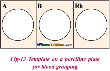
Write the procedure to identify your blood group with the help of kit. (As 3)
Answer:
Aim :
Identification of blood group.
Apparatus :
Blood identification kit, glass slide, wax pencil, disposable needle.
Materials used :
Cotton, 70/6 alcohol, toothpicks.
Procedure:
- Take one porcelain plate, clean and dry it.
- With a wax pencil, draw three circles on the plate to divide the surface into three parts and draw three circles.
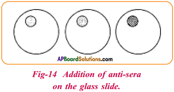
- Place one drop of the corresponding antiserum near the edge of the circles.
- Clean the fingertip with an alcohol and let it dry.
- Press on the bottom of the fingertip with the thumb and quickly prick the fingertip with the help, of a needle.
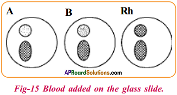
- Quickly, let one drop of blood get into each circle, but not touching the anti-serum.
- Apply gently pressure to the wound with cotton ball.
- Use a toothpick to mix the blood and anti-serum and stir gently.
- Watch to see if any of the samples show agglutination.
Result and Inference :
By using the following table determine the blood group.
| Anti – A | Anti – B | Type |
| Yes | No | A |
| No | Yes | B |
| Yes | Yes | AB |
| No | No | O |
If agglutination occurs in anti – RhD serum, the Rh factor is positive, and if it does not . the Rh factor is negative.
Note :
1. See the needle is sterile.
2. Usually choose left ring finger.
3. Don’t use same needle to other body.
![]()
Question 12.
Ramu felt weak. Ramu’s father took him to hospital. The doctor advised a blood test. The report says that he does not have the required levels of haemoglobin. What are its ill effects?
Answer:
Ill effects of haemoglobin :
- Blood is red in colour due to the presence of red coloured protein called haemoglobin.
- Haemoglobin helps in the transport of oxygen and carbon dioxide.
- Low haemoglobin is the main cause for anemia.
- If there is not enough haemoglobin in blood. The oxygen supply to various parts will be less, which causes shortness of breath.
- Low haemoglobin levels many aggravate extant heart problems.
- People with low haemoglobin levels get very tired as their cells do not get enough oxygen to perform their activities.
Question 13.
How blood test is useful to diagnose the disease? Explain with daily life situation. (AS 1)
Answer:
- Blood test is useful to diagnose diseases such as malaria, typhoid, cancer, HIV/AIDS, diabetes, anemia coronary heart disease, abnormalities in the functioning of kidney, liver, thyroid, etc.
- Abnormal red blood cell levels might be a sign of anemia. Dehydration, bleeding, and other disorder.
- Complete blood count with differential can measure the amounts of different types of white blood cells in our body.
- Abnormal white blood cell levels might be a sign of infection, blood cancer or an immune system disorder.
- Abnormal platelet levels might be a sign of a bleeding disorder or thrombotic disorder.
- Abnormal haemoglobin levels might be a sign of anemia, sickle cell anemia or thalassemia.
- Abnormal glucose levels in the blood might be a sign of diabetes.
- Abnormal calcium levels in the blood might suggest kidney problems, bone disease, thyroid disease, cancer, or malnutrition.
- Abnormal electrolyte levels might be a sign of dehydration, kidney disease, liver disease, heart failure or high B.P.
- Abnormal Blood Urea Nitrogen (BUN) and creatinine levels might suggest a kidney disease.
- High levels of enzymes like Troponin and creatine kinase is a sign of Heart attack.
- Abnormal cholestrol or triglyceride levels might be a sign of increased risk of coronary heart disease.
- Abnormal coagulation pannel test results might suggest risk of bleeding or developing clots in blood vessels.
- Existence of microorganisms or their antibodies in the blood suggest occurence of corresponding disease.
E.g. : Plasmodium – Malaria, HIV – AIDS etc.
Question 14.
Collect the old blood reports of your friends/relatives and prepare a project report on the contents of the blood.
Answer:
On collection and observation of old blood reports I came to know that the contents of blood should present in definite proportions such as.
| Content of blood | Lower and upper limits |
| WBC | 5.0 – 10.0 103 cells / ul |
| RBC | 3.5 -5.5 106 cells/ul |
| HgB | Men 12 -16 g/dL; Women 9.9 – 13 g/dL |
| PLT (Platelet count) | 1.0-3.0 105 cells/ ul |
| Neutrophil | 40 – 75% |
| Lymphocytes | 20 – 45% |
| Eosinophil | 1 – 6% |
| Basophil | 0-1% |
| Monocyte | 0-3% |
9th Class Biology 3rd Lesson Animal Tissues InText Questions and Answers
9th Class Biology Textbook Page No. 29
Question 1.
Why do old people shiver in winter when compared to youngsters? Is there any insulator like substance to prevent the escape of heat energy during winter?
Answer:
- Old people shivers in winter when compared to youngsters.
- They didn’t have enough fat storages below the skin.
- Fat storing adipose tissue is found below the skin and between internal organs.
- The cells of this tissue are filled with fat globules.
- Storage of fat also acts as insulator.
![]()
Question 2.
Which tissue gives definite shape to body of vertebrae?
Answer:
- Bone is one type of connective tissue.
- It forms the frame work that supports the body.
- It is a major component of the skeletal system of several vertebrae.
9th Class Biology Textbook Page No. 34
Question 3.
During winter, body shivers. Why?
Answer:
- When the body is exposed to cold air, we shiver.
- During shivering muscles contract and relax and produce large amount of heat.
- This keeps the body heat.
- It is one type of defensive mechanism of the body.
9th Class Biology Textbook Page No. 30
Question 4.
Blood is a type of connective tissue. Why is it called connective tissue?
Answer:
Blood is considered as connective tissue because of the following reasons.
- Blood connects different organs of our body together by carrying oxygen, nutrients, hormones, and other signaling molecules and removing the waste.
- It has all the three components of connective tissue i.e., cells, fibers, and matrix.
- Similar to other connective tissues, blood is rich in fibres like collagen fibers and blood clotting fibres.
- Blood originates from the mesodermal layer of the embryo from which ail other connective tissues also originate.
9th Class Biology 3rd Lesson Animal Tissues Activities
Lab Activity – 1
Question 1.
Aim:
Identification of tissue in collected sample.
Apparatus:
Microscope, slide, dilute HCl, forceps, brush.
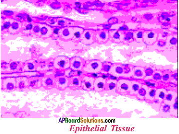
Procedure:
- Collect a small piece of chicken with bone from your nearby chicken centres or market.
- Put it in dilute HCl for two hours.
- Take the skin part of chicken piece.
- Place the material with forceps or brush on the slide
- Then keep the another slide on it and press both the slides gently.
- Place a cover slip tap on it and observe under microscope.
- Draw the diagram of what you have observed under microscope in your notebook.
- Compare your diagram with the given picture.
Answer these questions.
1. Are all the cells similar?
Answer:
Yes. All the cells are similar.
2. How are they arranged?
Answer:
They are arranged in layers. Each cell is round and nucleated. Observed diagram
3. Are these cells tightly packed and formed as continuous sheath?
Answer:
Yes. The cells are tightly packed and formed as continuous sheath.
4. Is there any intercellular space?
Answer:
No. There is no intercellular space.
5. Think, why these cells look like continuous sheath.
Answer:
These cells are look like continuous sheath because there is no intercellular space and the cells are tightly packed.
6. Does this tissue covering protect inside and outside of the animal body?
Answer:
Yes. This tissue covering protect inside and outside of the animal body.
![]()
Question 2.
Aim:
Identification of tissue in collected sample.
Apparatus:
Microscope, slide, blood sample, syringe, cotton.
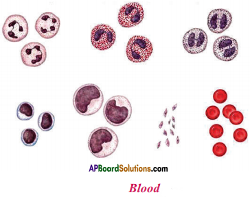
Procedure:
- Take a sterilized syringe needle.
- Collect one drop of blood from finger tip by pricking with syringe needle.
- Take a slide. Keep the finger on the slide to collect one drop of blood.
- Put another slide on it gently and press both :
- Observe under microscope.
- Draw the diagram of what you observe L microscope in your notebook. Compare diagram with the given picture.
In this procedure we can identify red blood
Question 3.
Aim:
Identification of tissue in collected sample
Apparatus:
Microscope, slide, dilute HCl, vinegar, forceps.
Procedure:
- Take a piece of muscle of chicken.
- Put in diluted HCl or vinegar and leave it for two hours.
- Next morning collect the piece of muscle on a slide with forceps.
- Press gently with another slide, put few drops of water and place a cover slip on it.
- Observe under microscope. Observed diagram
- Draw the diagram what you have observed under microscope in your notebook. Compare your diagram with the above picture.
Answer these questions.
1. How are the cells arranged?
Answer:
Cells are arranged in layers one above the other.
2. Do you find any difference between skin cells and muscle cells?
Answer:
Muscle cells are long and nucleated.
3. If you want to observe the bone tissue in the chicken bone, settle it in vinegar or diluted HCl over night. Then only the bone becomes soft. Take a piece from it by using knife. Do you find any relation among these tissues?
Answer:
Usually muscle tissue is attached to bones.
4. Is this tissue useful for movements in our body?
Answer:
Yes. This tissue is useful for movements in our body.
Activity – 1
Question 4.
1. Collect the substance lining of mouth by using wooden spoon and observe this under microscope.
2. Draw the diagram that you observed in the microscope, in your notebook.
a) How are the cells arranged?
Answer:
Cells are extremely thin and flat and form a delicate lining.
b) Are there any intercellular spaces?
Answer:
No. Intercellular spaces are absent.
c) Think, why are the epithelial cells in skin are arranged in the form of layers?
Answer:
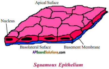
Because skin has to protect our body from cold, heat etc.
d) If you drink hot tea or chilled cool drink, how would you feel?
Answer:
Inner layers of our mouth cannot bear hot tea or chilled cool drink. We immediately spill hot or cold substances from our mouth.
e) If your skin burns or wounded, which tissue would effected ?
Answer:
Epithelial tissue.
Activity – 2
Question 5.
1. Take a permanent slide of cuboidal epithelium from your laboratory slide box and observe under microscope.
2. Draw the picture in your notebook.
3. How are the cells arranged?
Answer:
The cells are compactly arranged without intercellular spaces.
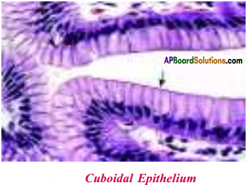
4. Conclusion :
These are the cuboidal epithelial cells which form the lining of organs or tubules or other parts and provide mainly mechanical support.
Activity – 3
Question 6.
1. Take a permanent slide of columnar epithelium from the slide box and observe under microscope.
2. Draw the figure that you observed under microscope. Observed diagram
3. How are the cells? Do you find any hair like projections on the outer surface of epithelial cells?
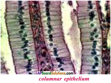
Answer:
a) The cells are long, compactly arranged without intercellular spaces.
b) Hair like projections are present on the outer surface of these cells.
c) These type of epithelial cells are present in the small intestine.
Activity – 4
Question 7.
1. Invite a scientist or doctor to your place.
2. Record an interview about blood structure and its functions.
3. It is important to make a questionnaire in order to conduct an interview.
4. After completion of interview, prepare a booklet about blood and display it on bulletin board or classroom library.
Booklet about blood.
- Blood is a fluid connective tissue.
- There are different types of cells in blood and each one has a different function.
- All the cells in the blood cells float freely in the plasma.
- Extracellular space is filled with fluid called plasma. There are no fibres in the blood.
- Normal adult human beings have about 5 litres of blood. A chief component in plasma is water.
- Besides water it also has several nutrients such as glucose, aminoacids, proteins, vitamins and hormones.
- Plasma also contain factors responsible for blood clotting. Heparine helps to prevent blood clotting in blood vessels.
- Cells present in blood are corpuscles. They are three types l.RBC, 2. WBC, 3. Blood platelets.
- Red blood cells are also known as erythrocytes. They are red in colour due to the presence of haemoglobin.
- haemoglobin helps in transport of oxygen and carbon dioxide.
- When we are in mother’s womb, RBC are formed in the liver and spleen. After birth RBC are generated from the bone marrow of long bone.
- RBC live for 120 days.
- The second type of cells present in blood are white blood cells, which do not have haemoglobin. Hence they are called leucocytes.
- There are two types of cells in WBC – granulocytes and agranulocytes.
- There are three types of cells in the granulocytes – Neutrophils, Basophils and Esinophils.
- These cells attack and destroy the microorganisms that enter the blood.
- There are two types of agranulocytes – lymphocytes and monocytes.
- Lymphocytes secret anti – bodies to guard against foreign material that enter into blood. So they are called microscopic policemen.
- Monocytes move like amoeba and along with granulocytes. The foreign materials are destroyed inside these cells. They are called as ‘scavengers’.
- Blood platelets are a separate group of cells which do not have a nucleus. They help in blood clotting.
Lab Activity – 2
Question 8.
Aim:
Identification of blood group.
Apparatus:
Blood identification kit, glass slide, wax pencil, disposable needle, cotton, tooth picks, 70% alcohol.
Kit components:
| Components | Quantity (100 tests) |
| 1. anti-A sera | 5 ml |
| 2. anti-B sera | 5 ml |
| 3. anti-RhD sera | 5 ml |
| 4. porcelaine white plate | 2 |
| 5. wax pencil | 1 |
| 6. needle (24G) | 100 |
| 7. instructional mannual | 1 |
Procedure:
- Take one porcelain plate, clean and dry it. The plate must be very clean so that it does not interfere with the reaction.
- With a wax pencil, draw three circles on the plate to divide the surface into three parts and draw three circles, one in each part as shown in figure.
- Place one drop of the corresponding antiserum near the edge but within each of the circles as shown in figure.
- Choose a left ring finger clean it with alcohol in a cotton ball and let it dry. Keep the cotton ball nearby, as it is needed again. Dangle the hand down to increase the amount of blood in the fingers.
- Press on the bottom of the finger tip with the thumb of the same hand and quickly prick the finger tip with the help of a needle.
- Quickly, let one drop of blood get into each circle but not touching the anti-sera.
- After putting three drops of blood, apply gentle pressure to the wound with cotton ball.
- Use a toothpick to mix the blood and antiserum and stir gently. Do it for each of the circles using a fresh toothpick every time.
- Watch to see if any of the samples show agglutination.
Result and inference :
Determine the blood type depending on the result. Following table can be used to determine the blood type :
| Anti – A | Anti – B | Type |
| Yes | No | A |
| No | Yes | B |
| Yes | Yes | AB |
| No | No | 0 |
If agglutination occurs in anti-RhD serum, the Rh factor is positive; and if it does not, the Rh factor is negative.
Result should be noted in the given table :
| Name | Blood group |
| Ramu | O |
| Gopal | B |
| Krishna | AB |
| Apparao | A |
| Gupta | B |
Activity – 5
Question 9.
Collect three types of muscle slides (Striated muscles, Non-striated muscles, Cardiac muscles) from slide box. Then observe these under microscope. Write your findings in the following table.
Activity – 6
Question 10.
Collect the slide of nerve cells from the slide box. Observe it under microscope. Write your findings.
Answer:
- We can identify three distinct parts in nerve cells.
- They are
1. Cell body or cyton,
2. Axon and
3. Dendrites - Cell body or cyton has a large nucleus and cytoplasm. The cytoplasm contains granular structure called Nissal’s granules.
- Projections arising from the cell body are called dendrites. They are sharp, branched, more in number.
- One projection of the cyton is somewhat longer than remaining projections. This is called axon.
- Nerve cell is covered with myeline sheath. Nodes of Ranvier are present in myelin sheath.